Journal List > Investig Clin Urol > v.58(Suppl 1) > 1088198

Lee, Gee, and Shin: Genetics of vesicoureteral reflux and congenital anomalies of the kidney and urinary tract

Abstract

The definition of congenital anomalies of the kidney and urinary tract (CAKUT) is the disease of structural malformations in the kidney and/or urinary tract containing vesicoureteral reflux (VUR). These anomalies can cause pediatric chronic kidney disease. However, the pathogenesis of CAKUT is not well understood, because identifying the genetic architecture of CAKUT is difficult due to the phenotypic heterogeneity and multifactorial genetic penetrance. We describe the current genetic basis and mechanisms of CAKUT including VUR via approaching the steps and signaling pathways of kidney developmental processes. We also focus on the newly developed strategies and challenges to fully address the role of the associated genes in the pathogenesis of the disease.

INTRODUCTION

The definition of congenital anomalies of the kidney and urinary tract (CAKUT) refers to the disease of structural malformations in the kidney and/or urinary tract [1]. CAKUT covers renal agenesis, renal hypodysplasia, multicystic dysplastic kidney, hydronephrosis, ureteropelvic junction obstruction, megaureter, ureter duplex, posterior urethral valves and vesicoureteral reflux (VUR) [1]. It's frequency and morbidity is relatively high because it can occur in about 1:500 live born children and account about 40%–50% of children with chronic kidney disease [23]. Among the whole feature of CAKUT, VUR has been traditionally considered the most important phenotype for urinary tract infection (UTI) and kidney damage, with possibly serious long-term consequences, such as hypertension and decreased renal function causing reflux nephropathy [4]. It is controversial, however, that the pathogenesis of reflux nephropathy is “congenital” or “acquired.” Some studies hypothesized that patients with VUR showed congenital renal scar before birth [56]. It could be caused by the progression to “congenital” reflux nephropathy and renal failure, although UTI and associated kidney inflammation is absent [56]. Others believe that the pathogenesis of “acquired” reflux nephropathy is the high pressure caused by the refluxing urinary stream and recurrent UTI which injure the kidney parenchyma [78]. Like VUR or reflux nephropathy, the pathogenesis of CAKUT remains unclear, and it is not well understood why only some patients progress to develop chronic kidney disease. The cause of most CAKUT cases is unknown, therefore, many studies searching for the genes are attempted. In this article, we will review on genetic causes of VUR and CAKUT.

CURRENT KNOWN CAKUT-CAUSING GENES RELATED TO KIDNEY DEVELOPMENT

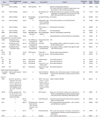
Understanding the genetic architecture of CAKUT is difficult due to the phenotypic heterogeneity and multifactorial genetic penetrance [9]. Until recently, more than 20 human single-gene causes for CAKUT have been identified [10111213]. Understanding the genetic basis of CAKUT can be helpful to know about the kidney development. Kidney development consists of the following developmental stages: ureteric budding, metanephric kidney, mesenchymalto-epithelial transition (MET), and nephron patterning and elongation (which include proximal and distal tubule morphogenesis and glomerulogenesis) [11415161718]. These developmental stages are controlled by a large number of genes and signaling pathways and defects in these steps can lead to the clinical phenotypes of CAKUT (Table 1).

“BUD THEORY” RELATED GENES OF VUR AND CAKUT

VUR can be induced by multiple birth defects affecting the urinary tract development: ureteric budding, ureter differentiation and elongation, peristalsis, UVJ formation, and bladder and urethra development [19]. The ectopic ureteric orifice is related to “Bud theory” stage, which is abnormal development of the embryonic ureteric bud [20]. This theory hypothesizes that the site at which the ureteric bud grows out from the mesonephric duct is essential for development of normal kidney and urinary tract. Actually, many early development genes involved in the ureteric budding stage lead to VUR and CAKUT. This hypothesis is closely correlated to analysis of duplex kidneys which showed that a more severe hypoplasia and dysplasia were closely associated with mal-displacement of the ureteral orifice.
In fact, ureteric budding is related to signaling mechanisms including transcription factors such as RET, PAX2, EYA1, SALL1, SIX1, SIX2, BMP4, and GATA3 [2122232425262728293031323334353637]. They have been recognized to be mutated with CAKUT and present several extrarenal manifestations [2122232425262728293031323334353637].
The RET proto-oncogene provides instructions for producing a protein that is involved in signaling within cells and mutations in the RET gene cause several diseases [38]. For example, translocations in the gene were correlated with papillary thyroid cancer [21] and multiple endocrine neoplasia (MEN) 2A and 2B syndromes [22]. RET mutation is also frequently showed in humans with renal diseases, because ureteric budding is promoted through receptor RET by Glial cell-derived neurotrophic factor (GDNF) signaling [23]. Therefore, RET mutations were subsequently reported to develop CAKUT in fetuses with bilateral renal hypodysplasia/agenesis [24].
Paired box gene 2 (PAX2) plays a key role in the development and proliferation of multiple cell lines and development of organs [39]. Therefore, PAX2 mutations in kidney can develop some disease spectrum of CAKUT, including VUR, renal hypodysplasia, renal cysts, and multicystic dysplastic kidneys [25].
Eyes absent homolog 1 (EYA1) encodes the eyes absent (EYA) family of proteins. The protein may play important roles in the development of eye, ear, branchial arches and kidney. EYA1 mutations have been related with branchiootic (BO) syndrome and congenital cataracts [2627].
Sal-like 1 (SALL1) is one of the human orthologues of the spalt (sal) gene known in Drosophila [40]. SALL1 mutations are a cause of Townes-Brocks syndrome (TBS) and branchio-oto-renal (BOR) syndrome [2829]. TBS is an autosomal dominant syndrome containing following features: renal abnormalities (multicystic kidneys, dysplastic kidney, hypoplastic kidneys), abnormalities of the external ears (dysplastic ears, sensorineural hearing loss or deafness), anorectal malformations (rectovaginal fistula, anal stenosis, imperforate anus/absence of an anal opening), heart abnormalities (ventricular septum defects and Tetralogy of Fallot) and hand and foot abnormalities (hypoplastic or fingerlike thumbs, syndactyly, fusion of the wrist bones, overlapping foot and/or toe bones) [2829].
SIX1 and SIX2 genes are similar genes each other known as the SIX gene family. Genes in this family encode proteins that bind to DNA and control the activity of other genes [3031]. When the SIX1 is mutated, it impairs the normal development of tissues at fetal stage and develops the major signs and symptoms of BOR/BO syndrome similar to phenotypes resulting from EYA1 or SALL1 mutations [3031].
Bone morphogenetic protein 4 (BMP4) produces a member of the transforming growth factor-β superfamily protein [32]. Some studies hypothesize about the role of BMP4 that it implicates in several aspects of embryonic development by regulating cell proliferation, differentiation, and apoptosis [323334]. BMP4 is further continuously expressed beyond the stage of ureteric budding throughout the embryonic development of the kidney and urinary system [323334].
Trans-acting T-cell-specific transcription factor GATA-3 (GATA3) gene encodes a transcription factor of the Gata protein family that is expressed in the ureteric buds. It is regulated by the PAX2 gene and involved in the Wolffian duct morphogenesis [35]. GATA3 mutation can cause hydroureter, ectopic ureteric budding, vas deferens hyperplasia, duplex kidney, uterine agenesis as well as Hypoparathyroidism, sensorineural Deafness, and Renal disease syndrome (including renal dysplasia, unilateral kidney agenesis, and VUR) [3637].

THE METANEPHRIC KIDNEY RELATED GENES

The metanephric kidney is formed after interaction between the ureteric bud and the metanephric mesenchyme. It starts at 4 weeks of gestation in humans, and it is regulated by Slit homolog 2 (SLIT2) and Roundabout homolog 2 (ROBO2) when the genes are expressed as a ligand and transmembrane receptor [41]. SLIT2-ROBO2 signaling has been shown to play a role after initiating ureteric budding [41].
ROBO2 encodes a protein as an “immunoglobulin receptor” for the Slit protein and functions in axon guidance and cell migration. ROBO2 mutations are related with VUR [41].
SLIT2 encodes Slit protein which is a member of secreted glycoproteins. This protein acts as “ligands” for the Robo family. SLIT2 may associated with guiding commissural axons in the forebrain by acting as a repulsive signal preventing inappropriate midline crossing of axons projecting from the olfactory bulb [42]. Therefore, SLIT2 can affect the formation and maintenance in the nervous system [43]. In the uretero-renal system, SLIT2 mutations also can cause duplicated collecting system, unilateral renal agenesis, and cystic dysplastic kidneys [4144].
SLIT-ROBO Rho GTPase-activating protein 1 (SRGAP1), as the gene name shows, is a small GTPase activating protein associated with the pathway mediating the repulsive signaling of Robo and Slit proteins in the cell migration process [45]. In 2015, Hwang et al. [44] identified 2 heterozygous mutations in SRGAP1 in 2 unrelated families and described it as a novel monogenic candidate gene as causes of CAKUT in humans.

GENES INVOLVED IN OTHER DEVELOPMENTAL STAGES

After metanephric kidney stage, there are 3 steps left to complete the whole kidney development. Several genes involved in CAKUT have been described for the following process: (1) WNT4 and FGF20 for MET, (2) AGT, REN, ACE, and AGTR1 for renin-angiotensin system, and (3) Uromodulin (UMOD) for nephron patterning and elongation.

1. WNT4 and FGF20 for MET

WNT oncogene analog 4 (WNT4) is on chromosome 1 and structurally related to secreted signaling proteins. WNT4 regulates kidney tubule induction and the MET within the WNT signaling pathway [141546]. Because it influences both the cortical and medullary stroma during development, mutations of WNT4 can cause kidney malformation related to Müllerian aplasia and hyperandrogenism [47]. WNT4 is activated by BMP4, a known smooth muscle differentiation factor, therefore, the absence of WNT4 can result in reduced smooth muscle [48].
Fibroblast growth factor (FGF) family possesses broad mitogenic and cell survival activities, and is associated with embryonic development, cell growth, morphogenesis, and tissue repair [49]. Especially FGF20 is an important key player in MET and can cause renal hypodysplasia or aplasia [50].

2. AGT, REN, ACE, and AGTR1 for renin-angiotensin system

Renal tubular dysgenesis, one of important phenotypes of CAKUT, is manifested by anuria, hypotension, and oligohydramnios, which eventually lead to Potter syndrome [51]. Several gene mutations of the renin-angiotensin system have been related to the distinct severe phenotype of CAKUT of renal tubular dysgenesis: Angiotensinogen (AGT), renin (REN), angiotensin-converting enzyme (ACE), and angiotensin II receptor type 1 (AGTR1) [52]. Gribouval et al. [53] analyzed 48 cases of renal tubular dysgenesis, and mutations in ACE accounted for 64.4%. Mutations in REN, AGT, and AGTR1 were seen in 20.8%, 8.3%, and 6.3% of cases, respectively.

3. Uromodulin (UMOD) for nephron patterning and elongation

The Uromodulin (UMOD) gene encodes the Tamm-Horsfall protein, which is the most abundant urinary protein in humans, and is related to the nephron patterning and elongation. Its mutation causes glomerulocystic kidney disease, familial juvenile hyperuricemic nephropathy, medullary cystic kidney disease type 2 [5455].
Hepatocyte nuclear factor 1B (HNF1B) is a homeodomain-containing transcription factor for embryogenesis of the liver, pancreas, and very early developmental stage of kidney which is expressed in the Wolffian duct [56]. HNF1B mutations have been reported as the cause of the Renal Cysts and Diabetes Syndrome and reported as CAKUT spectrum malformations, such as single kidney, multicystic dysplastic kidney, renal hypodysplasia, cystic kidney disease, and autosomal recessive polycystic kidney disease (ARPKD) [575859].

OTHER CAKUT ASSOCIATED GENES WITH UNKNOWN RELATIONSHIP OF KIDNEY DEVELOPMENT

Not only genes described above, following genes are also known as a leading cause of CAKUT in several studies: TRAP1, PKHD1, KAL1, HOXA13, and NIPBL. Their pathogenesis of CAKUT especially associated with kidney development process, however, are not clearly revealed.
Tumor Necrosis Factor Receptor-associated Protein 1 (TRAP1) encodes a mitochondrial chaperone protein that is member of the heat shock protein 90 family. The protein has ATPase activity and may function in regulating cellular stress responses [60]. Mutations of TRAP1 can develop many kinds of CAKUT phenotypes or CAKUT in VACTERL syndrome (combination of at least three of the following congenital anomalies: vertebral defects (V), anorectal malformations (A), cardiac defects (C), tracheoesophageal fistula with or without esophageal atresia (TE), renal malformations (R), and limb defects (L) [61].
Polycystic kidney and hepatic disease 1 (PKHD1) provides instructions for making a protein called fibrocystin. Fibrocystin exists on the cell membrane of kidney cells as a receptor and interacts with molecules and signals outside of the cell [62]. More than 270 mutations in the PKHD1 gene have been identified in human with autosomal recessive polycystic kidney disease (ARPKD) [59].
Kallman syndrome 1 (KAL1) gene encodes a protein called anosmin-1. Anosmin-1 is expressed in the fetal certain regions of the brain, respiratory tract, digestive system and kidneys [63]. But the precise function of anosmin-1 in kidney is not well known. It is also unclear how KAL1 gene mutations lead to signs of Kallmann syndrome, including unilateral renal agenesis [6465].
Homeobox protein Hox-A13 (HOXA13) is a part of homeobox gene family, which appears to play a key role for the formation and development of the limbs (particularly the hands and feet), urinary tract, and reproductive system. At least 14 HOXA13 gene mutations have been found in hand-foot-genital syndrome, which can have features of VUR [6667].
Nipped-B-like protein (NIPBL) makes a protein named delangin, which helps the activity of chromosomes during cell division and controls human development [68]. NIPBL gene mutations have been identified in people with Cornelia de Lange syndrome which is a developmental disorder affecting many parts of the body. In this syndrome, sometimes VUR is also found [6970].

EFFORTS TO FIND OUT ADDITIONAL CAKUT RELATED GENES

Nowadays, there are many attempts and technological developments to find out additional CAKUT related genes: linkage studies, targeted next-generation sequencing (NGS), copy number variation (CNV) analysis, whole genome sequencing (WGS) and genome-wide association studies (GWAS).
Linkage analyses have been successful for mapping genetic markers by analyzing a disease in a family-based approach in insulin-dependent diabetes mellitus, Alzheimer disease, breast cancer [717273]. The majority of published linkage studies on CAKUT have been about familial VUR, which occurs in ~1% of children [74]. However, linkage analysis is not sufficient tool to detect small effect sized loci [9].
Analysis of multiple genes using NGS in patients with CAKUT has demonstrated that <10% patients with CAKUT carry variants in previously implicated genes, such as HNF1B, PAX2, EYA1, SIX5, and RET. Therefore, the causative genes are not yet identified in most of CAKUT cases even though NGS technique was applied [757677].
Another aspect of CAKUT is CNVs [78]. It can be identified by high-resolution microarrays, such as the array comparative genomic hybridization (array-CGH) and single nucleotide polymorphism (SNP) microarrays [79].
Currently, genetic studies in unexplained diseases are shifting from whole exome sequencing or CNV analysis to WGS [9]. Also, GWAS, which is known as a high-throughput genotyping technologies, makes the rapid genotyping of >1 million SNPs throughout the whole genome. GWAS can also identify associated loci that confer risk of CAKUT with large sample size [8081].

CONCLUSIONS

Because morbidity of CAKUT may not manifest until later in life, early detection of these patients is important. CAKUT is a genetically heterogeneous group of disorders that are caused by mutations in genes related to the kidney development process. The malformation phenotypes due to gene mutations diverse from normally structured kidneys with intact kidney function to severe hypodysplasia and end stage renal failure. Although primary VUR is one of the most common phenotype of CAKUT, its prevalence can be underestimated until it shows symptoms such as fever.
However, some studies have pointed that CAKUT always has to be considered when the patient develops small phenotypes of anomalies related to urinary tract and kidney [910111213]. The advancement in sequencing and bioinformatics technologies will show us the additional CAKUT-related genes and more relevant etiologies of disease entities than can be identified by imaging study tools or histopathology alone. Newly advancing bioinformatics technologies will show us the additional CAKUT-related genes and more relevant etiologies of disease entities. Moreover, these genetic findings will provide an opportunity to develop an ideal diagnostic CAKUT genetic test in clinical practice to facilitate early diagnosis, better management of the disease, and familial genetic counselling [9]. When the price of GWAS gets lower than that of these days and it becomes more popular, we will be able to apply genetic tests to suspected patients. Using these new tools, we could identify the patients with high risks of having the deformities in kidneys before it is too late. Understanding of CAKUT and VUR genetic bases will help the management of this condition in children.

Figures and Tables

Table 1

Human genes implicated in CAKUT

icu-58-S4-i001
Gene Kidney developmental stage Location Signals Gene product Disease Inheritance type #Gene OMIM Reference number
Ureteric budding
RET Ureteric budding 10p13 GDNF signaling RTK Bilateral renal hypodysplasia/agenesis AR 191830 23,82
VUR, ureteral obstruction, megaureter, duplex kidney, renal abnormalities, as well as Hirschsprung's disease
PAX2 Ureteric budding 10q24.31 (-) PAX (paired box domain) Papillorenal syndrome that comprises renal defects (oligomeganephronia, hypodysplasia, VUR, and cysts) and ocular defects affecting the optic nerve and/or the retina AD 120330 39,83
EYA1 Ureteric budding 8q13.3 RTK signaling pathway Eyes absent homolog 1 BO syndrome AD 602588 26,27
SALL1 Ureteric budding 16q12.1 (-) Sal-like protein 1 (also known as spalt-like transcription factor 1) Townes-Brocks syndrome, as well as BOR syndrome AD 107480 28,29
SIX1 Ureteric budding 14q23.1 (-) SIX homeobox 1, 2 and 5 BOR syndrome, BO syndrome AD 608389 30
SIX2 Ureteric budding 2p21 WNT-pathway SIX homeobox 2 Renal hypodysplasia NA 604994 31
BMP4 Ureteric budding 14q22.2 BMP-BMPR signaling system Bone morphogenetic protein 4 Syndromic microphthalmia, orofacial cleft AD 112262 31
GATA3 Ureteric budding 10p14 (-) (-) Hypoparathyroidism, sensorineural deafness, renal dysplasia AD 146255 84
Development of the metanephric kidney
ROBO2 Development of the metanephric kidney 3p12.3 SLIT2-ROBO2 signaling pathway Receptor for the Slit2 ligand VUR NA 610878 41
SLIT2 Development of the metanephric kidney 4p15.31 SLIT2-ROBO2 signaling pathway Slit protein Cystic dysplastic kidneys, unilateral renal agenesis, and duplicated collecting system NA 603746 41,44
SRGAP1 Development of the metanephric kidney 12q14.2 SLIT2-ROBO2 signaling pathway SRGAP1(small GTPase activating protein) Cystic dysplastic kidneys, unilateral renal agenesis, duplicated collecting system NA 606523 44
MET
WNT4 MET 1p36.12 WNT-pathway Protein Wnt-4 Müllerian aplasia, hyperandrogenism AD 158330 47
FGF20 MET 8p22 (-) Fibroblast growth factor 20 Renal hypodysplasia/aplasia AR 605558 50
RAS
AGT RAS 1q42.2 RAS pathway Angiotensinogen Renal tubular dysgenesis AR 106150 53
REN RAS 1q32.1 RAS pathway Renin Renal tubular dysgenesis AR 179820 53
ACE RAS 17q23.3 RAS pathway Angiotensin-converting enzyme Renal tubular dysgenesis AR 106180 53
AGTR1 RAS 3q24 RAS pathway Angiotensin II receptor type 1 Renal tubular dysgenesis AR 106165 52
Nephron patterning and elongation
Uromodulin (UMOD) Nephron patterning and elongation 16p12.3 (-) Tamm-Horsfall protein (uromodulin) Medullary cystic kidney disease type 2, familial juvenile hyperuricemic nephropathy, and glomerulocystic kidney disease NA 191845 54,55
Unknown
TRAP1 Unknown (possibly antiapoptotic and endoplasmic reticulum-stress signaling in proximal tubules and in thick medullary ascending limbs of Henle's loop.) 16p13.3 (-) TRAP1 heat shock protein 75 (also known as TNF receptor associated protein 1-mitochondrial HSP90 protein) CAKUT or CAKUT in VACTERL NA 606219 61
HNF1B Unknown (possibly wolffian duct from a very early developmental stage of the kidney) 17q12 (-) HNF1B protein Renal cysts and diabetes syndrome also referred to as MODY5, renal hypodysplasia, multicystic dysplastic kidney, cystic kidney disease, single kidney, oligomeganephronia AD 189907 83
PKHD1 (-) 6p12.3-p12.2 (-) Protein fibrocystin Autosomal recessive polycystic kidney disease, polycystic kidney and hepatic disease AR 606702 59
KAL1 (-) Xp22.31 FGF signaling, that there is an interaction between anosmin-1 and FGFR1 Anosmin 1 VUR, hypogonadotropic hypogonadism 1 with or without anosmia (Kallmann syndrome) XL 300836 64,65
HOXA13 (-) 7p15.2 (-) Homeobox protein VUR, hand-foot-genital syndrome, Guttmacher syndrome(preaxial deficiency, postaxial polydactyly, and hypospadias) AD 142959 67
NIPBL (-) 5p13.2 (-) Delangin VUR, Cornelia de Lange syndrome AD 608667 68

CAKUT, congenital anomalies of the kidney and urinary tract; OMIM, Online-Mendelian inheritance in man; GDNF, Glial cell line-derived neurotrophic factor; RTK, receptor tyrosine kinase; BO, branchio-otic; BOR, branchio-oto-renal; WNT, wingless and integration site growth factor; VUR, vesicoureteral reflux; AR, autosomal recessive; AD, autosomal dominant; NA, not applicable; MET, mesenchymal-to-epithelial transition; RAS, renin-angiotensin system; VACTERL, combination of at least three of the following congenital anomalies: vertebral defects (V), anorectal malformations (A), cardiac defects (C), tracheoesophageal fistula with or without esophageal atresia (TE), renal malformations (R), and limb defects (L); TNF, tumor necrosis factor; HSP, heat shock protein; FGF, fibroblast growth factor; FGFR1, FGF receptor 1; MODY, maturity onset diabetes of the young; (-): unknown.

Notes

CONFLICTS OF INTEREST The authors have nothing to disclose.

References

1. Schedl A. Renal abnormalities and their developmental origin. Nat Rev Genet. 2007; 8:791–802.
2. Loane M, Dolk H, Kelly A, Teljeur C, Greenlees R, Densem J, et al. Paper 4: EUROCAT statistical monitoring: identification and investigation of ten year trends of congenital anomalies in Europe. Birth Defects Res A Clin Mol Teratol. 2011; 91:Suppl 1. S31–S43.
3. North American Pediatric Renal Transplant Cooperative Study (NAPRTCS). 2008 Annual report. Rockville (MD): The EMMES Corporation;2008.
4. Agostiniani R, Mariotti P. The natural history of vesicoureteral reflux. J Matern Fetal Neonatal Med. 2011; 24:Suppl 1. 2–3.
5. Cendron M. Reflux nephropathy. J Pediatr Urol. 2008; 4:414–421.
6. Peters C, Rushton HG. Vesicoureteral reflux associated renal damage: congenital reflux nephropathy and acquired renal scarring. J Urol. 2010; 184:265–273.
7. Coulthard MG, Keir MJ. Reflux nephropathy in kidney transplants, demonstrated by dimercaptosuccinic acid scanning. Transplantation. 2006; 82:205–210.
8. Toffolo A, Ammenti A, Montini G. Long-term clinical consequences of urinary tract infections during childhood: a review. Acta Paediatr. 2012; 101:1018–1031.
9. Nicolaou N, Renkema KY, Bongers EM, Giles RH, Knoers NV. Genetic, environmental, and epigenetic factors involved in CAKUT. Nat Rev Nephrol. 2015; 11:720–731.
10. Yosypiv IV. Congenital anomalies of the kidney and urinary tract: a genetic disorder? Int J Nephrol. 2012; 2012:909083.
11. Chen F. Genetic and developmental basis for urinary tract obstruction. Pediatr Nephrol. 2009; 24:1621–1632.
12. Renkema KY, Winyard PJ, Skovorodkin IN, Levtchenko E, Hindryckx A, Jeanpierre C, et al. Novel perspectives for investigating congenital anomalies of the kidney and urinary tract (CAKUT). Nephrol Dial Transplant. 2011; 26:3843–3851.
13. Sanna-Cherchi S, Caridi G, Weng PL, Scolari F, Perfumo F, Gharavi AG, et al. Genetic approaches to human renal agenesis/hypoplasia and dysplasia. Pediatr Nephrol. 2007; 22:1675–1684.
14. Dressler GR. Advances in early kidney specification, development and patterning. Development. 2009; 136:3863–3874.
15. Reidy KJ, Rosenblum ND. Cell and molecular biology of kidney development. Semin Nephrol. 2009; 29:321–337.
16. Faa G, Gerosa C, Fanni D, Monga G, Zaffanello M, Van Eyken P, et al. Morphogenesis and molecular mechanisms involved in human kidney development. J Cell Physiol. 2012; 227:1257–1268.
17. Vainio S, Lin Y. Coordinating early kidney development: lessons from gene targeting. Nat Rev Genet. 2002; 3:533–543.
18. Ichikawa I, Kuwayama F, Pope JC 4th, Stephens FD, Miyazaki Y. Paradigm shift from classic anatomic theories to contemporary cell biological views of CAKUT. Kidney Int. 2002; 61:889–898.
19. Nino F, Ilari M, Noviello C, Santoro L, Rätsch IM, Martino A, et al. Genetics of vesicoureteral reflux. Curr Genomics. 2016; 17:70–79.
20. Mackie GG, Stephens FD. Duplex kidneys: a correlation of renal dysplasia with position of the ureteral orifice. J Urol. 1975; 114:274–280.
21. Grieco M, Santoro M, Berlingieri MT, Melillo RM, Donghi R, Bongarzone I, et al. PTC is a novel rearranged form of the ret proto-oncogene and is frequently detected in vivo in human thyroid papillary carcinomas. Cell. 1990; 60:557–563.
22. Hofstra RM, Landsvater RM, Ceccherini I, Stulp RP, Stelwagen T, Luo Y, et al. A mutation in the RET proto-oncogene associated with multiple endocrine neoplasia type 2B and sporadic medullary thyroid carcinoma. Nature. 1994; 367:375–376.
23. Skinner MA, Safford SD, Reeves JG, Jackson ME, Freemerman AJ. Renal aplasia in humans is associated with RET mutations. Am J Hum Genet. 2008; 82:344–351.
24. Jeanpierre C, Macé G, Parisot M, Morinière V, Pawtowsky A, Benabou M, et al. RET and GDNF mutations are rare in fetuses with renal agenesis or other severe kidney development defects. J Med Genet. 2011; 48:497–504.
25. Bower M, Salomon R, Allanson J, Antignac C, Benedicenti F, Benetti E, et al. Update of PAX2 mutations in renal coloboma syndrome and establishment of a locus-specific database. Hum Mutat. 2012; 33:457–466.
26. Weber S, Moriniere V, Knüppel T, Charbit M, Dusek J, Ghiggeri GM, et al. Prevalence of mutations in renal developmental genes in children with renal hypodysplasia: results of the ESCAPE study. J Am Soc Nephrol. 2006; 17:2864–2870.
27. Abdelhak S, Kalatzis V, Heilig R, Compain S, Samson D, Vincent C, et al. A human homologue of the Drosophila eyes absent gene underlies branchio-oto-renal (BOR) syndrome and identifies a novel gene family. Nat Genet. 1997; 15:157–164.
28. Yoshida Y, Yamagishi S, Matsui T, Nakamura K, Imaizumi T, Yoshimura K, et al. Increased levels of pigment epithelium-derived factor in aqueous humor of patients with uveitis. Br J Ophthalmol. 2007; 91:149–150.
29. Kohlhase J, Wischermann A, Reichenbach H, Froster U, Engel W. Mutations in the SALL1 putative transcription factor gene cause Townes-Brocks syndrome. Nat Genet. 1998; 18:81–83.
30. Ruf RG, Xu PX, Silvius D, Otto EA, Beekmann F, Muerb UT, et al. SIX1 mutations cause branchio-oto-renal syndrome by disruption of EYA1-SIX1-DNA complexes. Proc Natl Acad Sci U S A. 2004; 101:8090–8095.
31. Weber S, Taylor JC, Winyard P, Baker KF, Sullivan-Brown J, Schild R, et al. SIX2 and BMP4 mutations associate with anomalous kidney development. J Am Soc Nephrol. 2008; 19:891–903.
32. von Bubnoff A, Cho KW. Intracellular BMP signaling regulation in vertebrates: pathway or network? Dev Biol. 2001; 239:1–14.
33. Hogan BL. Bone morphogenetic proteins in development. Curr Opin Genet Dev. 1996; 6:432–438.
34. Miyazaki Y, Oshima K, Fogo A, Ichikawa I. Evidence that bone morphogenetic protein 4 has multiple biological functions during kidney and urinary tract development. Kidney Int. 2003; 63:835–844.
35. Grote D, Souabni A, Busslinger M, Bouchard M. Pax 2/8-regulated Gata 3 expression is necessary for morphogenesis and guidance of the nephric duct in the developing kidney. Development. 2006; 133:53–61.
36. Grote D, Boualia SK, Souabni A, Merkel C, Chi X, Costantini F, et al. Gata3 acts downstream of beta-catenin signaling to prevent ectopic metanephric kidney induction. PLoS Genet. 2008; 4:e1000316.
37. Benetti E, Murer L, Bordugo A, Andreetta B, Artifoni L. 10p12.1 deletion: HDR phenotype without DGS2 features. Exp Mol Pathol. 2009; 86:74–76.
38. Alberti L, Carniti C, Miranda C, Roccato E, Pierotti MA. RET and NTRK1 proto-oncogenes in human diseases. J Cell Physiol. 2003; 195:168–186.
39. Eccles MR, He S, Legge M, Kumar R, Fox J, Zhou C, et al. PAX genes in development and disease: the role of PAX2 in urogenital tract development. Int J Dev Biol. 2002; 46:535–544.
40. Jürgens G. Head and tail development of the Drosophila embryo involves spalt, a novel homeotic gene. EMBO J. 1988; 7:189–196.
41. Grieshammer U, Le Ma, Plump AS, Wang F, Tessier-Lavigne M, Martin GR. SLIT2-mediated ROBO2 signaling restricts kidney induction to a single site. Dev Cell. 2004; 6:709–717.
42. Niclou SP, Jia L, Raper JA. Slit2 is a repellent for retinal ganglion cell axons. J Neurosci. 2000; 20:4962–4974.
43. Itoh A, Miyabayashi T, Ohno M, Sakano S. Cloning and expressions of three mammalian homologues of Drosophila slit suggest possible roles for Slit in the formation and maintenance of the nervous system. Brain Res Mol Brain Res. 1998; 62:175–186.
44. Hwang DY, Kohl S, Fan X, Vivante A, Chan S, Dworschak GC, et al. Mutations of the SLIT2-ROBO2 pathway genes SLIT2 and SRGAP1 confer risk for congenital anomalies of the kidney and urinary tract. Hum Genet. 2015; 134:905–916.
45. Li X, Chen Y, Liu Y, Gao J, Gao F, Bartlam M, et al. Structural basis of Robo proline-rich motif recognition by the srGAP1 Src homology 3 domain in the Slit-Robo signaling pathway. J Biol Chem. 2006; 281:28430–28437.
46. Park JS, Ma W, O'Brien LL, Chung E, Guo JJ, Cheng JG, et al. Six2 and Wnt regulate self-renewal and commitment of nephron progenitors through shared gene regulatory networks. Dev Cell. 2012; 23:637–651.
47. Vivante A, Mark-Danieli M, Davidovits M, Harari-Steinberg O, Omer D, Gnatek Y, et al. Renal hypodysplasia associates with a WNT4 variant that causes aberrant canonical WNT signaling. J Am Soc Nephrol. 2013; 24:550–558.
48. Itäranta P, Chi L, Seppänen T, Niku M, Tuukkanen J, Peltoketo H, et al. Wnt-4 signaling is involved in the control of smooth muscle cell fate via Bmp-4 in the medullary stroma of the developing kidney. Dev Biol. 2006; 293:473–483.
49. Koga C, Adati N, Nakata K, Mikoshiba K, Furuhata Y, Sato S, et al. Characterization of a novel member of the FGF family, XFGF-20, in Xenopus laevis. Biochem Biophys Res Commun. 1999; 261:756–765.
50. Barak H, Huh SH, Chen S, Jeanpierre C, Martinovic J, Parisot M, et al. FGF9 and FGF20 maintain the stemness of nephron progenitors in mice and man. Dev Cell. 2012; 22:1191–1207.
51. Uematsu M, Sakamoto O, Nishio T, Ohura T, Matsuda T, Inagaki T, et al. A case surviving for over a year of renal tubular dysgenesis with compound heterozygous angiotensinogen gene mutations. Am J Med Genet A. 2006; 140:2355–2360.
52. Gribouval O, Gonzales M, Neuhaus T, Aziza J, Bieth E, Laurent N, et al. Mutations in genes in the renin-angiotensin system are associated with autosomal recessive renal tubular dysgenesis. Nat Genet. 2005; 37:964–968.
53. Gribouval O, Morinière V, Pawtowski A, Arrondel C, Sallinen SL, Saloranta C, et al. Spectrum of mutations in the renin-angiotensin system genes in autosomal recessive renal tubular dysgenesis. Hum Mutat. 2012; 33:316–326.
54. Tamm I, Horsfall FL Jr. Characterization and separation of an inhibitor of viral hemagglutination present in urine. Proc Soc Exp Biol Med. 1950; 74:106–108.
55. Hart TC, Gorry MC, Hart PS, Woodard AS, Shihabi Z, Sandhu J, et al. Mutations of the UMOD gene are responsible for medullary cystic kidney disease 2 and familial juvenile hyperuricaemic nephropathy. J Med Genet. 2002; 39:882–892.
56. Coffinier C, Thépot D, Babinet C, Yaniv M, Barra J. Essential role for the homeoprotein vHNF1/HNF1beta in visceral endoderm differentiation. Development. 1999; 126:4785–4794.
57. Lindner TH, Njolstad PR, Horikawa Y, Bostad L, Bell GI, Sovik O. A novel syndrome of diabetes mellitus, renal dysfunction and genital malformation associated with a partial deletion of the pseudo-POU domain of hepatocyte nuclear factor-1beta. Hum Mol Genet. 1999; 8:2001–2008.
58. Edghill EL, Bingham C, Ellard S, Hattersley AT. Mutations in hepatocyte nuclear factor-1beta and their related phenotypes. J Med Genet. 2006; 43:84–90.
59. Hiesberger T, Bai Y, Shao X, McNally BT, Sinclair AM, Tian X, et al. Mutation of hepatocyte nuclear factor-1beta inhibits Pkhd1 gene expression and produces renal cysts in mice. J Clin Invest. 2004; 113:814–825.
60. Felts SJ, Owen BA, Nguyen P, Trepel J, Donner DB, Toft DO. The hsp90-related protein TRAP1 is a mitochondrial protein with distinct functional properties. J Biol Chem. 2000; 275:3305–3312.
61. Saisawat P, Kohl S, Hilger AC, Hwang DY, Yung Gee H, Dworschak GC, et al. Whole-exome resequencing reveals recessive mutations in TRAP1 in individuals with CAKUT and VACTERL association. Kidney Int. 2014; 85:1310–1317.
62. Onuchic LF, Furu L, Nagasawa Y, Hou X, Eggermann T, Ren Z, et al. PKHD1, the polycystic kidney and hepatic disease 1 gene, encodes a novel large protein containing multiple immunoglobulin-like plexin-transcription-factor domains and parallel beta-helix 1 repeats. Am J Hum Genet. 2002; 70:1305–1317.
63. Choy C, Kim SH. Biological actions and interactions of anosmin-1. Front Horm Res. 2010; 39:78–93.
64. Duke V, Quinton R, Gordon I, Bouloux PM, Woolf AS. Proteinuria, hypertension and chronic renal failure in X-linked Kallmann's syndrome, a defined genetic cause of solitary functioning kidney. Nephrol Dial Transplant. 1998; 13:1998–2003.
65. Hardelin JP, Levilliers J, del Castillo I, Cohen-Salmon M, Legouis R, Blanchard S, et al. X chromosome-linked Kallmann syndrome: stop mutations validate the candidate gene. Proc Natl Acad Sci U S A. 1992; 89:8190–8194.
66. Goodman FR, Bacchelli C, Brady AF, Brueton LA, Fryns JP, Mortlock DP, et al. Novel HOXA13 mutations and the phenotypic spectrum of hand-foot-genital syndrome. Am J Hum Genet. 2000; 67:197–202.
67. Bower M, Eccles M, Heidet L, Schimmenti LA. Clinical utility gene card for: renal coloboma (Papillorenal) syndrome. Eur J Hum Genet. 2011; 19.
68. Tonkin ET, Wang TJ, Lisgo S, Bamshad MJ, Strachan T. NIPBL, encoding a homolog of fungal Scc2-type sister chromatid cohesion proteins and fly Nipped-B, is mutated in Cornelia de Lange syndrome. Nat Genet. 2004; 36:636–641.
69. Ramos FJ, Puisac B, Baquero-Montoya C, Gil-Rodríguez MC, Bueno I, Deardorff MA, et al. Clinical utility gene card for: Cornelia de Lange syndrome. Eur J Hum Genet. 2015; 23.
70. Jackson L, Kline AD, Barr MA, Koch S. de Lange syndrome: a clinical review of 310 individuals. Am J Med Genet. 1993; 47:940–946.
71. Coon KD, Myers AJ, Craig DW, Webster JA, Pearson JV, Lince DH, et al. A high-density whole-genome association study reveals that APOE is the major susceptibility gene for sporadic late-onset Alzheimer's disease. J Clin Psychiatry. 2007; 68:613–618.
72. Julier C, Hyer RN, Davies J, Merlin F, Soularue P, Briant L, et al. Insulin-IGF2 region on chromosome 11p encodes a gene implicated in HLA-DR4-dependent diabetes susceptibility. Nature. 1991; 354:155–159.
73. Hall JM, Lee MK, Newman B, Morrow JE, Anderson LA, Huey B, et al. Linkage of early-onset familial breast cancer to chromosome 17q21. Science. 1990; 250:1684–1689.
74. Williams G, Fletcher JT, Alexander SI, Craig JC. Vesicoureteral reflux. J Am Soc Nephrol. 2008; 19:847–862.
75. Hwang DY, Dworschak GC, Kohl S, Saisawat P, Vivante A, Hilger AC, et al. Mutations in 12 known dominant disease-causing genes clarify many congenital anomalies of the kidney and urinary tract. Kidney Int. 2014; 85:1429–1433.
76. Saisawat P, Tasic V, Vega-Warner V, Kehinde EO, Günther B, Airik R, et al. Identification of two novel CAKUT-causing genes by massively parallel exon resequencing of candidate genes in patients with unilateral renal agenesis. Kidney Int. 2012; 81:196–200.
77. Chatterjee R, Ramos E, Hoffman M, VanWinkle J, Martin DR, Davis TK, et al. Traditional and targeted exome sequencing reveals common, rare and novel functional deleterious variants in RET-signaling complex in a cohort of living US patients with urinary tract malformations. Hum Genet. 2012; 131:1725–1738.
78. Weber S, Landwehr C, Renkert M, Hoischen A, Wühl E, Denecke J, et al. Mapping candidate regions and genes for congenital anomalies of the kidneys and urinary tract (CAKUT) by array-based comparative genomic hybridization. Nephrol Dial Transplant. 2011; 26:136–143.
79. Alkan C, Coe BP, Eichler EE. Genome structural variation discovery and genotyping. Nat Rev Genet. 2011; 12:363–376.
80. Köttgen A, Glazer NL, Dehghan A, Hwang SJ, Katz R, Li M, et al. Multiple loci associated with indices of renal function and chronic kidney disease. Nat Genet. 2009; 41:712–717.
81. Köttgen A, Pattaro C, Böger CA, Fuchsberger C, Olden M, Glazer NL, et al. New loci associated with kidney function and chronic kidney disease. Nat Genet. 2010; 42:376–384.
82. Emison ES, McCallion AS, Kashuk CS, Bush RT, Grice E, Lin S, et al. A common sex-dependent mutation in a RET enhancer underlies Hirschsprung disease risk. Nature. 2005; 434:857–863.
83. Madariaga L, Morinière V, Jeanpierre C, Bouvier R, Loget P, Martinovic J, et al. Severe prenatal renal anomalies associated with mutations in HNF1B or PAX2 genes. Clin J Am Soc Nephrol. 2013; 8:1179–1187.
84. Ali A, Christie PT, Grigorieva IV, Harding B, Van Esch H, Ahmed SF, et al. Functional characterization of GATA3 mutations causing the hypoparathyroidism-deafness-renal (HDR) dysplasia syndrome: insight into mechanisms of DNA binding by the GATA3 transcription factor. Hum Mol Genet. 2007; 16:265–275.
TOOLS
Similar articles