

 PDF
PDF ePub
ePub Citation
 Print
Print


INTRODUCTION
The lifetime prevalence of kidney stone disease is estimated at 1% to 15%, with the probability of having a stone varying according to age, gender, race, and geographic location [1]. Most patients with renal calculi of less than 1 cm can be treated satisfactorily with extracorporeal shock wave lithotripsy (ESWL) [2]. The lower efficacy of ESWL with increasing stone size, however, and the promising role of retrograde intrarenal surgery and mini-percutaneous nephrolithotomy have made ESWL a less popular choice for stones sized 1 to 2 cm. Many advancements and methods have been tried to increase the stone-free rate of ESWL for larger calculi. One such method is the use of double J (DJ) stents.
The insertion of DJ stents during ESWL for renal calculi is debatable. On one hand, some studies support a role for DJ stents in facilitating stone passage in a dilated ureter and also in preventing renal colic and steinstrasse, whereas other reports claim that stent presence causes significant lower urinary tract symptoms (LUTS), hematuria, urinary tract infection (UTI), and a lower stone-free rate [34].
The present study utilized a novel model in which a DJ stent was inserted at least 7 days before the ESWL and was then removed on the morning of the procedure, just prior to the procedure. In this way, the advantages of prior DJ stenting such as a dilated ureter favoring stone passage could be accessed along with adequate water-urine interface in the pelvis for effective fragmentation. Absence of stenting would also provide a better targeting of the stone and reduced stent-related LUTS. This model was then compared with nonstented and stented patients undergoing ESWL.
MATERIALS AND METHODS
1. Study population
The study included 88 adult patients with renal stone disease with stone size between 15 and 20 mm who presented during the study period from February 2013 to December 2015 at a single center. This prospective study was approved by the Ethics Committee of the R G Kar Medical College and Hospital (approval number: RGK/EC/13-14/551). Sample size was calculated by using G*Power version 3.1.9.2 (Heinrich Heine Universität Düsseldorf; http://www.gpower.hhu.de/en.html) by selecting ‘Goodness of fit test: contingency tables.’ Noncontrast computed tomography kidney-ureter-bladder (KUB) imaging was done routinely in all patients to assess stone size, location, density, and skin-stone distance. Patients with elevated creatinine (>1.5 mg% or 132.6 µmol/L), unresolved UTI, hydronephrosis, coagulopathy, morbid obesity (body mass index [BMI]>40 kg/m2), pregnancy, urinary tract anomalies, or stones elsewhere in the urinary tract were excluded from the study.
2. Randomization and procedures
After providing informed consent, patients were assigned to 1 of 3 treatment groups via the block randomization method (blocks with equal size of 9) with the help of Random Allocation Software (ver. 1.0.0). The first group received ESWL without any stenting. In the second group, DJ stenting was done 1 week before the ESWL and the procedure was accomplished with the DJ stent in situ. The stent was kept until the completion of 3 sittings, done 4 weeks apart, or it was removed earlier upon clearance of the stones. In the third group, DJ stenting was done 1 week before ESWL and the stent was removed on the morning of the day of the procedure.
Dornier compact sigma under fluoroscopic guidance was utilized for the lithotripsy. Voltage ramping was utilized in all cases. Detailed documentation of the procedure, including number of shocks, sittings, energy level, pain score during the procedure (0, no pain; 1, minimal pain; 2, mild pain; 3, moderate pain; 4, severe pain; 5, unbearable pain), and analgesic requirement after the procedure (in number of days), were noted. Follow-up KUB X-rays were done every 4 weeks after the session. A repeat session was given in case of persistent calculi, at 4-week intervals and to a maximum of 3 sessions. Complications were recorded by use of the modified Clavien-Dindo (MCD) classification.
3. Assessment and statistical analysis
Results of the procedure were measured in terms of fragmentation and clearance. Fragmentation was categorized as complete (<4-mm fragment), partial (>4-mm fragment), and no fragmentation (intact stone). This was reported immediately after the ESWL. Clearance was categorized as complete (no residual fragment), partial (clinical insignificant residual fragment <4 mm), and no clearance (>4-mm residual fragment). This was assessed at 4 weeks after the procedure. Final outcome was reported as either success or failure. The success group was defined by either complete clearance or a clinically insignificant residual fragment (CIRF) of less than 4 mm. Failure was defined as a residual fragment of more than 4 mm even after completion of 3 sittings of ESWL.
Descriptive analysis, chi-square test, and analysis of variance test were used with the help of SPSS ver. 16.0 (SPSS Inc., Chicago, IL, USA). Bonferroni adjustment was made for post hoc analysis. The level of statistical significance was kept at p<0.05 and the confidence interval was set at 95%.
RESULTS
1. Baseline characteristics
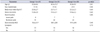
Most of the patients were in their 4th or 5th decade of life. The patients' mean age was 37.9±10.9 years (range, 20–67 years). The sex ratio was skewed slightly towards males (47 vs. 41). Most patients had a BMI between 18.5 and 28 kg/m2, with mean of 23.6±2.2 kg/m2. Symptom prevalence, sex ratio, BMI, and stone parameters among the 3 groups were comparable (Table 1).
2. Procedural details
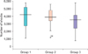
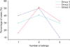
Group 3 received a fewer mean number of shocks (mean, 3,155) than did group 1 (mean, 3,859; p=0.05) or group 2 (mean, 3,872; p=0.04) (Fig. 1). All patients tolerated a similar voltage level during ESWL (p=0.06). Group 2 required a greater number of sittings (mean, 2.2) than did group 1 (mean, 2.0; p=0.39) or group 3 (mean, 1.7; p=0.01) (Fig. 2).
3. Stone fragmentation, clearance, and outcome
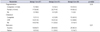
The compete stone fragmentation rate in groups 1, 2, and 3 was 18.5%, 16.1%, and 33.3%, respectively, whereas partial fragmentation was seen in 63%, 71%, and 63.3% of cases, respectively (p=0.24). The overall clearance rate (complete+CIRF) was higher in group 3 (83.3%) than in groups 1 (63%) and 2 (64.5%) (p=0.02). Successful outcome was found in 83.3% of cases in group 3, whereas in groups 1 and 2 this percentage was 66.7% and 64.5%, respectively (Table 2).
4. Effect of stone size, density, and location
When the groups were divided by stone size into 2 categories of 10–15 mm and 16–20 mm, the fragmentation rate was not significantly affected (p=0.59 and p=0.25, respectively). However, a better complete and overall (complete + partial) clearance rate was discovered in group 3 in the 10–15 mm subgroup (50% and 83%) than in group 1 (11.1% and 72.2%) or group 2 (10.0% and 65.0%) (p=0.02). In the large size subgroup, the clearance rate was similar (p=0.42). Outcome was not significantly affected by size categories (p=0.44 and p=0.35).
Stone density was categorized into 3 groups: <800 Hounsfield units (HU), 800–1,200 HU, and >1,200 HU. A higher rate of partial fragmentation was observed in the >1,200 HU subgroup in group 3 (3/3) than in group 1 (4/8) or group 2 (0/4) (p=0.03). A better clearance (complete + CIRF) was found in the 800–1,200 HU subgroup in group 3 (16/16) than in group 1 (5/9) or group 2 (9/14) (p=0.008). Similarly, a better outcome was discovered in the 800–1,200 HU subgroup in group 3 (100%) than groups 1 (66.7%) or 2 (64.3%) (p=0.03).
Stone location was categorized into 2 groups: lower pole (L) and nonlower pole (NL). The fragmentation rate was similar in the groups at both locations (p=0.19 and p=0.66). Clearance of lower pole calculi (complete + CIRF) was improved in group 3 (6/7), compared with group 1 (4/8) and group 2 (4/7) (p=0.03). Clearance in the NL group was similar (p=0.31). Higher successful outcome was found in group 3 in the L subgroup (85.7%) than in group 1 (50.0%) or group 2 (57.1%), but this was not statistically significant (p=0.32).
5. Effect of obesity
As most patients were within a BMI range of 18.5 to 28.0 kg/m2, they were divided into normal and overweight categories. No significant difference in fragmentation was observed in either group (p=0.06 and p=0.48). Clearance and outcome were better in the normal BMI population in group 3 (complete, 42.9%; CIRF, 47.6%; p=0.03).
6. Intraprocedural pain assessment and postprocedural analgesic need
Pain scores recorded during ESWL were similar among groups (mean: group 1, 2.6; group 2, 2.5; group 3, 2.4; p=0.75). However, the analgesic requirement after the procedure differed. Group 2 patients took analgesics for a longer time (3.2±2.0 days) than did patients in group 1 (1.9±1.5 days) or group 3 (1.7±1.5 days, p=0.00).
7. Complication rate
MCD grade I complications (requirement of analgesic, antipyretic, antiemetic, LUTS, or hematuria <48 hours) occurred in 67% of all patients and were distributed uniformly among all groups (p=0.87). Grade II complications were encountered in 6 cases. Two cases in group 1 and 1 case in group 2 developed a UTI and required intravenous antibiotics. One case each in group 1 and group 3 had hematuria for >48 hours and ethamsylate tablets (500 mg, 3 times a day) were prescribed. One case in group 3 had persisting vomiting, and intravenous fluids and antiemetic injections were administered.
Grade IIIa complications were seen in 3 cases. DJ stenting under local anesthesia was done in 1 case in group 1 for persisting pain and hydronephrosis. In another 2 cases in group 2, the DJ stent had to be removed owing to fever and severe LUTS. Grade IIIb complications developed in 2 cases (one each from groups 1 and 2) and required ureteroscopic lithotripsy for steinstrasse.
DISCUSSION
The role of ESWL as a prime modality for large renal calculi is being challenged by miniaturizing endourological procedures, even though ESWL remains a safe and effective noninvasive alternative. Its utilization for large calculi has been hampered by incomplete fragmentation, incomplete clearance, long duration of treatment, renal colic, steinstrasse, and a lower stone-free rate [5]. The success rate of this treatment modality is in the range of 60% to 90% in various series [678]. Shouman et al. [9] reported an 83.3% stone-free rate for renal stones >25 mm in children.
Mobley et al. [10], Thomas [11], and Mustafa and Ali-El-Dein [12] in their studies found that placement of a ureteral stent had no effect on stone-free rates or passage of stones at any ureteral location. In our study, overall fragmentation (complete+partial) was marginally better, but nonsignificant, in group 3 (96.6%) than in group 1 (81.5%, p=0.12) or group 2 (87.1%, p=0.16). A significant improvement in clearance was noted in group 3 (83.3%) compared with group 1 (63.0%, p=0.02) and group 2 (64.5%, p=0.02). Thus, removal of the stent in group 3 not only had a slightly favorable effect on fragmentation but also improved clearance in a dilated ureter.
In the subgroup analysis, improvement in clearance in group 3 was more evident in the 10–15 mm subgroup (p=0.02) than in the 16–20 mm group (p=0.42). Similarly, clearance (p=0.00) and outcome (p=0.03) were significantly better in the 800–1,200 HU density subgroup. A high clearance rate was also observed for lower pole calculi in group 3 (p=0.03). Group 3 patients also required a fewer number of shocks (p=0.04) and a fewer number of sittings (p=0.01) compared with other groups.
Pryor and Jenkins [13] and Ouzaid et al. [14] showed that the success rate is inferior if ESWL is given with a DJ stent because of poor localization and inferior fragmentation. Mohayuddin et al. [15] suggested that the stent does not alter the outcome of ESWL but increases the cost of treatment. Our study's final outcome, in terms of success or failure, was marginally better in group 3 (83.3%) than in group 1 (66.7%, p=0.14) or group 2 (64.5%, p=0.09), but this difference was not significant statistically.
Shen et al. [3] and El-Assmy et al. [16] found no improvement in the stone-free rate or requirement for auxiliary treatment with prior stenting. They also reported higher LUTS in the stented group. We also observed a significantly higher number of days of analgesic requirements in group 2 (stented) than in groups 1 and 3 (p=0.00). However, the presence of the stent did not make the ESWL more painful, as suggested by similar intraprocedural pain scores.
Chandhoke et al. [17] found fewer hospital readmissions and emergency room visits in the stented group during ESWL. A significant advantage of stenting for preventing steinstrasse was also suggested by Shen et al. [3]. However Bierkens et al. [18] reported significant complications in one-third of the stented group, including fever, pyelonephritis, and steinstrasse [18]. In our study, grades I and II complications were encountered in 67% and 6.8% of cases, respectively, and no significant difference in occurrence among groups was observed. However, group 2 patients had more grade IIIa (2/3) and IIIB (1/2) complications. A total of 3 cases developed steinstrasse (group 1, 1; group 2, 2); one case was resolved after removal of the stent under local anesthesia (group 2) and the other two cases ultimately required ureterolithotripsy under general anesthesia.
A limitation of our study was the small sample size and lack of evaluation of the cost-effectiveness of treatment among the groups.
CONCLUSIONS
Use of DJ stents, in our opinion, neither increases clearance nor prevents against steinstrasse or colic. It also makes the postprocedural period uncomfortable owing to LUTS. Our novel model of a temporary period of stenting for 1 week followed by ESWL not only had a slightly better effect on fragmentation but also resulted in a marked improvement in clearance, especially of 10- to 15-mm, middensity, lower pole stones. The total number of shocks, number of sittings, and analgesia requirements were also reduced with the new model, along with no incidence of steinstrasse. However, the benefit on the overall success rate was only modest. We suggest the model as a safer alternative to stenting for selected, large renal calculi.
 XML Download