

 PDF
PDF Citation
 Print
Print


INTRODUCTION
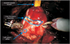
Robot-assisted radical prostatectomy (RARP) has progressively gained popularity amongst urologists and is now the dominant surgical approach for localized prostate cancer [1]. Robotic surgery provides certain inherent advantages, including high definition 3-dimensional vision, magnification, tremor filtration, movement scaling, wristed instrumentation with 6 degrees of freedom and improved surgeon ergonomics [2]. These features refine the surgeon's dexterity, particularly when dissecting in a narrow space within the male pelvis during radical prostatectomy [3]. The approach to the bladder neck (BN) is one of the critical steps of RARP. Clear identification of the BN can be difficult in some cases including excessive adipose tissue, previous surgery, thickened bladder wall, enlarged prostate, and those with protruding median lobe. Furthermore, a surgical mistake at this point can compromise oncological principles with dissection into the prostate, the integrity of the trigone with injury of the ureteric orifices and finally the BN with necessity of reconstruction for an optimal, leak-free anastomosis [4]. In this paper, with over 2,000 case experience, we demonstrate our current surgical technique of performing BN dissection during RARP and the management of difficult cases with variant anatomy.
ANATOMICAL−SURGICAL CORRELATIONS
As explained previously, the BN is composed of three layers of detrusor muscle and has in its luminal portion, the mucosa, which continues together with the inner longitudinal fibers into the prostatic urethra. Six regions around the BN can be identified on a 3-dimensional view. Anterior to the BN we find the superficial branch of the dorsal venous complex (DVC), the puboprostatic ligaments the endopelvic fascia and the Retzius space. Proximally, the BN is related to the bladder lumen and the trigone. Lateral and posterior-lateral to the BN are the lateral part of the seminal vesicles, the neurovascular bundles of the prostate, and the prostatic pedicles. Posterior to the BN we find, under the retrotrigonal and the Denonvillier's fascia, the ducts vasa deferentia and the medial part of the seminal vesicles. A well-defined midline strip called the retrotrigonal layer or posterior bladder apron is located posterior to the muscular BN, that extends from the trigone (superiorly) to the base of the prostate (inferiorly) [45]. The retrotrigonal layer serves as a key anatomical landmark to facilitate posterior prostatic dissection, particularly in men with large prostates, prominent median lobes, or previous transurethral prostatic surgery. Further, in our practice, this layer marks the posterior limit of dissection in which electrocautery is still used. Finally, the retrotrigonal layer serves to buttress the posterior layer of the urethrovesical anastomosis [6].
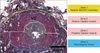
In order to facilitate understanding the BN anatomy and it's correlation to surgical approach we divided the BN to 4 anatomical layers from anterior to posterior include: (1) the DVC and peri-vesicle fat (yellow), (2) the anterior bladder muscle and mucosal wall (pink), (3) the posterior bladder mucosa and muscle wall (pink) and finally (4) the retrotrigonal fascia (blue). These anatomic layers are summarized in (Fig. 1).
SURGICAL TECHNIQUE
Our patient selection, preoperative preparation, and standardized surgical steps of performing RARP has been previously described [78]. In this paper, we focus on the 4-step approach to BN dissection to optimize BN diameter, tissue visualization and avoidance of complication.
After obtaining distal suture control of the DVC with a 0 Vicryl suture, and gently retracting the bladder using the fourth robotic arm to create a little tension on the BN, the robotic instruments are used to define the junction between the prostate and the bladder. Different maneuvers have been described to determine the correct plane of dissection:
Visual identification of the point of transition of the prevesical fat to the anterior prostate can serve as a guide.
Intermittent and repetitive caudal retraction of the urethral catheter balloon by the bedside assistant can help identify and confirm the transition between BN and prostate (Foley jiggle test)
Using the 4th arm Prograsp forceps to retract the dome of the bladder in a cephalad direction results in “tenting” of the BN at its attachment to the prostate.
Bimanual “palpation” or “pinch” of the BN using the tips of two robotic instrument.
1. Step 1
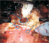
Once the optimal plane of dissection is visually identified, the first step starts with horizontal dissection of the first layer which is composed of DVC branches and fat. Because of the rich venous content of this layer, the bipolar forceps is initially used to isolate and fulgurate the fanning tributaries in the midline. The monopolar scissors with tip electrocautery can then be used to gently dissect through this midline BN fat down to the bladder muscle. Care must be made to stay in the midline to avoid the lateral branches of the DVC (Fig. 2A). Once the correct avascular plane is identified, the bipolar instrument can be used to laterally and bluntly dissect to allow secured Hemolock placement by the assistant (Fig. 2B). Care should be made to control any venous back-bleed oozing, particularly on the proximal bladder side with either Hemolock or cautery. If not attended to, ongoing slow bleeding and downward gravitation into the BN tissues can obscure visualization and hamper the later aspects of the BN dissection (Video clip 1, Supplementary material).
2. Step 2
After completing anterior exposure of the muscular BN (second layer), we always perform the Foley catheter jiggle maneuver to verify and confirm the localization of the BN. Occasionally, due to thick anterior tissues, the initial marking of the BN may be off by a small margin. At this point, the plane of entry has not been committed and the surgeon can readjust and migrate the next layer of dissection according to the funneling point of the catheter balloon at the thinned BN. The second step then is performed by slow dissection of the anterior BN muscle fibers using the monopolar tip cauterization until the urethral catheter is identified (Fig. 3). The Foley catheter balloon is then deflated, and an inspection of the posterior BN is made in order to identify the ureteral orifices as well as the presence of a median lobe. This is easily done with the upward Foley traction by the assistant, the left robotic instrument in the BN and placing traction towards 7-o'clock direction and the assistant suction tip placing traction at 5-o'clock at the BN. This triangulation allows safe assessment of the trigone (Video clip 2, Supplementary material).
3. Step 3
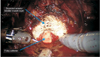
The posterior, muscular BN is of great importance to avoid inadvertent dissection in to the prostate or along the surgical capsule of the transition zone. With bedside assistant to create counter traction externally on the penile meatus Foley catheter, the prostate is then suspended anteriorly towards the abdominal wall by grasping the tip of the catheter creating upward traction. The anterior BN is gently pushed downward by the tip of the suction instrument creating counter traction tension on the tissue (Fig. 4A). The mucosa and posterior bladder muscle (third layer) is dissected carefully to assure proper dissection in the correct space and not to dissect into the prostate tissue. Similarly, inadvertent migration towards the bladder trigone and ureteral orifices (Fig. 4B) could also occur. The key elements in this dissection are the avoidance of tissue spreading with the bipolar instrument. The tip of the monopolar scissor should be used to hemostatically dissect layer by layer through the posterior wall. One should aim to paint over the tissue in a methodical and continuous fashion from left to right rather than staying in the same location and attempting in a small area to dissection all the way through the muscle wall. Attention to the thickness of the anterior bladder muscle should be observed to allow the surgeon with the appreciation of the thickness for the posterior muscle layer. Once the posterior bladder muscle is dissected, the retrotrigonal fascia is exposed. Care should be made to avoid dissecting too laterally to avoid entry into the vascular pedicles or neurovascular bundles (Video clip 3, Supplementary material).
4. Step 4
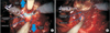
The retrotrigonal fascia is a key anatomic landmark during BN dissection and is easily identified by the well-defined midline fascial strip located posterior to the BN. It has a white appearance with longitudinal fibers (Fig. 5A). Blunt puncture, most often with the left-hand bipolar instrument, is performed to enter into the previously dissected seminal vesical space. In our practice, initial dissection of the vas deferens and seminal vesicles is routinely performed. We feel that this allows a greater area to dissect such tissues, minimize thermal energy in its dissection (especially at the tip of the seminal vesicles adjacent to the neurovascular bundle [NVB]) and offers a safer, more reliable BN dissection. Once the retrotrigonal fascial plane has been slightly opened with the monopolar scissors, both vas deferens and seminal vesicles are collectively grasped, pulled through the open BN and handed to the assistant for Microfrance upper traction to initiate Denovillier's incision and prostate pedicle/NVB control (Fig. 5B). In our, this layer marks the posterior limit of dissection in which electrocautery is still used. Furthermore, the proximal, cut retrotrional layer serves as the posteriorreconstruction support for the Rocco sutures during vesicourethral anastomosis (Video clip 4, Supplementary material).
Piechaud and Annino [4] have described their technique of performing BN dissection during RARP. Similarly, they follow the same approach of identifying the BN and dissecting the first layer, then with the use of combined blunt and sharp dissection the anterior muscular fibers of the BN. Thereafter, the dissection follows the plain at 12 o'clock and laterally to the BN at 2 and 10 o'clock, until the inner longitudinal fibers of the BN is identified. Then, the dissection continue lateral and posterior to the urethra with the aiming to preserve the proximal urethral sphincter, the urinary catheter is then removed, the urethra is transected and the remaining part of the posterior wall and retrotriognal fascia are dissected, followed by dissecting the seminal vesicles.
When compared to our technique, 3 few differences that are noteworthy. These include the traction maneuvers in our technique allow for clear visualization of the plain with safe dissection, initial dissection of the vas deferens and seminal vesicles provide a greater area of dissection with minimal thermal energy adjacent to the NVB, and finally our preservation of BN is not priority because of the greater possible risks positive margins.
MEDIAN LOBE
The normal anatomy can be modified in the case of a large median lobe, where usually the posterior relations are changed. In fact, the presence of a voluminous median lobe pushes the BN cranially, reducing its distance from the ureteral orifices and separating the ducts and the seminal vesicles, which in some cases can be placed far away from the BN, with consequent difficult identification of the right plane of dissection. This specific situation does not modify the anterior relations of the BN, but it can compromise the right identification of its position during the procedure which might prolonged the surgery completion time. Alenizi et al. [9] observed that prostate volume and median lobe were independent risk factors for prolonged surgery time for BN dissection completion. For this reason, the presence of a median lobe should be well investigated before the surgery [49]. While cystoscopy is not a standard practice for localized prostate cancer evaluation, the urologist should take the time to re-evaluate the transrectal ultrasound (TRUS) volume and dimension to recognize median lobe presence.
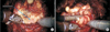
In our practice, we again will apply the 4-layer technique as previously noted. Upon the Foley jiggle test, the surgeon will likely note an off-centered balloon impression through the BN. After performing the anterior dissection of the BN, the posterior wall is carefully inspected. From our experience, use of the 4th arm Prograsp to hold the median lobe often leads to unwanted mucosal bleeding and slippage. As such, upon the immediate observation of the median lobe upon opening of the bladder mucosa, we will always switch instruments to the needle drivers to place a holding suture. Once this has been accomplished with upward traction on the suture (rather than the Foley), further exposure to assess the trigone and ureteric orifices can be performed. Suture suspension of the median lobe is quickly performed with a single 15-cm 3-0 VLoc suture with several passes through the median lobe mucosa and stroma. This will allow a strong hold of the median lobe, avoid tearing through the tissue and achieve good hemostasis for the dissection of the posterior BN. The upward lift by the assistant's laparoscopic Microfrance grasper on the suture rather than the Foley catheter (Fig. 6) provides safe, reliable exposure for the robotic surgeon to comfortably dissect through the mucosa and muscle with full awareness of the trigonal anatomy (Video clip 5, Supplementary material).
PREVIOUS TURP
In case of a prior classic or laser transurethral resection of the prostate (TURP), the BN margin is less evident and often distorted as a result of prior resection and scarring. Careful inspection of the posterior BN (step 3) is of greatest importance, paying specific attention to the location of the ureteral orifices. Due to tissue scarring and contraction, they are often found much closer to the posterior BN margin (Fig. 7).
In cases of recent TURP, we advise to wait 2–3 months following the endoscopic surgery to book RARP. Tips for intraoperative improved outcomes include starting with posterior dissection of vas deferens and seminal vesicles. This approach allows for larger space in addition to safer and more confidence with BN dissection. Fear of entering into the prostate base is of concern and enabling the surgeon to simply pop through the posterior plane into a previously dissected space greatly facilitates this posterior BN dissection. Once the anterior BN is dissected and the trigone is exposed, we must again highlight the importance of careful inspection of both ureteric orifices jets before commencing the posterior wall dissection. If their locations are not apparent or there is any doubt, the anesthesiology team should be asked to administer intravenous indigo carmine to help identify the orifices location. After identification, the mucosal floor can be scored with the monopolar scissor. We also suggest that the posterior bladder muscle be approached from a more lateral location so as to identify the normal tissue planes rather than the 6'oclock area of the BN where post-TURP tissue scarring may be at its greatest. In the event that the ureteric orifices are injured or are too close to the cut BN, use of temporary ureteric JJ stents can be placed intraoperatively to minimize their obstruction or urinary leakage. We have previously reported on our technique of transabdominal JJ stent placement while docked [10].
Moreover, it should also be emphasized that such post-TURP RARP cases can be along longer procedures. Gupta et al. [11] reported on a longer operating time, greater operative difficulty and potentially compromised oncological or continence outcomes in RARP after TURP.
Such details should be taken into consideration during patient counseling.
CONCLUSIONS
BN dissection is complex, difficult particularly during learning curve. Appreciation of the anatomic tissues and layers that comprise the BN coupled with a standardized approach to RARP will help optimize patient outcomes. Key clinical features related to BN dissection include surgeon attention to preoperative International Prostate Symptom Score, TRUS prostate biopsy ultrasound details (volume and median lobe presence), previous endoscopic prostate surgery, intraoperative catheter jiggle test (midline vs. lateral deviation of balloon) and anterior bladder wall thickness (to assist with posterior dissection).


 XML Download