Journal List > Investig Clin Urol > v.57(Suppl 1) > 1088106

Teo and Lim: Robotic assisted adrenalectomy: Is it ready for prime time?

Abstract

Adrenal surgery is undergoing continuous evolution and minimally invasive surgery is increasingly being used for the surgical management of adrenal masses. With robotic-assisted surgery being a widely accepted surgical treatment for many urological conditions such as prostate carcinoma and renal cell carcinoma, the use of the robot has been expanded to include robotic-assisted adrenalectomy, offering an alternative minimally invasive platform for adrenal surgery. We performed a literature review on robotic-assisted adrenalectomy, reviewing the current surgical techniques and perioperative outcomes.

INTRODUCTION

Adrenalectomy is generally performed for both benign and malignant indications. Prior to the first description of laparoscopic adrenalectomy by Gagner et al. [1] in 1992, adrenalectomy was traditionally performed via the open approach. In 1999, Piazza et al. [2] and Hubens et al. [3] described the first robotic assisted adrenalectomy using the AESOP 2000, a commercially available robotic platform in Europe at that time.
The war between man (conventional laparoscopy) and the robot has been waging over the last decade with the introduction of the da Vinci robotic system (Intuitive Surgical, Sunnyvale, CA, USA). Robots are seemingly emerging victorious in the frontiers of prostatectomy [4] and partial nephrectomy [5] and are beginning to make major headways along the frontlines of radical cystectomy [6] with intracorporeal urinary diversion and nephroureterectomy [7]. Along the battlefronts of extirpative surgeries like radical nephrectomy where the benefits of the robot is less pronounced, many robotic centres of excellence are starting to relegate conventional laparoscopy to the reserves. One main reason is as urologists get better in robotic assisted partial nephrectomies for increasing complex tumours, radical nephrectomies are now reserved for the most complex and largest of renal tumours and robot assistance in such cases are increasingly preferred. Adrenalectomy appears to be the final frontier of the robot's foray into urological surgeries. But is it ready for prime time yet?
This review aims to study the available evidence comparing the techniques and surgical outcomes of robotic assisted adrenalectomy and laparoscopic adrenalectomy.

METHODS

A literature review was performed using PubMed to identify relevant studies. Searches were performed with the following keywords: laparoscopic adrenalectomy and/or robotic and searches were restricted to publications in English. There were a total of 26 studies identified, reporting the techniques and perioperative outcomes of robotic assisted adrenalectomy, robotic assisted partial adrenalectomy or single port robotic assisted adrenalectomy (SPRA). The principles of the Helsinki declaration were followed in this review.

SURGICAL TECHNIQUES

Surgical management of adrenal disorders has seen a paradigm change in its approach. Many centres have performed robotic assisted adrenalectomy successfully, establishing it as a safe, feasible and effective approach. With the use of the da Vinci robotic system, challenges and limitations associated with pure laparoscopic surgery are alleviated while preserving the benefits of minimally invasive surgery. The superior ergonomics, 3-dimensional magnification of the operative field, tremor filtration and the Endowrist technology of robotic instruments providing a greater range of motion as compared to the human hand has allowed for easier handling of the fragile adrenal gland surrounded by major vessels and viscera in a confined space.
The lateral transperitoneal and the posterior retroperitoneal approaches are the commonest approaches adopted by most centers during robotic assisted adrenalectomies.
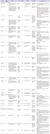
The operative details of studies reporting their techniques on robotic assisted adrenalectomy are detailed in Table 1 [89101112131415161718192021222324252627282930313233].

Patient positioning

Robotic assisted or conventional laparoscopic adrenalectomy can be performed via a transperitoneal or retroperitoneal approach. The transperitoneal approach provides greater working space, facilitates orientation by providing readily identifiable anatomical landmarks and better visualisation of surrounding anatomical structures. It also provides greater versatility in the angles of approach of laparoscopic trocars and instruments. In the lateral approach, peritoneal contents fall medially to give greater surgical exposure. In the supine position, both adrenal glands can be accessed without the need for intraoperative repositioning.
For robotic assisted transperitoneal adrenalectomy, most centres describe a lateral transperitoneal technique where patients are usually positioned in the lateral decubitus or modified lateral position with varying degrees of tilt of between 30 to 60 degrees.
Adrenalectomy can also be performed via the retroperitoneal approach. This approach mimics open surgery with its avoidance of the peritoneal cavity. This becomes the main advantage of this approach, as the adrenal gland is right against the thoracic cage when accessed from the back. There is also no entry into the peritoneal cavity and complications associated with intraperitoneal access such as intraperitoneal visceral injury, problems associated with pneumoperitoneum and adhesion formation are reduced. As such, it may be the preferred approach in patients requiring access to bilateral adrenal glands and in patients with multiple previous abdominal surgeries where intraperitoneal surgery may be more challenging due to previous adhesion formation. The greatest limitation with retroperitoneal adrenalectomy, however, is the limitation in working space which increases the technical difficulties of the operation.
Eight centres have described their techniques for robotic assisted posterior retroperitoneal adrenalectomy [81517192022252829]. In these centres, the patients are positioned in a prone position with the table flexed into a jack knife position.

Port placement

Port placement and the choice of port size is surgeon dependent. Most techniques describe a port placement conf iguration of between 3–5 ports f or lef t sided adrenalectomy with one additional port required for right sided adrenalectomy to aid in liver retraction. More details regarding port placement are shown in Table 1. In comparison to laparoscopic surgery, 4 ports are typically used in the transperitoneal approach with an option for an additional port inserted to aid in difficult dissection. Three ports are usually utilized in adrenalectomy performed using the retroperitoneal approach.
In recent years, laparoendoscopic single site (LESS) adrenalectomy has been described based on the principle that with a smaller number of incisions and ports, enhancement of cosmesis and reduction of associated port site complications can be attained. Both the retroperitoneal and transperitoneal approaches have been described for LESS adrenalectomy with variable strategies in terms of patient positioning, incision sites and ports placement. Usually a 2- to 3-cm incision is required for the insertion of a multiport device, typically described to be placed at the umbilicus for cosmetic benefits. Careful preoperative assessment and patient selection are imperative in minimizing challenges during surgery, reducing complications and ensuring quality outcome.
The disadvantages of LESS adrenalectomy include that of reduced distance between ports and loss of instrument triangulation resulting in cross over and paradoxical movement of instruments, as well as suboptimal approach to the adrenal gland and inadequate counter-traction. Nozaki et al. [34] described their technique of intraumbilical access to solve the problem associated with crossover instrumentation during LESS adrenalectomy. This involves a longitudinal incision of the umbilicus and a wider area of subcutaneous tissue dissection to accommodate multiple ports. The incision length remains within the depression of the umbilicus therefore preserving normal umbilical appearance.
Few centres have reported their experience with robotic assisted single port adrenalectomy [26272829] performed via both the transperitoneal and the retroperitoneal approaches. Park et al. [28] reported their initial experience with robotic single site posterior retroperitoneal approach, demonstrating its safety and feasibility. In their described technique, the operation is performed in the prone jack knife position, with a 3-cm transverse skin incision made just below the lowest tip of the 12th rib. For the transperitoneal approach, the patient is placed in a flexed lateral decubitus position, with a ipsilateral middle quadrant incision made for the single site port.

TRANSPERITONEAL VERSUS RETROPERITONEAL ADRENALECTOMY

Some retrospective comparisons of laparoscopic retroperitoneal and transperitoneal approaches tend to favour the retroperitoneal approach. Several operative parameters have been found to favour adrenalectomy performed via the retroperitoneal approach. These include shorter hospital stay [35363738], faster resumption of oral intake [3538], decreased analgesic requirement and postoperative pain which in turn leads to earlier ambulation [3739], shorter operative time [3739], blood loss [3839], and morbidity [40] associated with the procedure. The major benefit of the retroperitoneal approach is that with the adrenal against the ribcage at the back, there was no need to move any other organs out of the way. By mimicking open surgery, the peritoneal cavity is avoided, eliminating bowel handling and potential for injury to the intra-abdominal viscera. Walz et al. [41] reported that out of 142 patients who had posterior retroperitoneal adrenalectomy, half the patients did not require any postoperative analgesia and only five required pain medication for more than 24 hours postoperatively. Faster resumption of oral intake, together with decreased analgesia requirement and postoperative pain, may all contribute towards a shorter convalescence and hospital stay. While patients with smaller tumours, lower body mass index and bilateral adrenal pathologies and having significant prior abdominal surgery tend to benefit from retroperitoneal approach, patients with a higher body mass index with larger tumours and no prior abdominal surgeries tend to benefit more from the lateral transperitoneal approach [42]. These 2 approaches were found to be complementing and not competitive to each other when certain patient selection criteria are followed.
There have been descriptions of robotic assisted posterior retroperitoneal adrenalectomy [17192022] including descriptions of robotic assisted single port retroperitoneal adrenalectomy [2829]. In a comparison between robotic assisted posterior retroperitoneal adrenalectomy and laparoscopic posterior retroperitoneal adrenalectomy, it was found that beyond the initial learning curve, robotic assisted posterior retroperitoneal adrenalectomy shortens the skin to skin operative time by 28 minutes when compared with the laparoscopic approach. However, this may be nullified should there be additional intraoperative time used for transportation of the robotic unit to the operating room, starting up of the system, calibration of the robotic cameras and draping of the robotic arms. There was also lower immediate postoperative pain level for patients who underwent robotic assisted posterior retroperitoneal adrenalectomy [22]. Nevertheless, more randomised controlled trials need to be performed to study more meaningful outcomes and measures before this procedure can be justified.

ROBOTIC ASSISTED PARTIAL ADRENALECTOMY

Robotic assisted laparoscopic partial adrenalectomy has been described in various studies [30313233] to be safe and technically feasible with excellent short term functional and oncologic outcomes. Current indications for partial adrenalectomy include bilateral benign adrenal lesions, a solitary adrenal gland or unilateral tumours in patients with hereditary syndromes. Partial adrenalectomy has also been shown to be feasible in excision of adrenal metastasis in patients with a solitary adrenal gland. While adrenal sparing surgery offers selected patients a substantially better quality of life without the need for lifelong hormonal supplementation, the use of minimally invasive procedures in the treatment of malignant adrenal lesions have always been controversial in view of the potential problems of incomplete resection and risk of recurrence.
Certain technical modifications have been described by Asher et al. [31] to facilitate successful completion of partial adrenalectomy. Extreme flank positioning with axis of robotic ports directed at ipsilateral clavicle allows for easier access to the adrenal gland and for better visualisation of the upper retroperitoneum. The liver must also be well mobilised to allow access to the supra-adrenal vena cava on the right side so that short hepatic veins can be appreciated. Dissection should also be carried out between the pseudocapsule of the lesion and normal adrenal gland to minimise bleeding. The advantage of the robotic platform over traditional laparoscopy may best be appreciated during tumour resection whereby the articulation of the robotic instruments allows easier dissection around the tumour deep within the adrenal gland, taking care to minimize handling of normal adrenal tissue and to reduce the use of cautery to preserve adrenal blood supply.

LAPAROSCOPIC AND ROBOTIC SURGERY FOR LARGE ADRENAL TUMOURS

Minimally invasive resection of large adrenal tumours can be challenging due to a higher risk of complications and greater concerns of malignancy. The transperitoneal approach has been shown to provide greater exposure for resection of larger tumours, with studies showing preference for this approach when tumours are larger than 5 cm [43]. Resection of adrenal masses larger than 6 cm can be challenging when performed via a restricted retroperitoneal space [44]. Comparing to laparoscopic adrenalectomies for large adrenal tumour, the use of robotic assistance has been shown to shorten operative time and decreased the rate of open conversion when compared to laparoscopic adrenalectomy for tumours larger than 5 cm [8]. This can be due to the fact that the robotic instruments are wristed whereas laparoscopic instruments are rigid as well as a three dimensional view which made dissection faster and more accurate in robotic assisted adrenalectomy.

LESS ADRENALECTOMY

Meta-analysis comparing LESS adrenalectomy versus conventional laparoscopic surgery showed no significant difference in estimated blood loss, time to oral intake resumption, complications, conversion and transfusion rates between the 2 groups [45464748]. However, patients who have undergone LESS adrenalectomy have a significantly lower postoperative visual analog pain score [46] or had less analgesia demand [47]. Hospital stay was also found to be similar [46] or shorter [454748] when LESS adrenalectomy was performed. Operative time was however, found to be longer, though Ishida et al. [48] noted that when LESS adrenalectomy was performed, adjustment of the roticulator took an addition of 14.5±8.1 minutes, and in their retrospective case control study, when this time was taken off operative time for LESS adrenalectomy, the operative time was more comparable when compared to conventional adrenalectomy (76.7 minutes vs. 74.3 minutes, p=0.880). LESS adrenalectomy has been proven to be a safe and feasible alternative. Apart from superior cosmesis, current evidence appears to also offer an advantage of shorter convalescence and decreased postoperative pain when compared to conventional laparoscopic adrenalectomy.
SPRA has also been described using both the transperitoneal and retroperitoneal approach [26272829]. While most perioperative outcomes of SPRA were comparable to laparoscopic adrenalectomy, it was found that operative times were shorter for unilateral adrenalectomy (130±8 minutes for SPRA and 188±12 minutes for laparoscopic adrenalectomy) and there was also statistically significant lower narcotic use in the first 24 hours after surgery. While length of hospital stay as well as cost trended to be lower for robotic single port adrenalectomy, these results were not found to be statistically significant [27].

SURGICAL OUTCOMES

Systematic reviews and meta-analyses of current evidence available have demonstrated the safety and efficacy of robotic assisted adrenalectomy when compared to laparoscopic adrenalectomy.

COMPARING ROBOTIC AND LAPAROSCOPIC ADRENALECTOMY

In 2004, Morino et al. [13] in a prospective randomized controlled study comparing robotic and laparoscopic adrenalectomy concluded that laparoscopic adrenalectomy was superior to robotic assisted adrenalectomy in terms of feasibility, morbidity and cost in view of longer operative time, higher 30-day complication rate and a similar length of hospital stay. However, since then, many subsequent retrospective studies and meta-analyses have been performed comparing the outcomes of robotic versus laparoscopic adrenalectomy which demonstrates equivalence if not superior outcomes for robotic assisted adrenalectomy.
Perioperative outcomes of the robotic assisted adrenalectomy studies are presented in Table 2 [89101112131415161718192021222324252627282930313233]. Perioperative outcomes for laparoscopic adrenalectomy within the same study were also included.

Operative times

There is a wide range of operative times reported by different centres with a mean reported time of between 98 to 234.4 minutes. Brunaud et al. [12] identified several criteria that had an impact on operative time such as surgeon experience, first assistant training level as well as tumour size, with tumours less than 4.5 cm having a shorter operative time. Longer operative times were typically demonstrated in the initial part of the learning curve. This can be partly attributed to time spent docking the robot. Once the ports are placed in traditional laparoscopic surgery, the operation commences. However, in robotic surgery, after the ports are placed, the robot tower must then be docked with instruments inserted, and this has been found to increase operative time by between 15–40 minutes [24] with initial docking time to be reported to be as long as 1 hour [12]. While these can be streamlined with increasing experience, this is still an extra step when compared to laparoscopic surgery. However, beyond the initial learning curve, Agcaoglu et al. [22] reported a significant improvement in operative time after the 10th procedure, and the difference in operative time can be eliminated from as early as the 20th operative case [12]. They reported that the mean operative time decreased 134 minutes in the last 45 cases compared with first 50 cases and by multiple regression analysis, surgeons experience, first assistant level and tumour size were independent predictors of operative time. Brandao et al. [49] in a meta-analysis comparing robotic and laparoscopic adrenalectomy found no statistical difference between the operative times between the 2 procedures. Karabulut et al. [19] also found that the time spent for individual steps of procedure was similar between the laparoscopic and the robotic group and even though the tumour size was larger in the robotic groups.

Duration of hospital stay

The duration of hospital stay in the robotic assisted studies reported a mean range of 1.1 to 6.4 days. Perioperative outcome studies have reported a shorter hospital stay when robotic assisted adrenalectomy is performed when compared to laparoscopic adrenalectomy [151949]. Karabulut et al. [19] found that in their cohort of patients, the main reasons for hospital stay in the robotic group was for nausea, atelectasis and the need for pain control, and all patients were discharged within 2 days. This is in comparison to their patients who underwent laparoscopic adrenalectomy who stayed between 1–4 days. This shorter hospital stay is possibly the result of a combination of various improved outcomes such as a shorter operative time and lesser blood loss, though hospital stay can be an unreliable outcome parameter for comparison as it can be confounded by many factors.

Blood loss

One other significant outcome in favour of robotic surgery was the lower estimated blood loss. Reported mean blood loss ranged from less than 50 mL to 576 mL, with most centres reporting mean blood loss of less than 100 mL. Bilateral adrenalectomies tend to result in greater blood losses. Lee et al. [26] reported a mean of 1,698 mL (150–6,140 mL) in their 5 cases of bilateral robotic assisted single site adrenalectomy that were performed. Pineda-Solís et al. [50] in their retrospective study found that the blood loss tended to be lower in the robotic group versus the laparoscopic group (30±5 mL vs. 55±74 mL, p=0.07) though this was not statistically significant. Other studies also reported equivalence in terms of intraoperative blood loss [1421]. Brandao et al. [49] in their meta-analysis comparing outcomes between robotic assisted adrenalectomy and laparoscopic adrenalectomy, found that 7 out of 9 studies reported less bleeding for the robotic group with a statistically significant difference between the 2 groups. However, this difference may not be clinically significant and that both techniques can be performed with minimal associated blood loss.

Conversion rates

In current literature, low conversion rates have been reported for both robotic and laparoscopic adrenalectomy [49]. Conversion rate for robotic assisted adrenalectomy were reported to range between 0% to 40% for laparoscopic conversion and 0% to 10% for open conversion while open conversion rates in the laparoscopic studies ranged between 0% to 10.5%. Of note, in both groups, there were many studies which reported a 0% conversion rate. Common reasons for conversion in robotic cases cited included adherence of the tumour to surrounding structures or adhesions (5 cases) or bleeding (4 cases). Other reasons included poor visualisation of structures (2 cases), technical difficulties resulting in incomplete isolation, camera malfunction and failure to progress (1 case each). Conversion rate for robotic cases was found to decrease with increasing surgical experience [13].

Complication rates

Studies comparing robotic and laparoscopic adrenalectomy reported same or superior results for the robotic group in postoperative complications rate (7% vs. 11%) [11]. Meta-analysis [49] performed comparing these outcomes also showed a nonstatistically significant difference in a higher complication rate in the laparoscopic group (6.8% vs. 3.6%, p=0.05) There were more reported severe complications in the laparoscopic groups including grade 4 and 5 complications according to the Clavien-Dindo classification system grading system. Reported complications in the robotic group were generally of a lesser degree of severity [49]. Postoperative morbidity and mortality have been demonstrated to be comparable to conventional laparoscopy [13].

LEARNING CURVE

It is well known that being early in a surgeon's learning curve is associated with worse perioperative outcomes and increased complications. It has been estimated that the learning curve of laparoscopic adrenalectomy is between 20–40 cases [5152] while that of robot-assisted transperitoneal adrenalectomy is only about half, ranging between 10–20 cases [1222]. This is especially important for lower volume surgeons as the inherent advantages of the robotic platform may help surmount the initial learning curve faster, leading to better perioperative outcomes and reduced complications.

COST AND QUALITY OF LIFE ASSESSMENTS

One of the major points of criticism with robotic surgery has always been the higher cost factor. Brunaud et al. [53] found that when cost evaluation was performed using baseline cost in their hospital, robotic adrenalectomy was 2.3 times more costly than laparoscopic adrenalectomy. (4,102 euro vs. 1,799 euro). Total cost was found to be most affected by the total number of robotic cases per year and depreciation of the robotic system. Operative time, in contrast, was found to only play a minor role in the overall cost. This finding was also echoed by Morino et al. [13] who found a difference of $729 excluding the capital investment of the da Vinci robotic system. This increased expense was mainly due to the use of semidisposable robotic instruments and longer operative time. However, it is to be noted that these studies were performed in the earlier era of robotic assisted adrenalectomy. With increasing volumes and improved outcomes associated with robotic assisted adrenalectomy, more up to date cost analysis studies should be performed to evaluate this parameter. Arghami et al. [27] analysed the cost associated with single-port robotic adrenalectomy and found that in their health system, as there are no specific billing codes for robotic assisted adrenalectomy with similar reimbursement compared to the laparoscopic technique, a robotic procedure adds about $950 to the cost compared to laparoscopic adrenalectomy. However, they found that the total bill cost for single port robotic adrenalectomy was 16% lesser than laparoscopic adrenalectomy, which may be related to shorter hospital stay and an approximately 50% reduction in narcotic use. Probst et al. [54], in a recent paper comparing costs of robotic adrenalectomy and open adrenalectomy demonstrated that the additional costs of robotic surgery were equalized if at least 150 cases of robotic procedures were performed per year based on certain healthcare cost assumptions within the healthcare system.
In terms of quality of life assessment, no significant difference was observed for all Short Form 36 health survey scores between patients after laparoscopic or robotic adrenalectomy except for role limitations due to emotional problems. These were increased after 6 weeks in patients who underwent robotic adrenalectomy. There was also no significant difference regarding state and trait anxiety, postoperative pain, quality of sleep and sleep duration [53].

CONCLUSIONS

Current evidence has shown that robot assisted adrenalectomy can be performed safely and effectively with equivalent or even superior outcomes compared to laparoscopic adrenalectomy with potential advantages of shorter operative times in high volume centres, reduced blood losses, shorter hospital stay and decreased intraoperative blood loss.
However, there is still a paucity of reports on perioperative and long term outcomes these needs to be evaluated in well-designed prospective randomized controlled trials with adequate power and follow-up. Further more detailed cost analysis are also required to justify the higher costs associated with robotic assisted adrenalectomy.
And so it seemed that it is a stalemate in the war between robot and man along the frontlines of adrenalectomy at this point in time. As to who will ultimately emerge victor, only time will tell.

Figures and Tables

Table 1

Operative details of studies reporting their robotic assisted adrenalectomy techniques

icu-57-S130-i001
Study No. of patients Diagnosis Year Procedure Approach Positioning Number of ports
Robotic assisted adrenalectomy (RAA)
 Agcaoglu et al. [8] 25 Pheochromocytoma (8), cortical adenoma (7), complex cyst (5), others (5) 2000 to 2011 RAA Lateral transperitoneal (LT)/posterior retroperitoneal (PR) Lateral decubitus/prone jack knife LT – 4 ports for both right and left adrenalectomy. Placed under costal margin
PR – 3 ports – incisions below 12th rib. No assistant port
 Giulianotti et al. [9] 42 Cortical adenoma (19), pheochromocytoma (9), hemorrhagic cyst (6), aldosteronoma (2), bilateral hyperplasia (2), adrenal carcinoma (1), ganglioneuroma (1), myelolipoma (1), metastasis (1) November 2000 to February 2010 RAA Transperitoneal Supine with 20 degrees tilting 5 Ports
Right sided – 12-mm lateral side of pararectal line above umbilical transverse line, 8 mmx3 robotic ports forming a concave line curved towards adrenal fossa, 10/12-mm port near umbilicus
Left sided – 12-mm left upper quadrant, 8 mmx3 laterally in left side, right subcostal, approximate midline, 12-mm assistant port between optical
Bilateral – 6 ports. One additional port in subxiphoid position
 Undre et al. [10] 2 Cortical adenoma (2) December 2000 to June 2003 RAA Lateral transperitoneal Right lateral decubitus position with 45 degree side tilt
Table flexed
Left sided - 5 ports
2×8-mm robotic ports, 1x12-mm robotic camera port, 2×10-mm assistant ports
 Winter et al. [11] 30 Pheochromocytoma (11), aldosteronoma (9), glucocorticoid adenoma (5), adrenal adenoma (1), angiomyolipoma (1), combined aldosterone/cortisol secreting adenoma (1), metastatic carcinoma (1) April 2001 to January 2004 RAA Lateral transperitoneal Lateral decubitus flexed at the level of the kidney Left sided – 3 +/− 1
12-mm camera port midway between umbilicus and left costal margin. 8 mmx2 robotic ports 2 finger breadth from costal margin. 10-mm accessory port in left abdomen
Right sided – 4 +/− 1
Additional 5-mm liver retraction port midline in epigastrium.
 Brunaud et al. [12] 100 Nonfunctioning adenoma (19) cyst (2), pheochromocytoma (24), aldosteronoma (39), Cushing
adenoma (11), Cushing hyperplasia (5)
November 2001 to November 2007 RAA Not indicated Lateral decubitus 4 or 5 ports
12-mm robotic camera, 2x robotic ports, 1 or 2 accessory trocar.
 Morino et al. [13] 10 Nonfunctioning adenoma (3), aldosteronoma (3), pheochromocytoma (4) March 2002 to December 2002 RAA Lateral transperitoneal Lateral flank position. Robot at patients shoulder, 45 degrees from table axis Left – 5 ports
1x12-mm robotic camera port, 2x7-mm robotic port, subcostal margin side of lesion, 1x12-mm accessory port
Right – 6 ports
Additional 5-mm port
 Wu et al. [14] 5 Functioning adenoma (3), nonfunctioning adenoma (1), pheochromocytoma (1) January 2003 to February 2005 RAA Lateral transperitoneal 45 Degree flank position slight trendelenburg Left sided – 5 ports
12-mm camera port at umbilicus, 10-mm robotic port × 1, 2×5-mm robotic port midclavicular line 2 finger breadth below costal margin x1, anterior axillary line ×1, 10-mm assistant port mid axillary line umbilical region
Right sided – 6 ports
Additional 5-mm liver retraction port
 Aksoy et al. [15] 42 Cortical adenoma (10), Cushing syndrome (10), pheochromocytoma (8), aldosteronoma (60),
other (8)
2003 to 2012 RAA Lateral transperitoneal Posterior retroperitoneal Not indicated Not indicated Not indicated
Not indicated
 D'Annibale et al. [16] 30 Functioning adenoma (7), nonfunctioning adenoma (6), metastatic carcinoma (4),
pheochromocytoma (3), macro/micronodular hyperplasia (3), haemorrhagic cyst/pseudocyst (3), myelolipoma (2), cortical carcinoma (1), schwannoma (1)
July 2006 to October 2011 RAA Lateral transperitoneal approach 45 Degrees lateral decubitus with a 20 degree reverse trendelenburg
Table flexed at level of kidney
Left sided – 4 ports
12-mm camera port, 2x robotic port, 1x assistant port
Right sided – 5 ports
Additional 1x subxiphoid assistant port
 Ludwig et al. [17] 6 Adrenocortical adenomas (4) aldo- steronomas (2) September 2006 – not indicated RPRA Posterior retroperitoneal Prone 5
12-mm port 1.5-cm transverse incision below 12th rib,
 Zafar et al. [18] 1 Adrenocortical carcinoma (1) 2008 RAA Lateral transperitoneal Oblique 30 degree supine position Left sided -5 ports
1×10-mm camera port paramedian vertical incision midway between xiphoid and umbilicus, 2×8-mm robotic port 1 finger breadth below left costal margin in line with camera and 3 finger breadth below camera port midclavicular line, 2x 5-mm assistant port
 Karabulut et al. [19] 50 Pheochromocytoma (12), aldosteronoma (8), nonfunctioning ad- enoma (10), Cushing syndrome (8), metastasis (5), other (7) 2008 RAA RPRA Lateral transperitoneal
Posterior retroperitoneal
Lateral decubitus
Prone jack knife
4 ports - 3x robotic ports, 1x assistant port
3 Ports – 3x robotic ports
 Berber et al. [20] 8 Nonfunctioning adenoma (3), aldosteronoma (2), pheochromo- cytoma (1), Cushing syndrome (1), lymphangioma (1) December 2008 to February 2010 RPRA Posterior retroperitoneal Prone jack knife position 3 Ports
1×12-mm camera port 1-cm incision 2 cm inferior and parallel to 12th rib, 2x robotic 5-mm port 1 on each side of initial port
 You et al. [21] 23 Cortical adenoma (9), pheochromocytoma (4), diffuse medullary hyperplasia (1), myelolipoma (1) October 2009 to May 2012 RAA Lateral transperitoneal Lateral decubitus position Left sided – 3 ports
1×12-mm camera port anterior axillary line 1 finger breadth from left costal margin, 2×8-mm robotic port mid clavicular and mid axillary line along costal margin
Right sided – 4 ports
1x12-mm camera port mid axillary line 1 finger breadth from right costal margin, 3x8-mm robotic port posterior and anterior axillary line, subxiphoid in midline
 Agcaoglu et al. [22] 31 Cortical adenoma (8), pheochromocytoma (6), aldosteronoma (6), Cushing syndrome (5), other (6) 2009 to 2011 RPRA Posterior retroperitoneal approach Prone jack knife using Wilson frame 3- to 12-mm camera port 1 cm below 12th rib. 2x5- mm robotic port medial and lateral to camera port
 Brandao et al. [23] 30 Adrenocortical adenoma (18), pheochromocytoma (5), myelolipoma (2), metastasis (2), others (3) April 2010 to October 2013 RAA Lateral transperitoneal 60-degree flank position Left sided – 4 ports
1x12-mm camera port above and lateral to umbilicus at lateral border of rectus muscle across 12th rib. 1x8-mm robotic port, lateral border of ipsilateral rectus muscle 1 inch below costal margin, 1x8-mm robotic port 2-inch cephalad to anterior superior iliac spine, 1x12-mm assistant port lateral b order of rectus halfway between camera and lower robotic ports
Right sided – 5 ports
1 Additional 5-mm port subxiphoid in midline
 Akarsu et al. [24] 8 Cushing adenoma (4), nonfunctioning adenoma (4) January 2011 to February 2013 RAA Lateral transperitoneal Standard lateral decubitus 4
 Okoh et al. [25] 50 Not indicated Not indicated RAA
RPRA
Lateral transperitoneal
Posterior retroperitoneal
Lateral decubitus
Not indicated
4 Ports
Not indicated
Robotic assisted single port adrenalectomy (SPRA)
 Lee et al. [26] 33 Aldosteronoma (11), Cushing adenoma (11), pheochromocytoma (5), nonfunctioning adenoma (3), haemangioma (1), lymphoma (1), myelolipoma (1) January 2012 to October 2014 SPRA Lateral transperitoneal Modified flexed lateral decubitus position Left sided – single port
2.5-cm transverse incision 3-4 cm inferior to costal margin at midclavicular line.
Right sided – 1+5-mm retraction port
Additional 5-mm liver retraction port midline superior to single port site
 Arghami et al. [27] 16 Aldosteronoma (7), Cushings syndrome (4), pheochromocytoma (2), others (3) November 2012 to August 2013 SPRA Not indicated Flexed lateral decubitus position Left sided – single port
2.5-cm ipsilateral middle quadrant incision.
Right sided – 1+5-mm retraction port
 Park et al. [28] 1 Cortical adenoma (1) 2010 SPRA Posterior retroperitoneal Prone jack knife Left sided – single port
3-cm transverse incision below lowest tip 12th rib
 Park et al. [29] 5 Cortical adenoma (5) March 2010 to June 2011 SPRA Posterior retroperitoneal Prone jack knife with hip joint bent at right angle Single port
3-cm transverse incision below lowest tip 12th rib
Robotic assisted partial adrenalectomy (RAPA)
 Gupta et al. [30] 4 Pheochromocytoma (4) July 2006 to June 2009 RAPA Lateral transperitoneal Lateral decubitus position angled at 60 degrees with the floor. Left sided – 5
1× 12-mm robotic camera port paraumbilical, 1×
8-mm robotic port ipsilateral lower quadrant, 1×8- mm robotic port upper quadrant, 12-mm assistant port between camera and superior robotic port.
Right sided – 6
5 mm x1 additional subxiphoid liver retraction port
 Asher et al. [31] 15 Pheochromocytoma (15) 2007 to 2010 RAPA Lateral transperitoneal Extreme flank position with axis of shoulders close to a 90 degree angle to operating table Left sided – 4/5 ports
1× 12-mm robotic camera port superior and lateral to umbilicus, 1× 8-mm robotic port lateral to camera port, 1×8mm robotic port medial and superior to camera port, 12-mm assistant port between camera and lateral robotic arm.
Optional 5-mm port between camera port and medial robotic arm in obese patients
Right sided - 5/6 ports
Additional 5-mm subxiphoid liver retraction port
 Boris et al. [32] 10 Pheochromocytoma (17), adrenal cortical hyperplasia (2) July 2008 to January 2010 RAPA Lateral transperitoneal Modified flank position with robot docked over ipsilateral shoulder. Left sided – 4
1×12-mm robotic camera port superior and lateral to umbilicus, 1×8-mm robotic port lateral and infe- rior to camera port, 1×8-mm medial and superior to camera port, 12-mm assistant port between camera and lateral port
Right sided – 5
Additional 5-mm subxiphoid liver retraction port
 Kumar et al. [33] 1 Adrenal metastasis (1) 2009 RAPA Lateral transperitoneal Right lateral decubitus Left sided – 4
1×12-mm robotic camera port, 1×8-mm robotic port lateral left quadrant, 1×8-mm robotic port superior to assistant , 1×12-mm assistant port midline superior to camera port

RPRA, robotic posterior retroperitoneal adrenalectomy.

Table 2

Reported perioperative outcomes of robotic assisted adrenalectomy studies, including studies which provided a comparison to perioperative outcomes after laparoscopic adrenalectomy within the same study

icu-57-S130-i002
Study No. of patients Hospital stay (day) (range) Tumour size (cm) (range) Blood loss (mL) (range) Bill size Surgical time (min) (range) Conversion Mortality Blood transfusion Complications Unilateral / single / multiple
Giulianotti et al. [9]
 Robotic 42 Median 4 (1–22) 5.5 +/− 2.5cm Median 27 (10–400) Not indicated 118 +/− 46 Nil 1
Myocardial infarction with fatal arrhythmia
Nil Intraoperative 1 (9.0%)
Minimal capsular tear in large tumour ×1
Postoperative 1 (9.0%)
Clostridium difficile infection ×1
Unilateral- 41
Bilateral- 1
Undre et al. [10]
 Robotic 2 4 (3–5) 5.5×2×2.5 cm
13.9 cm
150 (0–300) Nil 149.5 (144–515) Nil Nil Not indicated Postoperative 1 (50%)
Pulmonary embolism
Unilateral - 2
Winter et al. [11]
 Robotic 30 Median 2 (1–5) Median
2.4 (1.1–8)
Not indicated USD $8,645 Median 185 (130–295) Nil Nil Not indicated Postoperative 2 (6.7%)
ileus × 1
Hypoxemia due to bronchitis and atelectasis x 1
Unilateral- 30
Brunaud et al. [12]
 Robotic 100 6.4 +/− 3.0 2.9 +/− 1.9 (0-12) Not indicated Not indicated 99 +/− 35 Laparoscopic -1/100 (1%)
Camera malfunction ×1
Open × 4/100 (4%)
Intraoperative bleeding × 3
Difficult visualisation of adrenal vein × 1
Nil Not indicated Intraoperative 1 (1%)
Cyst rupture ×1
Postoperative 10 (10%)
Grade 1 Wound infection × 2
Facial edema × 1
Grade II
Pneumonia × 3
UTI × 2, anemia ×1, hematoma × 1
Unilateral- 100
D'Annibale et al. [16]
 Robotic 30 5.2+/− 2.2 (2-11) 5.1 +− 2.4 <50 Not indicated 200 (180–255) Open 1 (3.3%)
Large liver causing incomplete isolation ×1
Nil Not indicated Intraoperative 2 (6.6%)
Capsular disruption with temporary hemostasis by sponge × 1
Marked arterial instability ×1
Postoperative 3 (10%)
Abdominal wall hematoma ×1
Pneumonia ×1
Myocardial infarction ×1
Unilateral- 30
Ludwig et al. [17]
 Robotic 6 23 h or less in 4 patients
2 d (2)
2.8 (1.1–4.3) <60 (<50 to 100) Not indicated 143 min
Docking 14
Laparoscopic 1/6 (16.7%)
Failure to progress
Nil Not indicated Nil Unilateral - 6
Zafar et al. [18]
 Robotic 1 2 8 20 Not indicated 138 Nil Nil Nil Nil Unilateral - 1
Berber et al. [20]
 Robotic 8 2 2.9 +− 1.7 24 +/− 35 Not indicated 214.8 +/− 40.8 Nil Nil Not indicated Nil Unilateral - 8
Akarsu et al. [24]
 Robotic 8 4.1 (2–11) 5.36 (2 Median-9) 50 3617.12 pounds 98 (55–175)
Docking duration 15–40
Nil Nil Not indicated Intraoperative 1 (12.5%)
Diaphragmatic injury x1
Unilateral - 8
Okoh et al. [25]
 Robotic 50 Not indicated LT 4.7
PR 2.7
Not indicated Not indicated LT 168
PR 166
Not indicated Not indicated Not indicated Not indicated Not indicated
Lee et al. [26]
 Robotic 33 2.8 (2–7) Not indicated Unilateral 159.5 (5–1,400)
Bilateral 1,698 (150–6,140)
Total 392.6 (5–6,140)
Not indicated 234.4 (136–630) Laparoscopic 5/33 (15.2%)
Bleeding, adhesions × 2
Limited visualisation ×2
Poor port placement ×1
Open 2/33 (6.1%)
Bleeding requiring nephrectomy ×2
Nil 2 Postoperative 3 (11%)
Ileus ×1
Adrenal insufficiency × 2
Unilateral - 28
Bilateral - 5
Park et al. [28]
 Robotic 1 4 1.5 Not indicated Not indicated 188 Nil Nil Not indicated Nil Unilateral - 1
Park et al. [29]
 Robotic 5 4.0+/− 2.23 (3–8) 1.48 +/− 0.28 (1.0–1.7) 46.0 +/− 56.8 (5–120) Not indicated 159.4+/−57.6 (103-245) Nil Nil Not indicated Nil Unilateral -5
Gupta et al. [30]
 Robotic 4 4 4.7 97.5 (50–160) Not indicated 77.5 (40–140) Nil Nil Not indicated Intraoperative 3 (75%)
Hypotension ×3
Postoperative 2 (50%)
Hypotension ×2
Single - 4
Asher et al. [31]
 Robotic 15 Not indicated Mean 2.7 Median 161 (50–300) Not indicated 163 (110–357 Open 1/15 (6.7%)
Severe adhesions with repeated surrounding structure injury
Not indicated 1 Intraoperative 1 (6.7%)
Clavien 3 – bile leak requiring drain
Multiple - 4
Single - 8
Boris et al. [32]
 Robotic 10 Not indicated Median 2.7 (1.3–5.5) 150 (25–1,000) Not indicated 200 (110–480) Open 1(10%)
Previous open partial adrenalectomy resulting in severe desmoplastic reaction obliterating plane
Not indicated 1 (7.5%) Intraoperative 1 (10%)
Bile leak requiring intraoperative drain × 1
Postoperative 1 (10%)
Ureteral stricture requiring ureteral stent × 1
Single – 10
Bilateral – 2
Bilateral multifocal - 1
Kumar et al. [33]
 Robotic 1 4 4 50 Not indicated 90 Nil Nil Nil Nil Single
Agcaoglu et al. [8]
 Robotic 24 1.4 +/− 0.2 6.5 +/− 0.4 (5–10.2) 83.6 +/− 59.4 Not indicated 159.4 +/− 13.4 (64–357) 1/24 (4%)
Adherence of tumour to renal hilum
0 Not indicated Not indicated Not indicated
 Laparoscopic 38 1.9 +/− 0.1 6.2 +/−0.3 (5-15) 166.6 +/− 51.2 Not indicated 187.2 +/− 8.3 (185–290) 4/38 (10.5%)
Bleeding ×2
Adherence of tumour to IVC ×1
Difficulty with dissection plane ×1
0 Not indicated Not indicated Not indicated
Morino et al. [13]
 Robotic 10 5.7 (4–9) 3.3 (1.4–6.5) Not indicated USD $3,466 169 (135–215) Laparoscopic – 4 (40%) Nil Not indicated Intraoperative 2 (20%)
Severe hypertension for pheochromocytoma ×2
Unilateral- 10
 Laparoscopic 10 5.4 (4–8) 3.1 (1.5–6) Not indicated USD $2,737 115.36 (95–115) Nil Nil Not indicated Nil Unilateral- 10
Wu et al. [14]
 Robotic 5 4 +/−0.7 5.1 +/− 1.0 9 +/− 54.8 Not indicated 188 +/− 305 Nil Nil Not indicated Nil Unilateral - 5
 Laparoscopic 7 3.4 +/−0.5 4.7+/−0.8 85.7+/−37.8 Not indicated 131.4+/−29 Nil Nil Not indicated Nil Unilateral - 7
Aksoy et al. [15]
 Robotic 42 1.3 +/− 0.1 (0–3) 4.0 +/− 0.4 (1–12) 50.3 +/− 24.3 (0–400) Not indicated 1,786.1 +/− 12.1 (106–380) Open - Nil Nil Not indicated Intraoperative 1 (2.4%)
Pneumothorax x1
Postoperative 1(2.4%)
Urinary tract infection × 1
Unilateral- 41
Bilateral 1
 Laparoscopic 57 1.6 +/−0.1 (1–5) 4.3 +/−0.3 (1–15) 76.6 +/− 21.3 (0–900) Not indicated 187.3 +/−1 (80–516) 3 (5.2%) 1/57 due to severe respiratory failure Not indicated Intraoperative 1(1.8%)
Pneumothorax ×1
Postoperative 1(1.8%)
Prolonged ileus × 1
Unilateral- 53
Bilateral - 4
Karabulut et al. [19]
 Robotic 50 1.1 +/− 0.3 (excluding those converted to open approach) LT 4.8 +/− 0.4
PR 2.7 +/− 0.3
41 +/− 10 Not indicated LT 165 +/− 10
Docking 21
PR 166 +/− 9
Docking 25
Open 1/50 (2%)
Adherence of pheochromocytoma to renal hilum
Not indicated Not indicated Postoperative 1 (2%)
Atelectasis × 1
Unilateral- 50
 Laparoscopic 50 1.5+/−0.9 LT 3.6+/−0.3 PR2.3+/−0.3 41+/−20 Not indicated LT 160+/−9
PR 170+/−15
Open 2/50 (4%)
Periadrenal invasion x1
Periadrenal inflammation x1
1(2%)
Respiratory insufficiency
Not indicated Postoperative 5 (10%)
Postoperative ileus ×2
COPD exacerbation ×1
Abnormally low cortisol levels ×1
Respiratory insufficiency ×1
Unilateral -50
You et al. [21]
 Robotic 15 5.86 (4–7) 2.57 (1–5.5) Not indicated Not indicated 207 (120–320) Nil Nil Not indicated Postoperative 2 (13.3%)
Wound seroma x1
Chylous ascites x1
Unilateral - 15
 Laparoscopic 8 6.71 (5–9) 2.8 (1–4.5) Not indicated Not indicated 183.13 (75–270) Nil Nil Nil Postoperative 2 (25%)
Acute kidney injury ×1
Cerebral infarction ×1
Unilateral - 8
Agcaoglu et al. [22]
 Robotic 31 Median 1 3.1 (0.2) 25.3 (10.3) Not indicated 163.2 (10.1) Nil Nil Not indicated Nil Unilateral -31
 Laparoscopic 31 Median 1 3.1 (0.2) 35.6 (9.6) Not indicated 165.7 (9.5) Nil Nil Not indicated Nil Unilateral - 31
Brandao et al. [23]
 Robotic 30 Median 2 (IQR, 1) Median 3 (IQR, 3 ) Median 50 (IQR, 50) Not indicated Median 120 (3) Nil Not indicated Intraoperative transfusion 1 (3.3%)
Postoperative transfusion 2 (6.7%)
Intraoperative 1 (3.3%)
Postoperative 6 (20%)
5 (16.7%) minor
Hyponatremia ×1, nausea and vomiting ×1, bleeding ×1, wound infection ×1, Atrial fibrillation ×1
1 (3.3%) major
Postoperative bleeding requiring surgery
Not indicated
 Laparoscopic 46 Median 2.5 (IQR, 1) Median 4 (IQR, 3) Median 100 (IQR, 288) Not indicated Median 120 (IQR, 60) 1/46 (2.3%) Not indicated Intraoperative transfusion 4 (8.7%)
Post op transfusion 2 (4.3%)
Intraoperative 6 (13%)
Postoperative 5 (10.9%)
4 (8.7%) Minor
Wound infection x1, bleeding x1, hyponatremia x1, pancreatic fistula ×1
1 (2.3%) Major
Postoperative bleeding requiring surgery
Not indicated
Arghami et al. [27]
 Robotic 16 2.3 +/− 0.5 3.6 +/−0.7 576+/−377 SPRA 0.84 relative cost to laparoscopic adrenalectomy 183 +/− 33 Open 1/16 (6.2%)
Bleeding
Laparoscopic 2/16 (12.5%)
Poor visualisation
Dense inflammatory reaction
Nil Not indicated Postoperative 2 (12.5%)
ICU admission ×1
Prolonged ileus ×1
Unilateral – 13
Bilateral – 3
 Laparoscopic 16 3.1+/−0.9 3.4+/−0.8 618+/−372 1+/−0.17 173+/−40 Open 1/16 (6.2%)
Bleeding
Nil Not indicated Postoperative 2 (12%)
Prolonged ileus ×1
Post op bleeding ×1
Unilateral - 13
Bilateral - 3

IQR, interquartile range; IVC, inferior vena cava; COPD, chronic obstructive pulmonary disease; ICU, intensive care unit.

Notes

CONFLICTS OF INTEREST The authors have nothing to disclose.

References

1. Gagner M, Lacroix A, Bolté E. Laparoscopic adrenalectomy in Cushing's syndrome and pheochromocytoma. N Engl J Med. 1992; 327:1033.
2. Piazza L, Caragliano P, Scardilli M, Sgroi AV, Marino G, Giannone G. Laparoscopic robot-assisted right adrenalectomy and left ovariectomy (case reports). Chir Ital. 1999; 51:465–466.
3. Hubens G, Ysebaert D, Vaneerdeweg W, Chapelle T, Eyskens E. Laparoscopic adrenalectomy with the aid of the AESOP 2000 robot. Acta Chir Belg. 1999; 99:125–127.
4. Allan C, Ilic D. Laparoscopic versus robotic-assisted radical prostatectomy for the treatment of localised prostate cancer: a Systematic Review. Urol Int. 2016; 96:373–378.
5. Benway BM, Bhayani SB, Rogers CG, Dulabon LM, Patel MN, Lipkin M, et al. Robot assisted partial nephrectomy versus laparoscopic partial nephrectomy for renal tumors: a multi-institutional analysis of perioperative outcomes. J Urol. 2009; 182:866–872.
6. Bak DJ, Lee YJ, Woo MJ, Chung JW, Ha YS, Kim HT, et al. Complications and oncologic outcomes following robot-assisted radical cystectomy: What is the real benefit? Investig Clin Urol. 2016; 57:260–267.
7. Lim SK, Shin TY, Rha KH. Current status of robot assisted laparoscopic radical nephroureterectomy for management of upper tract urothelial carcinoma. Curr Urol Rep. 2013; 14:138–146.
8. Agcaoglu O, Aliyev S, Karabulut K, Mitchell J, Siperstein A, Berber E. Robotic versus laparoscopic resection of large adrenal tumors. Ann Surg Oncol. 2012; 19:2288–2294.
9. Giulianotti PC, Buchs NC, Addeo P, Bianco FM, Ayloo SM, Caravaglios G, et al. Robot-assisted adrenalectomy: a technical option for the surgeon? Int J Med Robot. 2011; 7:27–32.
10. Undre S, Munz Y, Moorthy K, Martin S, Rockall T, Vale J, et al. Robot-assisted laparoscopic adrenalectomy: preliminary UK results. BJU Int. 2004; 93:357–359.
11. Winter JM, Talamini MA, Stanfield CL, Chang DC, Hundt JD, Dackiw AP, et al. Thirty robotic adrenalectomies: a single institution's experience. Surg Endosc. 2006; 20:119–124.
12. Brunaud L, Ayav A, Zarnegar R, Rouers A, Klein M, Boissel P, et al. Prospective evaluation of 100 robotic-assisted unilateral adrenalectomies. Surgery. 2008; 144:995–1001.
13. Morino M, Benincà G, Giraudo G, Del Genio GM, Rebecchi F, Garrone C. Robot-assisted vs laparoscopic adrenalectomy: a prospective randomized controlled trial. Surg Endosc. 2004; 18:1742–1746.
14. Wu JC, Wu HS, Lin MS, Chou DA, Huang MH. Comparison of robot-assisted laparoscopic adrenalectomy with traditional laparoscopic adrenalectomy: 1 year follow-up. Surg Endosc. 2008; 22:463–466.
15. Aksoy E, Taskin HE, Aliyev S, Mitchell J, Siperstein A, Berber E. Robotic versus laparoscopic adrenalectomy in obese patients. Surg Endosc. 2013; 27:1233–1236.
16. D'Annibale A, Lucandri G, Monsellato I, De Angelis M, Pernazza G, Alfano G, et al. Robotic adrenalectomy: technical aspects, early results and learning curve. Int J Med Robot. 2012; 8:483–490.
17. Ludwig AT, Wagner KR, Lowry PS, Papaconstantinou HT, Lairmore TC. Robot-assisted posterior retroperitoneoscopic adrenalectomy. J Endourol. 2010; 24:1307–1314.
18. Zafar SS, Abaza R. Robot-assisted laparoscopic adrenalectomy for adrenocortical carcinoma: initial report and review of the literature. J Endourol. 2008; 22:985–989.
19. Karabulut K, Agcaoglu O, Aliyev S, Siperstein A, Berber E. Comparison of intraoperative time use and perioperative outcomes for robotic versus laparoscopic adrenalectomy. Surgery. 2012; 151:537–542.
20. Berber E, Mitchell J, Milas M, Siperstein A. Robotic posterior retroperitoneal adrenalectomy: operative technique. Arch Surg. 2010; 145:781–784.
21. You JY, Lee HY, Son GS, Lee JB, Bae JW, Kim HY. Comparison of robotic adrenalectomy with traditional laparoscopic adrenalectomy with a lateral transperitoneal approach: a single-surgeon experience. Int J Med Robot. 2013; 9:345–350.
22. Agcaoglu O, Aliyev S, Karabulut K, Siperstein A, Berber E. Robotic vs laparoscopic posterior retroperitoneal adrenalectomy. Arch Surg. 2012; 147:272–275.
23. Brandao LF, Autorino R, Zargar H, Krishnan J, Laydner H, Akca O, et al. Robot-assisted laparoscopic adrenalectomy: step-by-step technique and comparative outcomes. Eur Urol. 2014; 66:898–905.
24. Akarsu C, Dural AC, Kankaya B, Çelik MF, Köneş O, Mert M, et al. The early results of our initial experience with robotic adrenalectomy. Ulus Cerrahi Derg. 2014; 30:28–33.
25. Okoh AK, Berber E. Laparoscopic and robotic adrenal surgery: transperitoneal approach. Gland Surg. 2015; 4:435–441.
26. Lee GS, Arghami A, Dy BM, McKenzie TJ, Thompson GB, Richards ML. Robotic single-site adrenalectomy. Surg Endosc. 2016; 30:3351–3356.
27. Arghami A, Dy BM, Bingener J, Osborn J, Richards ML. Single-port robotic-assisted adrenalectomy: feasibility, safety, and cost-effectiveness. JSLS. 2015; 19:e2014.
28. Park JH, Walz MK, Kang SW, Jeong JJ, Nam KH, Chang HS, et al. Robot-assisted posterior retroperitoneoscopic adrenalectomy: single port access. J Korean Surg Soc. 2011; 81:Suppl 1. S21–S24.
29. Park JH, Kim SY, Lee CR, Park S, Jeong JS, Kang SW, et al. Robot-assisted posterior retroperitoneoscopic adrenalectomy using single-port access: technical feasibility and preliminary results. Ann Surg Oncol. 2013; 20:2741–2745.
30. Gupta NP, Nayyar R, Singh P, Anand A. Robot-assisted adrenal-sparing surgery for pheochromocytoma: initial experience. J Endourol. 2010; 24:981–985.
31. Asher KP, Gupta GN, Boris RS, Pinto PA, Linehan WM, Bratslavsky G. Robot-assisted laparoscopic partial adrenalectomy for pheochromocytoma: the National Cancer Institute technique. Eur Urol. 2011; 60:118–124.
32. Boris RS, Gupta G, Linehan WM, Pinto PA, Bratslavsky G. Robot-assisted laparoscopic partial adrenalectomy: initial experience. Urology. 2011; 77:775–780.
33. Kumar A, Hyams ES, Stifelman MD. Robot-assisted partial adrenalectomy for isolated adrenal metastasis. J Endourol. 2009; 23:651–654.
34. Nozaki T, Ichimatsu K, Watanabe A, Komiya A, Fuse H. Longitudinal incision of the umbilicus for laparoendoscopic single site adrenalectomy: a particular intraumbilical technique. Surg Laparosc Endosc Percutan Tech. 2010; 20:e185–e188.
35. Mohammadi-Fallah MR, Mehdizadeh A, Badalzadeh A, Izadseresht B, Dadkhah N, Barbod A, et al. Comparison of transperitoneal versus retroperitoneal laparoscopic adrenalectomy in a prospective randomized study. J Laparoendosc Adv Surg Tech A. 2013; 23:362–366.
36. Rubinstein M, Gill IS, Aron M, Kilciler M, Meraney AM, Finelli A, et al. Prospective, randomized comparison of transperitoneal versus retroperitoneal laparoscopic adrenalectomy. J Urol. 2005; 174:442–445.
37. Bonjer HJ, Hazebroek EJ, Kazemier G, Giuffrida MC, Meijer WS, Lange JF. Open versus closed establishment of pneumoperitoneum in laparoscopic surgery. Br J Surg. 1997; 84:599–602.
38. Suzuki K, Kageyama S, Hirano Y, Ushiyama T, Rajamahanty S, Fujita K. Comparison of 3 surgical approaches to laparoscopic adrenalectomy: a nonrandomized, background matched analysis. J Urol. 2001; 166:437–443.
39. Chen W, Li F, Chen D, Zhu Y, He C, Du Y, et al. Retroperitoneal versus transperitoneal laparoscopic adrenalectomy in adrenal tumor: a meta-analysis. Surg Laparosc Endosc Percutan Tech. 2013; 23:121–127.
40. Terachi T, Yoshida O, Matsuda T, Orikasa S, Chiba Y, Takahashi K, et al. Complications of laparoscopic and retroperitoneoscopic adrenalectomies in 370 cases in Japan: a multi-institutional study. Biomed Pharmacother. 2000; 54:Suppl 1. 211s–214s.
41. Walz MK, Peitgen K, Walz MV, Hoermann R, Saller B, Giebler RM, et al. Posterior retroperitoneoscopic adrenalectomy: lessons learned within five years. World J Surg. 2001; 25:728–734.
42. Berber E, Tellioglu G, Harvey A, Mitchell J, Milas M, Siperstein A. Comparison of laparoscopic transabdominal lateral versus posterior retroperitoneal adrenalectomy. Surgery. 2009; 146:621–625.
43. Lezoche E, Guerrieri M, Feliciotti F, Paganini AM, Perretta S, Baldarelli M, et al. Anterior, lateral, and posterior retroperitoneal approaches in endoscopic adrenalectomy. Surg Endosc. 2002; 16:96–99.
44. Bonjer HJ, Lange JF, Kazemier G, de Herder WW, Steyerberg EW, Bruining HA. Comparison of three techniques for adrenalectomy. Br J Surg. 1997; 84:679–682.
45. Wu S, Lai H, Zhao J, Chen J, Mo X, Zuo H, et al. Laparoendoscopic single-site adrenalectomy versus conventional laparoscopic adrenalectomy: an updated meta analysis. Urol J. 2016; 13:2590–2598.
46. Wang L, Wu Z, Li M, Cai C, Liu B, Yang Q, et al. Laparoendoscopic single-site adrenalectomy versus conventional laparoscopic surgery: a systematic review and meta-analysis of observational studies. J Endourol. 2013; 27:743–750.
47. Hu Q, Gou Y, Sun C, Xu K, Xia G, Ding Q. A systematic review and meta-analysis of current evidence comparing laparoendoscopic single-site adrenalectomy and conventional laparoscopic adrenalectomy. J Endourol. 2013; 27:676–683.
48. Ishida M, Miyajima A, Takeda T, Hasegawa M, Kikuchi E, Oya M. Technical difficulties of transumbilical laparoendoscopic single-site adrenalectomy: comparison with conventional laparoscopic adrenalectomy. World J Urol. 2013; 31:199–203.
49. Brandao LF, Autorino R, Laydner H, Haber GP, Ouzaid I, De Sio M, et al. Robotic versus laparoscopic adrenalectomy: a systematic review and meta-analysis. Eur Urol. 2014; 65:1154–1161.
50. Pineda-Solís K, Medina-Franco H, Heslin MJ. Robotic versus laparoscopic adrenalectomy: a comparative study in a high-volume center. Surg Endosc. 2013; 27:599–602.
51. Brix D, Allolio B, Fenske W, Agha A, Dralle H, Jurowich C, et al. Laparoscopic versus open adrenalectomy for adrenocortical carcinoma: surgical and oncologic outcome in 152 patients. Eur Urol. 2010; 58:609–615.
52. Clavien PA, Barkun J, de Oliveira ML, Vauthey JN, Dindo D, Schulick RD, et al. The Clavien-Dindo classification of surgical complications: five-year experience. Ann Surg. 2009; 250:187–196.
53. Brunaud L, Bresler L, Ayav A, Zarnegar R, Raphoz AL, Levan T, et al. Robotic-assisted adrenalectomy: what advantages compared to lateral transperitoneal laparoscopic adrenalectomy? Am J Surg. 2008; 195:433–438.
54. Probst KA, Ohlmann CH, Saar M, Siemer S, Stoeckle M, Janssen M. Robot-assisted vs. open adrenalectomy: evaluation of cost effectiveness and perioperative outcome. BJU Int. 2016; 05. 12. [Epub]. DOI: 10.1111/bju.13529.
TOOLS
Similar articles