

 PDF
PDF ePub
ePub Citation
 Print
Print


INTRODUCTION
Lower urinary tract symptoms (LUTS), consisting of storage symptoms, voiding symptoms, and postmicturition symptoms, represents one of the most common urinary clinical complaints in men, and its prevalence increases continuously with age [12]. From the urodynamic point of view, it was long understood that the development of LUTS was clinically linked with overactivity bladder, detrusor underactivity (DU) and bladder outlet obstruction (BOO) as a result of benign prostatic hyperplasia [34]. DU and BOO are the two most prevalent conditions that affect the voiding phase of LUTS in elderly men [5678]. However, because of the clinical similarity of symptoms of DU and BOO, they are discriminated only by the pressure-flow component of an urodynamic study (UDS) [9], which is the standard for diagnosis. Nevertheless, the patient discomfort and potential complications from catheterization of UDS deter the patient regardless of its clinical utility. Indeed, the reported complication rate from UDS ranges from 4% to 45%, predominantly urinary tract infection and hematuria [10]. In addition, given its cost and time-consuming nature of procedure, UDS may not be an ideal option for a disease with a high prevalence rate.
To identify a reliable and noninvasive substitute for UDS, we focused on uroflowmetry, especially for the difference between maximal and average flow rate (DeltaQ). Our basic hypothesis is that DeltaQ would be lower in DU because of the diminished detrusor function decreasing the flow rate, both average and maximum, but higher in BOO, which has normal detrusor contraction during voiding phase. To test this hypothesis, we investigated urodynamic parameters associated with Delta Q, and its clinical usefulness.
MATERIALS AND METHODS
1. Data collection and patients enrolled
After approval of the Yeungnam University Medical Center (approval number: YUMC IRB 2016-07-030), the charts of 517 men who underwent both UDS and uroflowmetry from 2008 to 2014 were reviewed. Before UDS and uroflowmetry, all registered men underwent urinalysis and serum sampling, particularly for creatinine and electrolytes. A careful history and physical examination for routine neurologic evaluation were done.
The inclusion criteria for this series was the patient with (1) age over 50 years old who visited outpatient department complaining of weak stream, hesitation, intermittent urination and residual urine sense, (2) 8 points or more in International Prostatic Symptom Score (IPSS), (3) normal serum prostate-specific antigen (PSA) below 3.5 ng/dL, (4) no hematuria or pyuria, (5) and no alpha blocker, anticholinergics or beta agonists that could influence on detrusor function for a minimum eight weeks before evaluation.
The exclusion criteria includes (1) patients with central or peripheral neurogenic disease including cerebral vascular accident and spinal cord disease, (2) histories of urinary tract abnormalities or lithiasis, (3) surgeries of the pelvic floor or bladder, chronic pelvic pain, and cardiovascular disease, (4) patients unable to complete the voiding study.
2. Study design and statistical analyses
The UDS records were interpreted by a single experienced urologist who determined the UDS outcome: DU was defined as bladder contractility index (BCI; detrusor pressure at maximal flow rate [PdetQmax]+5Qmax) <100 cmH2O with bladder outlet obstruction index (BOOI; PdetQmax–2Qmax) <20 cmH2O, and BOO was defined as BOOI ≥40 cmH2O [11]. To get a clear contrast between these diagnostic groups, the men with other UDS criteria were excluded from this series. All patients received evaluations including measurements of IPSS and the quality of life, uroflowmetry, UDS, urinalysis, serum sampling and careful history.
UDS was performed according to the recommendations of the International Continence Society using a single system (UD-2000, Medical Measures Systems B.V., Enscheda, The Netherlands) [12]. The pressure flow study (PFS) was performed with infusion of normal saline (infusion rate: 30 mL/min). During the PFS examination, patients were instructed to void in a sitting position in a relaxed and silent environment.
Whole uroflowmetric values including maximum flow rate (Qmax), average flow rate (Qave), voiding volume (VV) and postvoid residual urine (PVR) were measured in the standard manner, then a novel index, DeltaQ, was calculated by Qmax minus Qave.
Statistical analysis used the SPSS ver. 17.0 (SPSS Inc., Chicago, IL, USA). Student t-test and Spearman correlation coefficient were used to confirm statistical differences between analyzed groups. Finally, the receiver operating characteristic (ROC) curve was calculated to identify important predictors and estimate operating characteristics. Null hypotheses of no difference were rejected if p-values were less than 0.05, or, equivalently, if the 95% confidence intervals of risk point estimates excluded 1.
RESULTS
Of 517 men initially selected, 197 were excluded by criteria, 24 men missed a routine evaluation, and 56 men had characteristics of DU and BOO by the UDS criteria. The final study sample included 240 men: DU (n=111) and BOO (n=129).
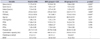
The characteristics of the study subjects are summarized in Table 1. The mean age (±standard deviation) was 65.3±9.2 years. There were no statistically differences between DU and BOO groups related to clinical variables. However, except for Qave, all uroflowmetry components were significantly different between groups. There was lower Qmax and larger PVR in DU group than the BOO group. DeltaQ was significantly smaller in DU group (8.71 mL/s vs. 5.26 mL/s, p<0.001) (Table 1). Multivariable logistic regression analysis demonstrated DeltaQ (Exp(B)=1.546, p<0.001) and PVR (Exp(B)=0.991, p<0.001, forward conditional method) as two independent predictors discriminating BOO from DU (Table 2).
In identifying DU using a single variable, the area under the curve (AUC) of ROC from DeltaQ (0.806) was significantly higher than that from Qmax (0.763, cutoff 11.05, sensitivity 69%, specificity 68.5%, p=0.0126) and Qave (0.574, p<0.0001) (Table 3). With a cutoff value of 6.65 mL/s, the sensitivity and specificity for DeltaQ were 71.3% and 70.3%, respectively.
DISCUSSION
A PFS is currently the only diagnostic test to differentiate between DU and BOO. BOO is described by increased detrusor pressure with decreased urinary flow, whereas DU is defined by decreased detrusor pressure with decreased urinary flow [13]. As a result, Qmax value of BOO is higher than that of DU or Qave value of DU is lower than that of BOO in uroflowmetry. This description is expected to investigate a DeltaQ for diagnosing DU and BOO. BOO is well characterized, and threshold values to distinguish between nonobstructed and obstructed bladders have been established by several researchers [11]. Combining reported parameters, several formulae and nomograms help physicians identify the degrees of BOO [11]. In contrast, for DU, the urodynamic definition of normal detrusor function and DU [14] is not diagnostically useful, besides being time consuming and invasive [1516].
Considering the high prevalence and similar clinical symptoms, several researchers have tried to develop alternative simple tools to distinguish DU from BOO, including IPSS and uroflowmetry parameters such as the amount of PVR. Rademakers et al. [17] reported a difference in IPSS, particularly using questionnaire 5, between nonobstructive patients with poor detrusor function and men with a normal detrusor function. Resnick et al. [18] suggested PVR over 50 mL as a potential indicator on DU in men without straining at voiding, BOO, and detrusor-sphincter dyssynergia. However, despite the clinical usefulness of these partial tools, there may be broader clinical parameters with better predictive characteristics, because interpretation with these tools is highly dependent on the clinical context [18]. Indeed, we found no difference in IPSS whether in obstructive or irritative symptoms. IPSS 5 was similar in this series. While the amount of PVR was significantly larger in DU group in this study as well, even in multivariable analysis, we do not believe this sole parameter to be a reliable predictor for DU, mainly because PVR itself usually is an effect rather than cause. These limitations lead us to focus on the equation using variables, not a single parameter from uroflowmetry. By combining the variables, our data demonstrated DeltaQ as a plausible parameter in discriminating DU and BOO. The basic concept of DeltaQ was originally addressed to identify a recurrence of urethral stricture. Tam et al. [19] reported an improved sensitivity using DeltaQ than Qmax. We adopted this concept to distinguish DU and BOO, and multivariable analysis demonstrated DeltaQ to be a significant predictor for DU. In ROC, the AUC from DeltaQ was significantly higher than Qmax. While static similarity in comparison with that from PVR, the highest AUC was obtained from DeltaQ. Indeed, the accuracy of PVR was similar to DeltaQ (sensitivity 70.3% and specificity 75.2% with PVR cutoff of 84.5 mL). However, because we cannot exclude certain diseases by using only PVR, this outcome let us invite expectation for the clinical usefulness of DeltaQ, in addition to current routine uroflowmetry variables.
The authors are aware of several limitations of this series. First, mainly because of innate limitation of the retrospective study design, the patient selection in this study may be biased. Because patient criteria for this study was divided into DU or BOO, the men with mixed urodynamic problems were excluded, although they are frequently met in clinical practice. While our data extracted a plausible surrogate of UDS, DeltaQ should be validated by a prospective trail. Second, there might be another condition affecting DeltaQ besides DU and BOO. Thus, further study of conditions that may affect DeltaQ should be undertaken. Third, while UDS was interpreted by a single experienced urologist, this should be cross-checked given the long time period of this series. Moreover, all of the data was collected by single institution.


 XML Download