

 PDF
PDF ePub
ePub Citation
 Print
Print


INTRODUCTION
The incidence of ureteral calculi is increasing, and Ureteroscopic lithotripsy (URS) plays a critical role in its treatment [1]. From the year 1997, ureteroscopy has remained the primary treatment modality for the management of symptomatic ureteral calculi, along with shock wave lithotripsy [2].
Postoperative hydronephrosis may be caused by fibrinous exudates produced by the mucosa that precipitate, or fragments of calculi remaining in the ureteral mucosa, causing inflammation and ischemia [3]. It is reported that the incidence of ureteral stricture formation, as a late complication after URS, occurs in 0% to 4% [45]. Several studies demonstrate that the development of ureteral perforations is related to a higher rate of postoperative ureteral stricture [6]. In addition, stricture may result from ureteral injury caused by surgical devices or from the presence of impacted stones. In this respect, passage of an oversized ureteroscope may cause stricture formation from mucosal trauma [7]. Ureteroscopic injury is the most common cause of ureteral stricture, involving both endoscopic and working ureteroscopic instruments [58]. In spite of significant technologic improvements, postoperative hydronephrosis as a late complication of URS still occurs in some patients, ultimately resulting in renal failure. Several studies have demonstrated the need for using imaging modalities, including renal ultrasonography (US), computed tomography (CT), and intravenous pyelography, routinely after URS in order to evaluate postoperative ipsilateral hydronephrosis, particularly those developing asymptomatically [91011]. However, data regarding the predictors of post-URS hydronephrosis are lacking. Therefore, we determined the predictors of ipsilateral hydronephrosis after URS for ureteral calculi.
MATERIALS AND METHODS
1. Study population
We retrospectively reviewed the data of patients who underwent URS for ureteral calculi between January 2010 and December 2014 at our institution. There were 204 patients who underwent URS for ureteral calculi. Patients with lack of clinical data (n=51), presence of ureteral rupture (n=2), and who underwent percutaneous nephrolithotomy at the same time (n=14) were excluded from the study. Based on these criteria, 137 patients were enrolled in the present study. Postoperative ureteral stricture was not reported in the medical records; therefore, we could not evaluate ureteral stricture following ureteroscopic stone removal. All procedures performed in studies involving human participants were in accordance with the ethical standards of the institutional and/or national research committee and with the 1964 Helsinki declaration and its later amendments or comparable ethical standards.
2. Data collection
All patients who were imaged using a CT scan or renal US 4 to 12 weeks after URS or ureteral stent removal were included in this study. Patients with hydronephrosis at that time were reevaluated consequently. Postoperative hydronephrosis was determined 6 months after URS with CT scan or renal ultrasound.
Operative details and patient and stone characteristics were investigated to identify variables associated with post-URS hydronephrosis. Preoperative patient characteristics acquired were sex, age, body mass index (BMI), history of hypertension, history of diabetes mellitus (DM), and history of ipsilateral stone procedure (including extracorporeal shock wave lithotripsy or ESWL, URS, and ureterolithotomy). The reasons for preoperative imaging were pain, gross hematuria, fever, anorexia and nausea, general edema, decreased urine output and incidental finding of ureteral stone.
Stone factors included stone location (upper ureter, midureter, lower ureter), maximum diameter of largest calculi, preoperative hydronephrosis, preoperative decompression procedure (indwelling ureteral stent or percutaneous nephrostomy), number of calculi, stone laterality (including 6 cases of bilateral ureteral stones), duration between time of stone diagnosis and time of operation, duration of postoperative ureteral stent placement, operative duration, and stone-free rate. Preoperative hydronephrosis grade and diameter were also evaluated. Regarding the preoperative hydronephrosis grade, cases without caliceal or pelvic dilation were classified as grade 0, cases with pelvic dilation only were classified as grade 1, cases with accompanying mild calix dilation were classified as grade 2, cases with severe calix dilation were classified as grade 3, and cases with calix dilation accompanied by renal parenchyma atrophy were classified as grade 4 [12]. The diameter of the hydronephrotic kidney was defined as the largest diameter of the ureter at the level of the ureteropelvic junction. Intraoperative findings, including presence of impacted stone, ureteral stricture, ureteral kinking or folding, simultaneously executed retrograde intrarenal surgery (RIRS), and ureterotomy or ureteral dilation, were acquired. Impacted stone is defined as the stone that has remained in the same position for follow period or not movable through the ureter with mucosal swelling as inspecting using the ureteroscopy in the operative field. Postoperative ipsilateral hydronephrosis was the outcome in this study. On assessing postoperative ipsilateral hydronephrosis, only presence or absence was recorded, excluding hydronephrosis grade and diameter, contrary to preoperative hydronephrosis. For the analysis of clinical variables, patients were distributed into 2 groups: patients without postoperative hydronephrosis and patients with postoperative hydronephrosis.
3. Statistical analysis
Data were analyzed using SPSS ver. 17.0 (SPSS Inc., Chicago, IL, USA). Demographic and clinical data were compared using the Student t-test for continuous variables and the chi-square test for categorical variables between the 2 groups. Logistic regression analysis (stepwise forward procedure) was used to quantify the adjusted odds ratios (ORs) for the presence of hydronephrosis on imaging. The factors included in the model were sex, age, BMI, ESWL history, URS history, ureterolithotomy history, preoperative symptom, stone laterality, stone location, stone size, stone number, preoperative hydronephrosis grade, preoperative diameter of hydronephrotic kidney, preoperative decompression procedure, operative findings, time from diagnosis to operation, operation time, ureteral stent duration, stone free. Among the factors, those with p<0.25 were selected (on univariable analysis) and included in the multivariable logistic regression analysis. In all cases, twotailed p<0.05 was considered statistically significant.
RESULTS
1. Baseline characteristics
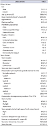
The data of a total of 137 patients were evaluated. The mean age of the patients was 58.8±14.2 years. Of the patients, 76 (55.5%) were male, while 61 (44.5%) were female. The mean BMI of the population was 24.5±3.6 kg/m2. Of the 137 patients, 45 (32.8%) were confirmed to have hypertension and 34 (24.8%) were confirmed to have DM. Eighty-seven patients underwent a prior ipsilateral stone procedure, including 69 patients (50.4%) with a prior ipsilateral ESWL, 14 patients (10.2%) with a prior ipsilateral URS, and 4 patients (2.9%) with a prior ipsilateral ureterolithotomy. The reasons for performing preoperative imaging for patients were pain for 93 patients (67.9%), gross hematuria for 11 (8.0%), fever for 2 (1.5%), decreased urine output for 2 (1.5%), anorexia and nausea for 1 (0.7%), and general edema for another patient (0.7%). In addition, 27 patients (19.7%) were diagnosed with ureteral stone incidentally (Table 1).
Mean stone size was 10.0±4.6 mm. Thirty-nine patients (28.5%) had multiple stones and 3 patients (2.2%) had steinstrasse. The stone-free rate on follow-up imaging was 85.4% (117 of 137 patients). The stone was located in the upper part of the ureter in 61 patients (44.5%), in the midureter in 29 patients (21.2%), and in the lower part of ureter in 47 patients (34.3%). When considering the laterality of the stone, 69 patients underwent an operative procedure for a right ureter stone, while 68 patients underwent an operation for a left ureter stone. Of the 137 patients, 121 patients were proven to have hydronephrosis before URS. Of the 121 patients with preoperative hydronephrosis, 39 patients were graded 1 (mean diameter, 12.1 mm), 35 patients were graded 2 (mean diameter, 15.7 mm), 28 patients were graded 3 (mean diameter, 22.5 mm), and 19 patients were graded 4 (mean diameter, 28.0 mm). Preoperative ureteral stenting or percutaneous nephrostomy was employed in 20 patients (14.6%). The mean operation time was 73.1±32.0 minutes (Table 1).
2. Comparison between nonhydronephrosis group and hydronephrosis group
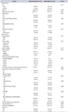
Overall, postoperative imaging revealed hydronephrosis in 44 of 137 patients (32.1%) between 4 and 12 weeks following stent removal. Among them, 6 patients (13.6%) had symptoms and 2 patients (4.5%) required an ancillary procedure, which is reureteral stenting. Clinical factors, such as sex, age, BMI, prior ipsilateral stone procedure and preoperative symptoms, were not different between the 2 groups (Table 2).
Regarding stone factors, the location of the stone (p=0.017) and the preoperative hydronephrosis (p=0.001, both grade and diameter) were significantly different between the groups (Table 2).
In addition, significant differences were noted between the hydronephrosis and nonhydronephrosis group in terms of impacted stones (p=0.003), operation time (p=0.049), and ureteral stent duration (p=0.001). Meanwhile, there was no difference in other intraoperative factors, such as presence of ureteral stricture, presence of ureteral kinking or folding, simultaneously executed RIRS, or ureterotomy, or ureteral dilation (Table 2).
3. Predictors of postoperative hydronephrosis
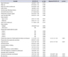
Univariable analysis showed that stone location (upper ureter: OR, 2.95; 95% confidence interval [CI], 1.17–7.40; p=0.021 and midureter: OR, 3.96; 95% CI, 1.37–11.3; p=0.011), preoperative diameter of hydronephrotic kidney (OR, 1.13; 95% CI, 1.06–1.17; p=0.001), impacted stone (OR, 3.24; 95% CI; 1.48–7.05, p=0.003) and ureteral stent duration (OR, 1.04; 95% CI, 1.02–1.08; p=0.002) were associated with postoperative hydronephrosis. The independent predictive value of increasing preoperative diameter of the hydronephrotic kidney (adjusted OR, 1.21; 95% CI, 1.12–1.31; p=0.001) and the presence of an impacted stone (adjusted OR, 3.01; 95% CI, 1.15–7.61; p=0.031) persisted after multivariable logistic regression analysis (Table 3).
DISCUSSION
In this study, we retrospectively reviewed the results of 137 patients who underwent URS for ureteral stones, with postoperative imaging 4 to 12 weeks after operation or stent removal, in order to identify the factors for the development of postoperative hydronephrosis. The incidence of hydronephrosis was 32.1%. An increasing preoperative diameter of the hydronephrotic kidney and the presence of an impacted stone were detected as independent predictors of postoperative hydronephrosis.
Studies on postoperative hydronephrosis after URS were investigated by several groups, but results were conflicting because a consensus for imaging after URS has not been established. Weizer et al. [9] investigated 241 patients who underwent URS with 3-year follow-up. On the basis of their results, the authors recommended routine postoperative imaging, including excretory urography, US, or spiral CT, within 3 months after URS. However, Bugg et al. [13] recommended that functional imaging studies are essential for patients who complain of postoperative pain and reveal obstruction. They investigated 87 patients with follow-up radiographic studies. In their study, 96% of patients without complaints of pain postoperatively and without obstruction preoperatively did not develop postoperative obstruction or present with residual stone fragments. Additionally, Karadag et al. [14] revealed that ureteral stricture did not develop in patients with no symptoms who underwent uncomplicated URS. Therefore, they recommended that imaging follow-up for evaluation of postoperative ureteral stricture or obstruction is not essential following complete stone removal via uncomplicated URS.
Newly developed hydronephrosis after URS may be caused by ureteral obstruction resulting from residual stone fragments or ureteral edema damage to the ureteral mucosa [1516]. Ureteral injury during URS may be a reason for postoperative hydronephrosis. Also, irrigation solution during operation may cause hydronephrosis, which can be exacerbated by a long operation time [16]. Embedding of stone fragments within the wall of the ureter may result in stone granuloma and stricture resulting in postoperative hydronephrosis [5]. Barbour and Raman [4] reported that prior ipsilateral URS, the diameter of the largest stone, the duration of operation and colic symptoms are independent predictors for the development of postoperative ipsilateral hydronephrosis, and are indications for imaging. In addition, Gokce et al. [17] reported that a history of ipsilateral URS, ureteral injury during operation, and presence of impacted stones are predictors of ipsilateral postoperative hydronephrosis in pediatric patients. In our study, the degree of preoperative hydronephrosis and presence of an impacted stone are independent predictors of postoperative hydronephrosis. When considering patients with ureteral calculi with higher grades of preoperative hydronephrosis, structural modifications in the ureter may develop, caused by persistent irritation and resulting in fibrosis or hyperplasia of the ureteral mucosa. Following URS, this irreversible modification would persist, since a larger hydronephrosis suggests a longer duration of modification. The presence of an impacted stone probably causes inflammation and edema of the ureteral wall and these changes may affect the tissues. Ureteral edema and fibrosis may arise from diminished blood flow secondary to chronic pressure or from an immunological reaction to the stone material [18]. In this study, if ureteral stricture, ureteral mucosal swelling, mucosal folding, or impacted stone were observed throughout the URS, postoperative ureteral stent longer maintained. These operative findings mentioned above account for the significant relationship with the occurrence of postoperative ipsilateral hydronephrosis. Roberts et al. [19] reported that 24% of patients with stones impacted for more than 2 months presented with ureteral stricture. Additionally, Adiyat et al. [20] reported that ureteral strictures were found in 27.2% of patients with impacted stones. However, in our study, postoperative ureteral stricture or cause of postoperative hydronephrosis was not investigated. We presumed that ureteral edema develops into temporary hydronephrosis, while ureteral fibrosis develops into persistent hydronephrosis; both of these mechanisms may explain why stone impaction influences the development of postoperative hydronephrosis. Barbour and Raman reported 49 of 324 patients (15%) had hydronephrosis following URS in 4 to 12 weeks [413]. In our study, 32.1% of patients had postoperative hydronephrosis. The clinical characteristics of patients in our study may have been somewhat different from those in previous study such as relatively high proportion of patients with impacted stone and preoperative hydronephrosis.
Ureteral obstruction or stricture formation following URS would have a negative impact on renal function [21]. Hydronephrosis resulting from obstruction has been acknowledged to harm the parenchyma of the kidney, resulting in renal failure [22]. Impaired renal function would become irreversible, while hydronephrosis improves after treatment [23]. Given this perspective, we should be concerned about risk of developing hydronephrosis following URS.
The present study is not without drawbacks. First, our study is retrospective. Second, the patients who were evaluated using renal US were included in the review. Renal US is a less sensitive modality for detecting postoperative residual f ragments and assessing stone-f ree rates, compared with CT [242526]. However, the patients follow-up were examined using renal ultrasound mostly rather than postoperatively CT, but not checked hydronephrosis diameter. Further prospective studies using equivalent imaging modalities to assess preoperative and postoperative hydronephrosis could resolve this limitation. Third, we could not distinguish the effect of the type of lithotripter. Other limitations include different surgeons performing the surgical procedures, a relatively small sample of patients, no investigation of cause of postoperative hydronephrosis and the lack of long-term follow-up. In addition, the patients with presence of ureteral rupture were excluded in our study. Therefore, the complication during URS was not evaluated validating for predicting factors for postoperative hydronephrosis.
Despite these limitations, this study remains important in evaluating the risk factors of ipsilateral hydronephrosis after URS. Therefore, the patients with these predictive factors should be considered to undergo imaging examinations and renal function follow-up routinely, to prevent renal insufficiency.
CONCLUSIONS
Large preoperative diameter of the hydronephrotic kidney and the presence of impacted stones were associated with postoperative hydronephrosis. Therefore, patients with these findings should receive more active imaging follow-up to avoid renal insufficiency. To confirm our findings, prospective and well-designed studies are needed.
 XML Download