

 PDF
PDF ePub
ePub Citation
 Print
Print


INTRODUCTION
Prostate cancer (PCa) has mostly affected the male population of the United States, and in 2012 US cases constituted approximately 29% (240,890) of the incidence of PCa [1]. In South Korea, the incidence of PCa has significantly increased while the mortality rate has steadily decreased. From 2007 to 2013, the incidence PCa in Korean men doubled (5,516 to 10,855 per year), while the prevalence tripled (18,830 to 51,411) during the same period. The mortality rate increased slightly, from 4.2 in 2000 to 5.9 in 2007 and 6.0 in 2013 (per 100,000, age-adjusted). PCa incidence increased significantly faster in men <70 years of age than in the older age group [2].
The emergence of nanotechnology has drawn much attention in the 21st century for applications in medicine such as "nanomedicine." Nanomedicine combines engineering, physics, biology, chemistry, mathematics, and medicine and strives to improve disease detection, imaging, and drug delivery through the use of nanodevices [3]. Nanotechnology encompasses the materials, devices, and delivery systems for disease diagnostics, prevention, and treatment. Both researchers and the pharmaceutical industry have shown particular interest in nanotechnology for medical applications with potential benefits for patients. Nanotechnology-based medicine can hurdle both physiological and biological barriers, such as the blood-brain barrier, endothelial barriers, cell membranes, and even nuclear envelopes, achieving both passive and active disease targeting [4]. Nanosized particles are normally composed of thousands of atoms and exhibit unique physical and biochemical properties with high surface area for therapeutic loads such as cancer drugs. However, challenges to nanomedicine exist, such as safety issues, bulk manufacturing issues, and compatibility with the human body of nano-sized drug delivery systems. Meeting these challenges is essential for successful application of drug-loaded nanoparticles (NPs) in the field of pharmaceutics [5].
Different approaches have been taken to conjugate NPs with targeting molecules that specifically bind to the surface markers expressed in various tumors [6], such as prostatespecific membrane antigen (PSMA) in PCa [7]. Prepared NPs are also used in real-time monitoring of treatment by attaching various imaging moieties that help in visualizing the distribution of NPs and the drugs [8]. NPs continue to be investigated for their potential to improve existing therapies and to develop novel therapies. At the length scale of less than 200 nm, NPs with encapsulated therapeutics can be injected intravascularly without concern for embolization. At this scale, NPs have the potential to permeate and traffic through different tissues, bind to cell surface receptors, enter target cells for intracellular delivery of their cargos, and influence the intracellular tracking pathways. Owing to these unique characteristics, a multitude of applications have been identified for NP-mediated drug delivery [9]. NPs can be used as probes with many advantages such as rapid clearance from the bloodstream by macrophages and depending on the size of the NPs renal clearance that helps them to remain in the circulation for a long time [1011]. Indeed, it is important that the NPs have a multivalency effect with more than one type of targeting ligand [1213]. Studies have shown that combined therapy and diagnosis (nanotheranostics), with both imaging and therapeutic capacity, can be achieved [14]. NP composition plays a critical role in drug delivery systems and determines the physicochemical nature of the particle surface for effective delivery. However, some factors of nano-formulated drugs create challenges for batch-to-batch manufacturing, reproducibility, and safety.
A thorough understanding of the therapeutic potential of nanodrugs is necessary for commercialization [15]. The following sections discuss the nanomedicines that have been explored in PCa in vitro and in vivo, respectively.
NANOMEDICINE DELIVERY FOR PROSTATE CANCER
Nanomedicines have emerged as promising treatments for cancer owing to their noninvasiveness. Live animal imaging technology and advances in nanotechnology are providing new research opportunities in the preclinical and clinical development of nanosized drug carriers in cancer therapy. Nanoparticulate-based medicine has been expanding and has attracted significant interest in recent years, with a primary focus on efficient delivery systems for various chemotherapy drugs [3]. The unique properties of nanomedicines are frequently used to improve the therapeutic value of various water-soluble and insoluble medicinal drugs and bioactive molecules by improving pharmacokinetics behavior, bioavailability, solubility, and retention time [16]. These NP-drug formulations reduce patient expenses and risks of toxicity [4]. Nanoencapsulation of chemotherapy drugs (nanomedicines) increases drug efficacy, specificity, tolerability, and the therapeutic index of corresponding drugs [51718192021].
Several disease-related drugs and bioactive molecules have been successfully encapsulated to improve bioavailability, bioactivity, and controlled delivery [222324]. Nanomedicines are being developed not only for cancer, but also in the area of acquired immunodeficiency syndrome, diabetes, malaria, prion disease, and tuberculosis [252627282930]. Hyaluronic acid has attracted interest for targeted delivery in cancer therapy mainly because of its specificity to bind cancer cells. Another nanomedicine being developed incorporates the well-known anticancer drug cisplatin. Currently, cisplatin is used in various solid tumor treatments [3132]. However, cisplatin has dose-limiting side effects such as neurotoxicity, gastrointestinal disturbance, and nephrotoxicity that limit its clinical use [33]. NP formulations have been developed for efficient delivery of cisplatin [34] and targeting of PCa stem cells by use of CD44 surface marker tagging with hyaluronic acid [3536]. With the use of a drug-induced ionic gelation method, cisplatin glyconanoparticles have been investigated as an efficient drug delivery system targeting PCa stem cells. One group showed that the nanomedicine formulation increases cancer stem cell suppression [37].
A proven concept for drug delivery applications in nanomedicine is the use of carriers such as nanovectors including liposomes, micelles, and dendrimers as well as metallic, ceramic, protein, and polymeric NPs and carbon-based nanostructures [3839]. In particular, carbon nanotubes possess unique features for biomedical applications, including the possibility of chemical functionalization with drugs and their internalization by various cell types through endocytosis and passive diffusion across cellular membranes. Carbon nanotubes have been shown to efficiently transport and release various anticancer drugs [40414243]. With the use of a carbon nanotube-based approach, the natural compound catechin was recently explored as a potential therapeutic nanomedicine for PCa treatment. Catechin is in the family of polyphenolic flavonoids, and catechin and its derivatives (2)-epigallocatechin-3-gallate, (2)-epigallocatechin, (2)-epicatechin-3-gallate, and (2)-epicatechin inhibit cancer cell proliferation, suppress tumor growth and metastasis, and sensitize tumors to various chemotherapy drugs and radiotherapy [444546]. Recently, a study showed that biocompatible catechin-loaded and gelatin-conjugated carbon nanotubes have good anticancer properties with the potential for targeting prostate PCa stem cells in combination with x-ray irradiation [47].
The various chemotherapy drugs currently in use have some drawbacks. For example, the semisynthetic chemotherapy drug known as docetaxel, which is used alone and in combination for various tumors, has limitations including poor aqueous solubility and low oral bioavailability. Moreover, docetaxel has intolerable side effects such as acute hypersensitivity reactions, fluid retention, neurotoxicity, and febrile neutropenia [4849]. To overcome these disadvantages of docetaxel, nanomedicine has been explored for passive and active targeting by enhancing permeability and retention (EPR) effect, avoidance of the reticuloendothelial system, prolonged blood circulation time, controlled release of drug, and providing an opportunity for surface modification for active drug targeting [5051]. To enhance the permeability and bioavailability of docetaxel, one researcher used D-α-tocopheryl polyethylene glycol succinate (TPGS, or Vit E TPGS), a Food and Drug Administration-approved adjuvant for drug delivery, and showed that cyclic peptide (cRGDfK) can be used to conjugate succinoyl TPGS nanomicelles for docetaxel delivery in a successful PCa-targeted drug delivery system [52].
Another interesting nanoparticulate system for cancer treatment is magnetic iron oxide NP clusters, which have been shown as promising drug delivery vehicles for both diagnostic and therapeutic applications. Magnetic NP clusters have been shown to be stable in a biological circulation system, to readily interact with cells or other biological units of interest, and to be capable of releasing the drug once the selected targeting is realized. In addition, the magnetic NP clusters are biocompatible, have been proven to have no toxicity in vivo, and have shown efficient contrast in magnetic resonance imaging (MRI) [53]. Magnetic NP clusters are excellent near-infrared photothermal mediators in the combination of photothermal therapy and chemotherapy. Recently, doxorubicin (DOX) when loaded into magnetic NP clusters achieved greater PCa cell cytotoxicity owing to both cluster-mediated photothermal ablation and cytotoxicity of light-triggered DOX release. Compared with photothermal therapy or chemotherapy alone, the chemophotothermal therapy approach with DOX in magnetic NP clusters showed synergistically higher therapeutic efficacy both in vitro and in vivo for treatment of the PCa cell line PC3 [54].
Natural gum arabic glycol protein has recently been explored for homogeneous dose distribution and efficient emission. Researchers have reported that NPs coated with gum arabic are stable for in vivo applications. One study showed that intralesional injection of gum Arabic-coated radioactive gold NPs resulted in minimal short-term systemic toxicity in a naturally occurring large animal model of PCa [55]. To achieve better therapeutic efficacy, the use of liposomal micelles to encapsulate dexamethasone was recently reported for sustained, antitumor effects versus free dexamethasone in metastatic bone disease from human PCa. These studies showed that intravenously administered liposomes localized efficiently to bone metastases in vivo and that treatment of established bone metastases with (liposomal) dexamethasone resulted in a significant inhibition of tumor growth up to 26 days after the initiation of treatment [56]. Furthermore, 1.0-mg/kg liposomal dexamethasone significantly outperformed 1.0-mg/kg free dexamethasone and was found to be well tolerated at clinically relevant dosages. Delivery of liposomal-loaded glucocorticoid dexamethasone was thus proposed for future clinical evaluation as a promising new treatment option for advanced, metastatic PCa [56].
In contrast to the overexpression of a membrane lectin, expression of the cation-independent mannose 6-phosphate receptor (M6PR) was specifically demonstrated in PCa cell lines and tissues. M6PR was proposed as a new, efficient target and alternative biomarker to PSMA for nanomedicine applications. M6PR is a ubiquitous receptor involved in several biological functions, mainly in addressing enzymes to lysosomes [575859]. In one study, a mannose-6-phosphate analogue was synthesized and grafted onto the surface of functionalized mesoporous silica NPs as a theranostic therapy for PCa. Mesoporous silica NPs can be used for targeting, imaging, and photodynamic therapy. Therefore, M6PR appears as a promising new target for nanomedicine applications in PCa, particularly for noninvasive and personalized therapy of small-sized prostate tumors [60].
In the area of novel drug inhibitors, histone deacetylase inhibitors (HDACIs) do not have significantly better outcomes in solid malignancies than current standard therapies [61]. Moreover, the efficacy of the first-generation HDACIs was limited in solid tumor indications owing to their suboptimal potency for specific HDAC enzymes and transient induction of histone acetylation in tumor tissue [62]. Considering the several drawbacks of HDACIs, nanomedicine applications have been designed with novel NP formulations of HDACIs, vorinostat and quisinostat, which release the drug in a slow and controlled fashion. The NPs vorinostat and quisinostat demonstrated higher therapeutic efficacy than small-molecule HDACIs in chemoradiotherapy in murine models of PCa. Vorinostat and quisinostat are promising cancer therapeutics with the potential to significantly improve chemoradiotherapy [63].
The chemotherapy drug docetaxel, a second-generation taxoid, has antimitotic chemotherapeutic properties and has been used for a variety of cancers, especially PCa. In response to drawbacks such as docetaxel solubility, toxicity issues, and inaccurate tumor targeting, researchers have successfully fabricated docetaxel as a nanomedicine for sustained drug release by use of a copolymer-based poly(styrene)-b-poly(DL-lactide) (PS-PDLLA) for intravenous injection. Interestingly, the carrier copolymer did not induce any serious cytotoxicity in PC3 cells. Moreover, improved inhibition of cancer cell growth was shown in the group treated with PS-PDLLA/docetaxel compared with the control group. Decreased in vivo drug clearance, elevated systemic exposure, and prolonged circulation of the drug were verified in a pharmacokinetic study in rats, which further supports the potential use of nanomedicines in PCa therapy [64].
In a similar study, researchers developed a novel nanoplatform based on N-(2-hydroxypropyl) methacrylamide (HPMA) copolymers that allows covalent bonding of two chemotherapeutics acting via different anticancer mechanisms and that can enter target cells by receptor-mediated endocytosis. In that study, DOX and 5-fluorouracil (5-Fu) were covalently conjugated to a nanosized HPMA copolymer via a pH-sensitive hydrazone bond and an enzymatically degradable oligopeptide Gly-Phe-Leu-Gly sequence. The conjugate was decorated with galectin-3 targeting peptide G3-C12 [P-(G3-C12)-DOX-Fu]. Interestingly, these two drugs showed similar release profiles in vitro, suggesting synergistic effects in the co-delivery system. Studies in the cellular system showed that overexpression of galectin-3 in human PC3 prostate carcinoma cells treated with a combination of the drug P-(G3-C12)-DOX-Fu surprisingly exhibited comparable cytotoxicity to free DOX at high concentrations by increasing cell internalization and exerting synergistic genotoxic effects of cell cycle arrest, caspase-3 activation, and DNA damage. In mice bearing PC3 tumor xenografts, the use of tumor-targeting ligand substantially enhanced the intracellular delivery of P-(G3-C12)-DOX-Fu with no adverse effects. Thus, potential exists for synergistic combination therapy using targeted nanomedicines for efficient treatment of PCa [65].
Like other imaging modalities such as computed tomography, transurethral ultrasound, and nuclear imaging, MRI cannot adequately detect small tumors [66]. Improvements in tumor imaging technologies are urgently required for early detection of disease, staging, and realtime assessment of response to therapy in PCa patients. Similar to other metal nanocarriers for nanomedicine, iron oxide magnetic NPs have emerged as powerful contrast agents for MRI [67]. One research group developed iron oxide magnetic NPs to enhance MRI of PCa with the PSMAtargeting antibody, J591, via a 1,2-distearoyl-sn-glycero-3-phosphoethanolamine-N-[amino(polyethylene glycol) (PEGDSPE) linkage. In this preclinical model, PSMA-targeted magnetic NPs effectively enhanced MRI of PCa [68].
Another interesting area of research on phototherapy is the quantum dot (QD) technique, and numerous studies have been done with QD-based fluorophores with high photostability for diagnostics and biomedical assays. QD-based systems can achieve cost-effective antigen detection. Recently, researchers designed a new multiplexed diagnostic system based on QD-encoded beads for simultaneous detection of free and total PSA for ultrasensitive, accurate, early PCa diagnosis [69].
In contrast to the nano-micelle area, other research groups have developed a carrier system for castrate-resistant PCa by using raloxifene-loaded poly(styrene-co-maleic acid) (SMA) micelles (SMA-raloxifene) to improve the biodistribution of raloxifene and increase its anticancer efficacy. The effect of SMA-raloxifene on apoptosis, reduction of cell proliferation, migration, and invasion of castration-resistant PCa (CRPC) cells was superior compared with free drug in vitro [70]. In addition, SMA-raloxifene induced changes in expression and localization of the estrogen receptors, as well as downstream effectors associated with cell proliferation and survival [70]. This study examined the cellular internalization and efflux of SMA-raloxifene compared with free raloxifene. The biodistribution and anticancer efficacy of SMA-raloxifene were then compared with free raloxifene in a CRPC mouse xenograft model [71].
In the area of radiation therapy for prostate tumors, researchers have developed gold NPs that can be targeted by using goserelin-conjugated gold nanorods. In this approach, megavoltage radiosensitization in vivo using tumor goserelin-targeted gold NPs showed independent mechanisms of tumor accumulation in vivo (EPR effect as a consequence of leaky tumor vasculature and active uptake by target cells). In combination with megavoltage radiation therapy, this led to delays in tumor growth in mice with heterotopic PCa. More importantly, significant radiosensitization to megavoltage radiation was not observed with unconjugated gold NPs despite their intratumoral concentration being only 3 folds less than that of conjugated gold NPs. This suggests that radiosensitization is improved by active targeting that leads to cellular internalization of NPs and the consequent increase in ionization density within the cytoplasm, rather than merely passive accumulation of NPs in the perivascular space by EPR. This report concluded that conjugation of NPs to tumor-specific antigens, which promotes internalization within cancer cells, causes radiosensitization and that goserelin-conjugated gold NPs can be effective PCa radiosensitizers when used with megavoltage radiation [72].
Another strategy being developed is to combine a chemotherapy drug and a natural drug as a hybrid nanomedicine for metastatic CRPC. One such study used lipid-polymer hybrid NPs to co-encapsulate DTX and curcumin (CUR). In mice bearing PC3 tumor xenografts, the DTX-CUR lipid-polymer NPs synergistically inhibited tumor growth to a greater extent with no adverse effects. Nanomedicine offers great promise for dual drug delivery to PCa cells, with the potential for synergistic combination therapy [73]
In summary, interesting nanomedicines have been developed for PCa in recent years. These namomedicines need to be further evaluated in higher animals in terms of their safety and toxicity profiles.
NANOGENE DELIVERY FOR PROSTATE CANCER
Gene therapy offers a solution to controlled and specific delivery of DNA, RNA, and protein to cells. Chitosan-based carriers are nonviral vectors that have gained popularity as a safe delivery system for gene materials including plasmid DNA, oligonucleotides, and small inhibitory RNA (siRNA). Chitosan has beneficial qualities such as low toxicity, low immunogenicity, and excellent biocompatibility [7475]. Our group reported a preliminary investigation with siRNA targeted to AGR2 conjugated with chitosan to form NPs. Secretion of AGR2 plays a predominant role in PCa progression and metastasis. The results of scanning electron microscopy and gel electrophoresis showed efficient condensation of siRNA for AGR2 [76]. To improve the transfection efficiency of the AGR2 siRNA, the complexes were tested in the human PC3 prostate cell line, which overexpresses AGR2 protein. In PC3 cells, the chitosan and siRNA AGR2 complex was found to be efficient and nontoxic to overall cells by 3-(4,5-dimethylthiazol-2-yl)-2,5-diphenyltetrazolium bromide assay. Thus, chitosan NPs have been shown to be a safe vector system for efficient gene delivery in cancer. Our study proposed further development of chitosan-based formulations for the delivery of AGR2 siRNA for clinical applications, which may represent a promising approach in PCa treatment [76].
Interestingly, another delivery strategy for siRNA in a vector involved coupling stearic acid to the N-terminus of HHHPKPKRKV. STR-HK formed stable complexes with siRNA via noncovalent interactions and mediated efficient delivery of siRNA to PC3 cells with minimal cytotoxicity compared with a gene-silencing chemical transfection agent like lipofectamine. These results proved that STR-HK is a promising vector for siRNA delivery [77]. In a similar fashion, research also demonstrated that intratumoral injections of siRNAs loaded on biodegradable chitosan NPs led to a downregulation of RXFP1 receptor expression and a dramatic reduction in tumor growth [78]. In the xenograft models treated with siRNA against RXFP1, the smaller tumor size was associated with decreased cell proliferation and increased apoptosis, which suggests that the suppression of RLN/RXFP1 might have potential therapeutic benefits in PCa [78]. Another interesting report showed that dicer complex of siRNA (dsiRNA), when delivered using a structurally flexible triethanolamine-core poly(amidoamine) dendrimer as the nanocarrier, gives rise to a much greater RNAi response than that with conventional siRNA. Dendrimer/dsiRNA complexes with a dual targeting peptide simultaneously promoted cancer cell targeting by interacting with integrins and cell penetration via the interaction with neuropilin-1 receptors, which led to improved gene silencing and anticancer activity. These findings suggest therapeutic applications of RNAi using dendrimer nanovector-based targeted delivery [79].
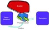
The role of nanomedicines and nanogene delivery in theranostics approaches for PCa are summarized in Fig. 1. Preclinical safety guidelines still need to be addressed.
CONCLUSIONS
This review has highlighted nanomedicine and nanogene therapy research in association with nanotechnology concepts for modern medicine, especially for improving the therapeutic outcomes of cancer. This review has also presented recent developments in nanomedicine for future clinical applications. Nanomedicine can effectively deliver medicine specifically to prostate tumor sites (Table 1). As the technology is perfected, nanomedicines can be used in the future in benign prostatic hyperplasia at the level of expression and location of proteins [80]. As image-guiding techniques such as magnetic iron oxide NPs (Fe3O4 NPs) are perfected, image-guided radiation therapy can be used for PCa [81]. Moreover, application of nanotechnology to extraperitoneal laparoscopic radical prostatectomy [82] can enable patients to have a better quality of life and improve the survival rate in the near future.

 XML Download