

 PDF
PDF ePub
ePub Citation
 Print
Print


INTRODUCTION
Bladder cancer (BC) is the eighth most common cancer in Korean men [1]. At the time of diagnosis, the majority of BCs are nonmuscle invasive bladder cancers (NMIBCs), with the remainder being muscle invasive bladder cancers (MIBCs) and metastatic disease. Although these cancer types originate from the same transitional epithelium (the so-called urothelium) in the urinary bladder, they have different clinical characteristics. Despite the fact that NMIBC has a better prognosis than MIBC or metastatic BC, frequent recurrence (50%–80%) and a tendency to progress to MIBC (10%–25%) after transurethral resection (TUR) are serious problems for both urologists and patients [2]. For example, while radical cystectomy is considered the gold standard treatment for MIBC, patients often have poor clinical outcomes [3]. Therefore, if we are to treat BC effectively, a precise and early diagnosis is critical.
Most patients with BC visit the hospital due to gross total painless hematuria [45]. However, hematuria is not a specific sign in BC patients. Urolithiasis, urinary tract infections, and many benign urological diseases can cause hematuria. Messing et al. [67] demonstrated that screening asymptomatic men aged 50 years and older, who self-test their urine repeatedly with a chemical reagent strip, can detect early stage BCs and other serious urologic diseases. They also determined whether patients with BCs detected by hematuria screening had earlier stage tumors and better outcomes than those whose tumors were detected in the absence of screening. However, the results were not promising in terms of early BC detection. Thus, there is still no "ideal" noninvasive method of discriminating between hematuria and BC.
Topoisomerases are enzymes belonging to the DNA gyrase family that are involved in many aspects of DNA metabolism, including DNA replication, transcription, and other critical cellular processes [8]. Topoisomerase-II alpha (TopoIIA ), a DNA gyrase isoform that plays an important role in the cell cycle, catalyzes the isomerization of DNA by facilitating the passage of one strand of DNA through a reversible break in the second strand [9]. Altered expression of TopoIIA occurs in both normal tissues and various human cancers [910111213]. We also previously reported that increased expression of TopoIIA is significantly related to a high rate of recurrence and progression of NMIBC. Thus, TopoIIA is a promising prognostic marker for NMIBC [14].
Historically, a diagnosis of BC depended on a combination of rigid or flexible cystoscopy and urinary cytology. However, the cystoscopic procedure is expensive, invasive, and uncomfortable. Urinary cytology is a convenient noninvasive method for diagnosing BC. However, although its specificity is high, its sensitivity is very low, which reduces its overall reliability. Therefore, novel and noninvasive diagnostic methods that can distinguish BC from noncancers are needed.
The aim of the current study was to measure the levels of TopoIIA cell-free DNA in urine samples from BC patients and controls (including normal controls and nonmalignant hematuric patients), and to assess its utility for the noninvasive diagnosis of BC.
MATERIALS AND METHODS
1. Study populations and samples
All primary tumor samples obtained from patients who underwent TUR or radical cystectomy were histologically verified as urothelial carcinoma at Chungbuk National University in South Korea. Normal bladder mucosa was harvested from patients with benign diseases such as benign prostatic hyperplasia and stress urinary incontinence after informed consent. All urine samples were collected prior to surgery on the first morning postadmission and centrifuged at 25,000 rpm for 15 minutes. The supernatants were then stored at –20℃ until use. All tumors were macro-dissected, typically within 15 minutes of surgical resection. Each BC specimen was confirmed by pathological analysis of fresh frozen sections cut from TUR or cystectomy specimens; the remaining unsectioned tissue samples were then frozen in liquid nitrogen and stored at –80℃ until use. Tumors were staged and graded according to the 2002 TNM classification and the European Association of Urology guidelines based on the 1973 World Health Organization grading system [151617]. The study protocol was approved by the Ethics Committee of Chungbuk National University. All subjects provided written informed consent. Sample collection and analysis were approved by the Institutional Review Board of Chungbuk National University. The biospecimens used for this study were provided by the Chungbuk National University Hospital, a member of the National Biobank of Korea, which is supported by the Ministry of Health, Welfare and Family Affairs.
2. Extraction of cell-free DNA from urine
Urinary cell-free DNAs were extracted using the QIAquick gel extraction kit (Qiagen GmbH, Hilden, Germany). Each frozen urine sample (1 mL) was thawed at room temperature and treated with 500 µL of QG buffer (contained in the QIAquick gel extraction kit). After incubation for 10 minutes at 50℃, 500 µL of isopropanol was added to the sample and mixed. The sample was then transferred onto a QIAquick column, which binds cell-free DNA. The column was placed into a 2-mL collection tube and centrifuged for 1 minute at 13,000 rpm. The aqueous flow-through was discarded, and the QIAquick column was placed back into the same collection tube. After addition of 500 µL of QG buffer, the column was centrifuged for 1 minute at 13,000 rpm, and bound cell-free DNAs were washed with 750 µL of PE buffer (contained in the QIAquick gel extraction kit) prior to centrifugation for 1 minute. The aqueous flow-through was discarded, and the QIAquick column was centrifuged for an additional 1 minute at 13,000 rpm and then placed into a clean 1.5-mL microcentrifuge tube. Cell-free DNAs were eluted by addition of 50 µL of EB buffer (contained in the QIAquick gel extraction kit) to the center of the QIAquick membrane, followed by centrifugation for 1 minute at 13,000 rpm. The cell-free DNAs dissolved in EB buffer were stored at –20℃ until use. The quantity and integrity of urinary cell-free DNA were checked with the Quant-iT PicoGreen ds DNA Assay Kit (Molecular probes, Waltham, MA, USA) and an EnSpire Multimode Plate Reader (Perkin Elmer, Waltham, MA, USA).
3. RNA extraction from tissues, and synthesis of cDNA
RNA was extracted from tissues using TRIzol reagent (Invitrogen, Carlsbad, CA, USA), as described previously [18], and cDNA was synthesized from 1 µg of total RNA using the first strand cDNA synthesis kit (Amersham Biosciences Europe GmbH, Freiburg, Germany), according to the manufacturer's protocol.
4. Real-time PCR
mRNA and urinary cell-free DNA expression were measured by real-time polymerase chain reaction (PCR) using a Rotor Gene 6000 instrument (Corbett Research, Mortlake, Australia). Real-time PCR was performed in microreaction tubes (Corbett Research) using SsoFast EvaGreen Supermix (Bio-Rad Laboratories, Hercules, CA, USA). The primers used to amplify mRNA for TopoIIA (150 base pairs) and GAPDH (156 base pairs) from tissues were as follows: sense, 5'-ATGCTGCGGACAACAAACAA-3' and antisense, 5'-TGAGAGCTGGGACATACATCAA-3'; and sense, 5'-CATGTTCGTCATGGGTGTGA-3' and antisense, 5'-ATGGCATGGACTGTGGTCAT-3', respectively. The primers used to amplify TopoIIA (87 base pairs) from urinary cell-free DNAs were as follows: sense, 5'-GACTGTCTGTTGAAAGAATC-3' and antisense, 5'-ATTCCACAGAACCAATGTAG-3'. The real-time PCR conditions used to amplify TopoIIA from bladder tissue specimens were as follows: 1 cycle for 2 minutes at 98℃, followed by 45 cycles of 2 seconds at 98℃ for denaturation and 30 seconds at 63℃ for annealing and extension together. For GAPDH, the conditions were 1 cycle for 2 minutes at 98℃, followed by 45 cycles of 2 seconds at 98℃ for denaturation and 30 seconds at 60℃ for annealing and extension together. The PCR conditions used to amplify TopoIIA from urinary cell-free DNA were as follows: 1 cycle at 98℃ for 2 minutes, followed by 45 cycles of 2 seconds at 98℃ for denaturation and 30 seconds at 52℃ for annealing and extension together. The melting program was performed at 65℃–95℃, with a heating rate of 1℃ per 45 seconds. For the tissue study, the expression of TopoIIA was normalized to that of GAPDH (which was analyzed in parallel). For the urine study, TopoIIA urinary cell-free DNA was normalized to DNA level of each sample that was quantitated in a picogreen assay. All samples were run in triplicate.
5. Statistical analysis
The Mann Whitney U-test was used to examine urinary cell-free DNA levels and mRNA expression. Receiver operating characteristics (ROC) curves were used to identify the optimal cutoff point for each risk score that yielded the highest combined sensitivity and specificity. Statistical analysis was performed using IBM SPSS Statistics ver. 21.0 (IBM Co., Armonk, NY, USA), and p<0.05 was considered significant.
RESULTS
1. Characteristics of the patients and controls
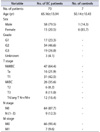
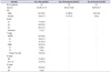
Cohort 1 (Table 1) comprised 73 BC patients (58 males and 15 females; average age, 65 years) and 7 controls (1 male and 6 females; average age, 50 years). Cohort 2 (Table 2) comprised 83 BC patients (65 males and 18 females; average age, 65 years), 54 nonmalignant hematuric patients (37 males and 17 females; average age, 64 years), and 61 normal controls (56 males and 5 females; average age, 68 years).
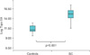
2. TopoIIA mRNA expression in bladder tissues
Expression of TopoIIA mRNA was signif icantly higher in samples from BC patients than in samples from noncancer patient controls (401.6 [142.6–945.1]×104 copies/µg vs. 6.7 [3.8–24.6]×104 copies/µg, respectively) (p< 0.001) (Fig. 1).
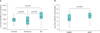
3. Expression of urinary TopoIIA cell-free DNA in BC, normal control, and hematuria samples
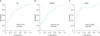
Expression of TopoIIA cell-free DNA in BC patients was significantly higher (40.4 [16.2–313.6]×103 copies/ng) than that in noncancer patient controls (11.1 [4.6–23.0]×103 copies/ng) (p<0.001) (Fig. 2A), and significantly higher than that in hematuria patients (4.0 [0.8–34.7]×103 copies/ng) (p<0.001). Expression of urinary TopoIIA cell-free DNA in hematuria patients was lower than that in normal controls (p=0.044). Expression of TopoIIA cell-free DNA was also significantly higher in MIBC (116.9 [34.2–556.7]×103 copies/ng) than in NMIBC (26.0 [8.5–224.8]×103 copies/ng) (p=0.002) (Fig. 2B). ROC analysis, a conventional tool for diagnostic test evaluations, was performed to determine how well TopoIIA cell-free DNA levels in urine discriminate BC patients from controls. ROC analysis revealed that the area under the ROC curve (AUC) for TopoIIA cell-free DNA in urine was 0.741, with a sensitivity of 73.8%, specificity of 68.3%, positive predictive value (PPV) of 64.2% and negative predictive value (NPV) of 78.6% for detecting BC at the cutoff value of 19,385.71. The AUC was 0.701 (cutoff value, 19,385.71; sensitivity, 63.3%; specificity, 70.4%; PPV, 47.7%; NPV, 81.8%) for detecting NMIBC and 0.838 (cutoff value, 24,992.39; sensitivity, 88.2%; specificity, 74.8%; PPV, 50.9%; NPV, 95.6%) for detecting MIBC (Fig. 3).
DISCUSSION
The results presented herein suggest that measuring the levels of TopoIIA cell-free DNA in urine samples from BC patients may be a useful noninvasive method for diagnosing BC. The levels of urinary TopoIIA cell-free DNA in BC samples were significantly higher than those in samples from normal controls or hematuria patients. In total, 73 bladder tissue specimens from BC patients (47 NMIBC and 26 MIBC patient tissues) and from 7 controls were analyzed. Data from the second cohort (83 urine specimens from BC patients, 54 from nonmalignant hematuric patients, and 61 from normal controls) support a correlation between TopoIIA cell-free DNA levels and cancer. To the best of our knowledge, this is the first clinical data set to describe the levels of urinary TopoIIA cell-free DNA in BC.
These patient-based results suggest that expression of TopoIIA is up-regulated in BC tissues, which is consistent with data obtained from urine samples. In general, the physiological behavior of human cancers is driven by abnormal proliferation and the potential to invade and metastasize. TopoIIA is a marker of cell proliferation in normal cells. The expression of TopoIIA increases markedly in late S phase before decreasing rapidly at the end of M phase; the variation in these levels is lower in tumor specimens [19]. It is suggested that because TopoIIA is present only during the late S and G2 phases, it might provide a better estimate of the number of actively cycling cells [19]. The finding herein that the levels of TopoIIA cell-free DNA are higher in BC patients than in normal controls and patients with hematuria suggests that it has utility as a urinary diagnostic marker for BC.
BC is usually diagnosed by cystoscopy. Although urinary cytology is noninvasive and detects BC with high specificity, its sensitivity is relatively low (40%–76%) [20]. Moreover, cystoscopy is highly invasive and expensive; thus, the majority of the patients suffer either mental or physical distress. Therefore, novel noninvasive diagnostic markers that can distinguish BC from noncancerous conditions with improved sensitivity and specificity are needed. A plethora of urine-based tests have been developed. These include tests based on bladder tumor antigen (BTA ), nuclear matrix protein 22 (NMP22 ), urine fibrin and fibrinogen degradation products, ImmunoCyt, and fluorescence in situ hybridization (UroVysion) [2122]. However, the diagnostic ability of all of these tests is not reliable, so they cannot replace cystoscopy or urinary cytology [23]. The diagnostic approach based on urinary TopoIIA described herein may be more promising, as it is clinically relevant, performs well, and is convenient for patient. The clinical relevance of this approach is based on the fact that urine is stored in the bladder and is in direct contact with bladder tissue. Thus, levels of TopoIIA cell-free DNA in the urine may closely reflect the status of the bladder tissue, making it more clinically relevant for BC diagnosis than more conventional urine-based tests. With regard to its diagnostic performance, the high sensitivity and specificity shown in this study suggest that the assay is reliable. The finding of high sensitivity (>70%) without compromising specificity suggests that the assay is comparable with (or better than) urinary cytology (which suffers from low sensitivity). The levels of TopoIIA cell-free DNA in urine were significantly higher in MIBC than NMIBC samples, suggesting that high levels of TopoIIA cell-free DNA may be associated with aggressive pathologic features. Additionally, there is an urgent need for methods that identify MIBC patients that are likely to experience disease progression or metastasis. The results presented herein demonstrate that measurement of urinary TopoIIA cell-free DNA levels can distinguish BC from normal controls, and MIBC from NMIBC.
Equally importantly, we compared samples from patients with nonmalignant hematuria with those from BC patients to ensure that urinary TopoIIA cell-free DNA levels could differentiate BC patients from patients with nonmalignant hematuria. BC patients usually present with hematuria, although hematuria can also be present in patients without cancer; thus, hematuria can be a serious confounding variable. The assay developed herein clearly distinguished patients with nonmalignant hematuria from BC patients. Therefore, the urinary TopoIIA cell-free DNA assay is likely to be reliable in a real clinical situation.
CONCLUSIONS
In summary, the results of this study provide evidence that TopoIIA is a biomarker for the diagnosis of BC. Although the present data are promising in terms of BC diagnosis (samples were carefully grouped as NMIBC or MIBC based on clinical evaluation), a larger prospective validation of our findings should be undertaken.
 XML Download