Abstract
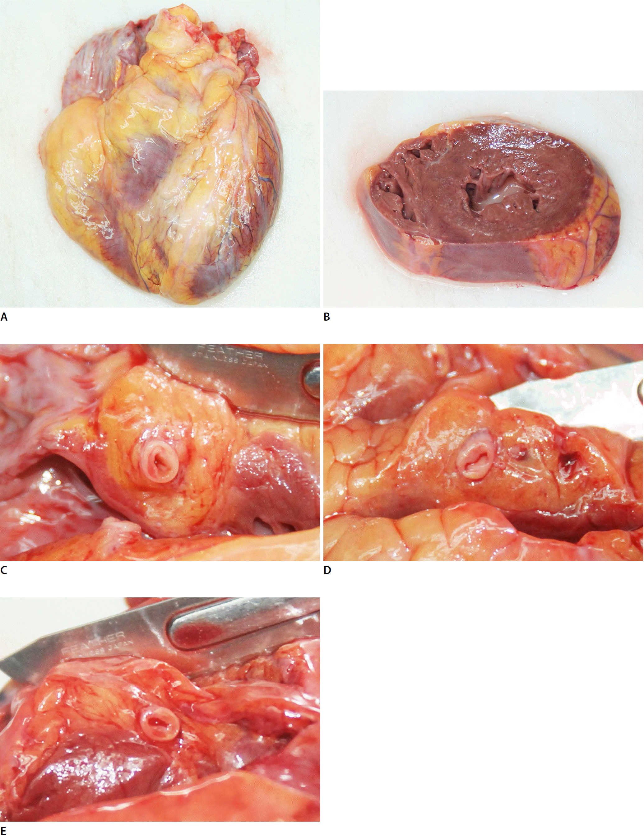
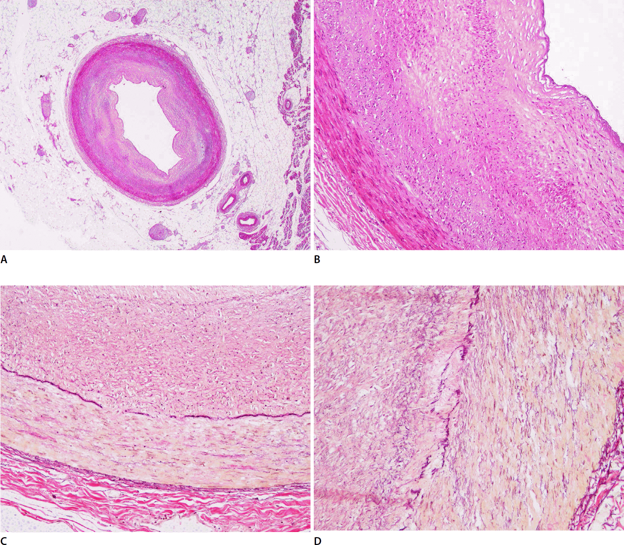
Fibromuscular dysplasia (FMD) of the coronary artery is a rare cause of sudden cardiac death; however, its prevalence and fatality may have been overlooked so far. A 47-year-old man complained of pain in his back and shoulder and became unconscious. Despite resuscitation, he died 3 hours after symptom onset. The heart weight was in the normal range; however, all three major coronary arteries showed intimal thickening without atherosclerosis or inflammatory cell infiltration. Fragmentations and duplications of the internal elastic lamina which are histologic features of intimal fibroplasia, a focal-type FMD, were observed. The prevalence of coronary FMD remains unknown, although it may be related to spontaneous coronary artery dissection and sudden death. The histopathologic confirmation of coronary FMD and exclusion of other possible coronary diseases through autopsy are essential to reveal the nature of the disease and therefore apply the information in dealing with legal problems after death.
REFERENCES
1. Park JH, Na JY, Lee BW, et al. A statistical analysis on forensic autopsies performed in Korea in 2015. Korean J Leg Med. 2016; 40:104–18.

2. Shivapour DM, Erwin P, Kim E. Epidemiology of fibromuscular dysplasia: a review of the literature. Vasc Med. 2016; 21:376–81.

3. Kadian-Dodov D, Gornik HL, Gu X, et al. Dissection and aneurysm in patients with fibromuscular dysplasia: findings from the U.S. Registry for FMD. J Am Coll Cardiol. 2016; 68:176–85.
4. Olin JW, Froehlich J, Gu X, et al. The United States Registry for Fibromuscular Dysplasia: results in the first 447 patients. Circulation. 2012; 125:3182–90.

5. Garcia RA, deRoux SJ, Axiotis CA. Isolated fibromuscular dysplasia of the coronary ostium: a rare cause of sudden death: case report and review of the literature. Cardiovasc Pathol. 2015; 24:327–31.

6. Zack F, Kutter G, Blaas V, et al. Fibromuscular dysplasia of cardiac conduction system arteries in traumatic and nonnatural sudden death victims aged 0 to 40 years: a histological analysis of 100 cases. Cardiovasc Pathol. 2014; 23:12–6.

7. Saw J, Bezerra H, Gornik HL, et al. Angiographic and intracoronary manifestations of coronary fibromuscular dysplasia. Circulation. 2016; 133:1548–59.

8. Olin JW, Gornik HL, Bacharach JM, et al. Fibromuscular dysplasia: state of the science and critical unanswered questions: a scientific statement from the American Heart Association. Circulation. 2014; 129:1048–78.
Fig. 1.
Gross findings of the heart and the coronary arteries of the deceased. (A,B) The heart was of normal weight without any abnormal features. (C-E) In the cross-sections of the major coronary arteries, the lumens were narrowed by wall thickening and there were no atherosclerotic changes (C, right coronary artery; D, anterior descending branch of the left coronary artery; E, circumflex branch of the left coronary artery).

Fig. 2.
Histologic findings of coronary arteries. (A) The cross-section of the coronary artery shows circumferential thickening of the vascular wall (H&E stain, ×4). (B) The intimal layer which showed neither noticeable inflammation nor lipid accumulation contributed mainly to the thickening (H&E, ×40). (C, D) Elastin stain highlighted the internal elastic lamina and both fragmentations and duplications of the internal elastic lamina were found (C, elastin stain, ×100; D, elastin stain, ×200). These findings were consistent in all three major coronary branches.


 PDF
PDF ePub
ePub Citation
 Print
Print


 XML Download