

 PDF
PDF ePub
ePub Citation
 Print
Print


Introduction
Postmortem examination can be divided into autopsy and postmortem inspection. Unlike autopsies, which examine the body of the deceased using invasive modalities such a dissection, postmortem inspection examines the body of the deceased using non-invasive modalities. The findings yielded by postmortem inspection of the body of the deceased are limited. In South Korea, the rate of autopsies is low [1]. To overcome this challenge, forensic radiology, such as postmortem computed tomography (PMCT), was introduced in the country. However, it is performed only at the National Forensic Service Seoul Institute [2]. Meanwhile, endoscopy is widely used in clinical medicine. The authors of the present study investigated the usefulness of endoscopic examination in postmortem inspection prior to autopsy. To the knowledge of the authors, this is the first study to investigate the usefulness of endoscopic examination in postmortem inspection in South Korea.
Materials and Methods
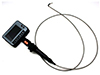
Endoscopic examination was performed on a total of 35 bodies before autopsies. The authors performed endoscopic examination before autopsy and photographed significant findings during endoscopic examination. The endoscopic findings were confirmed by subsequent autopsies. The endoscope was inserted through the nose or mouth. The upper respiratory tract, as well as the esophagus and other associated structure, were examined. Examination time lasted for 10 minutes. Endoscopy was performed using a 3.8-mm industrial video inspection endoscope with 4-way articulation (Fig. 1). This instrument can direct its camera tip in four directions, while capturing figures and recording.
Results
Endoscopic examination was performed on 27 males and eight females. The mean age was 51.0 years, with a range of 17 to 79 years. The manner of death was considered natural in 19 cases (54.3%) and unnatural in 16 cases (45.7%). The causes of death included ischemic heart disease in six cases, fire death in five cases, fatal injuries in four cases, and hanging and drowning death in three cases. The endoscope was inserted through the nose in 33 out of 35 cases. Endoscope insertion through the mouth was difficult in many cases due to the rigor mortis of the jaw joint and internal structures, such as the tongue.
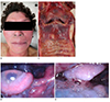
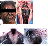
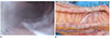
Out of the 35 cases on which endoscopic examination was performed, the cases in which significant endoscopic findings were confirmed are summarized in Table 1. Froth was detected by endoscopic examination in the airway in all three drowning cases even though no froth was detected during external examination (Fig. 2). In cases where death was caused by fire, thermal denaturation findings such as pharyngeal and laryngeal edema, and soot attached to and froth around the larynx, pharynx, and tracheal mucosa were detected during endoscopic examination (Fig. 3). In 14 of 19 natural death cases (73.7%), froth around the pharynx and larynx was detected. Froth in the tracheal cavity was detected in one out of three hanging cases, and petechiae and congestion of tympanic membrane were detected during endoscopic examination. Other substances were found in the airway in some cases. For example, agrochemical substances were detected in the airway in the agrochemical poisoning case (Fig. 4). Blood was found in the airway during endoscopic examination in the cases of fatal chest injuries due to sharp instrument. In addition, fly eggs were detected in the external auditory canal during endoscopic examination, although it was not detected during external examination.
Discussion
There are many methods used for postmortem examination, which provide information about the cause and manner of death. One of these methods is postmortem inspection. Since postmortem inspection is non-invasive, it has several limitations. For instance, previous studies have reported that postmortem inspection alone was inaccurate in determining the cause and manner of death [34]. Thus, several systematic improvements have been proposed to improve the effectiveness of postmortem inspection [45], and various methods, such as PMCT, have been studied [2].
Endoscopy is widely used in clinical medicine as a non-invasive diagnostic tool. Therefore, the authors of the present study preformed endoscopy before autopsy and investigated the effectiveness of endoscopic examination in the postmortem inspection. Endoscopic examination was performed on a number of bodies with various causes of death. Froth was detected in the airway in all three drowning cases upon endoscopic examination. In particular, the figure of froth was found to be different between the seawater drowning and freshwater drowning cases. In the seawater drowning cases, the froth bubbles showed a big difference in size, and were whitish. Froth bubbles were relatively constant in size and transparent in the freshwater drowning cases. These differences are postulated to be due to differences in the properties of the inhaled liquid. However, endoscopic studies involving a larger number of cases should be performed. In the fire death cases, soot and froth were found, along with thermal denaturation of structures in the airway. These were detected in endoscopic examination and later confirmed during autopsy. In some cases, these findings were not detected in external examination. Therefore, the results of the present study are significant because findings that could be confirmed only by autopsy were confirmed during postmortem inspection using endoscopic examination. In addition, endoscopes can be used to examine by magnifying the object prior to the interference of dissection. Therefore, more detailed findings can be obtained compared to conventional autopsies.
Froth in the pharynx and larynx was detected in 73.7% of natural cases during endoscopic examination, even though froth is a non-specific finding. The authors of the present study considered the possibility that these findings might be caused by pulmonary edema due to cardiac arrest in natural death, and that this finding was more clearly identified by endoscopic examination compared to gross examination through a routine autopsy. Froth was detected in the airway in one out of three hanging cases, and this finding suggests that the dying process due to hanging may be different between individual hanging cases. In addition, congestion of the tympanic membrane and mucosal bleeding can be detected in hanging cases during endoscopic examination. It is thought that these findings are vital reactions and may present in the same manner as periorbital and conjunctival petechial hemorrhage [6]. Blood was found in the case of fatal chest injury due to a sharp object whereas agrochemicals were found in the agrochemical poisoning case. These endoscopic findings suggest that material was aspirated in the airway during the process of dying. Therefore, causes of death can be ascertained through endoscopic examination performed prior to invasive procedures such as dissection. The presence of fly eggs, which was not detected during external examination on the body in the external auditory canal, was useful for estimating the postmortem interval and the location of the body.
Although studies on minimally invasive autopsies using less invasive methods, such as endoscopic examination are underway [7], it is difficult for minimally invasive autopsies to replace conventional autopsies. This is especially true at the legal level [8]. Minimally invasive autopsy modalities cannot replace conventional autopsies because the majority of autopsy performed in South Korea are forensic autopsies. However, this study demonstrated the effectiveness of less invasive examinations, such as endoscopic examination, as an adjunct to autopsy. In addition, it is thought that endoscopic examination is useful as one of the ways to improve the validation of postmortem inspection. However, the present study is limited because of the relatively small sample size and because industrial endoscopy was used for the study. Previous studies have performed postmortem inspection using endoscopy, thoracoscopy, or laparoscopy, which are clinically used [8]. However, if endoscopic equipment is modified to become more suitable for postmortem examination, endoscopic examination may be more useful for postmortem examination. Further studies on this topic should be conducted.
 XML Download