

 PDF
PDF ePub
ePub Citation
 Print
Print


Introduction
Cell-free fetal DNA (fDNA) in maternal serum generally exists in fragments of shorter lengths than fragments derived from the mother [1] and makes up 11.0%-13.4% of the total plasma cell-free DNA depending on gestational age [2]. Working with such minute quantities of DNA can be challenging for clinical DNA profiling. The feasibility of using massively parallel sequencing (MPS) for fDNA profiling with high enough sensitivity to diagnose aneuploidy, subchromosomal aberrations, and monogenic disorders has recently been demonstrated [3]. Single nucleotide polymorphism (SNP) panels have great potential for use in fDNA profiling by allowing analysis of fragmented DNA or low copy-number templates, particularly with MPS technology.
The HID-Ion AmpliSeq™ Identity Panel consisting of 124 SNPs is a commercial system developed for human identification and has been evaluated for coverage, heterozygote balance, and sensitivity in multiple validation studies [45678]. This panel can reliably provide genotype information given small amounts of template DNA, as well as allow quantitative analyses. These attributes make it possible to use serum DNA for genetic testing. Here, we report our findings on fDNA detection in maternal serum samples using the HID-Ion AmpliSeq™ Identity Panel. Because of the absence of reference information for fetuses and mothers, interpretation of the results was performed using criteria from previous studies [58].
Materials and Methods
Seven maternal serum samples were taken from anonymous pregnant women, and no detailed information about the fetuses, including sex, could be obtained because of the inclusion of samples from miscarried pregnancies. Genomic DNA (gDNA) was extracted from serum using a QIAamp DNA Mini Kit (Qiagen, Hilden, Germany), and the concentration of extracted gDNA was measured using a Qubit® fluorometer (Thermo Fisher Scientific, Waltham, MA, USA). One nanogram of gDNA from each sample was amplified with the 124 SNP primer sets in the HID-Ion AmpliSeq™ Identity Panel (Thermo Fisher Scientific), and a pooled library with a concentration of 20 pM was prepared. Emulsion polymerase chain reaction and enrichment were performed using the Ion PGM Template OneTouch™ System (Thermo Fisher Scientific) and the Ion OneTouch™ Enrichment System (Thermo Fisher Scientific), respectively, according to the manufacturer's instructions. Sequencing was performed using an Ion 316v2 chip on an Ion Torrent PGM™ (Thermo Fisher Scientific). Raw data was processed with Ion Torrent Suite v4.2.1 (Thermo Fisher Scientific), and SNP genotypes were analyzed using the HID_SNP_Genotyper plugin v4.2 (SNP_Genotyper) (Thermo Fisher Scientific). This study was approved by the ethical committee of the Institutional Review Board of Seoul National University Hospital Biomedical Research Institute (E-1309-023-517).
Results
1. Quality of sequencing data
The total number of reads generated for the tested samples was 3,651,420, of which 99.17% mapped to the reference sequence (genome build hg19). The mean read length was 79 bp with a quality score of AQ20. The number of mapped reads on average for each sample was 517,325 (range, 442,233 to 568,369), and the mean coverage per sample was 3,893× (3,394-4,437×).
2. Y chromosomal SNP detection in maternal serum
In two out of the seven samples obtained, 13 and six Y chromosomal SNP (Y-SNPs) were detected with a mean coverage of 82.3±63.0× (22-231×) and 47.5±21.0× (25-75×) for serum 6 and serum 7, respectively. No Y-short tandem repeats were amplified in any of the tested samples using the PowerPlex™ Y23 System (Promega, Madison, WI, USA) (data not shown). This could be because of the success rate of detection being dependent on amplicon size [9]. Detection of Y-SNPs in maternal/fetal DNA mixtures is potentially evidence for the mother carrying a male fetus, which could be supported by the finding that a single allele was amplified solely at a locus without any discordant calls such as a non-specific allele.
3. Autosomal SNP detection in maternal serum
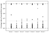
For interpreting the genotype results of autosomal SNP (A-SNP) loci, we assumed there were four possibilities for autosomal alleles given maternal (AA or AB) and fetal (aa or ab) genotypes: (1) AAaa, (2) AaaB indicating an imbalanced heterozygote, (3) AAab indicating a major homozygote with a minor contributor, and (4) AaBb. In addition, each allele was numbered for convenience in this study as first and second for two reference alleles at each locus, in order of their frequencies, and third and fourth for nonspecific alleles. In both cases 1 and 4, it was determined that the genotype profiles were identical to that of DNA from a single individual who is either homozygous (case 1) or heterozygous (case 4), and so these loci could not provide significant information. For case 2, the heterozygote balance was estimated from the allele ratio of first/[first+second], and the estimates were evaluated with a threshold of 0.65 [8]. Twelve loci were identified that had a ratio >0.65, and this included five loci reported as underperformed [8]; the five underperforming loci were rs7520386 (serum 1), rs4530059 (serum 3), rs1493232 (serum 3), rs4847034 (serum 4), and rs2046361 (serum 6) (Fig. 1). The rest of the loci (rs1413212, rs717302, rs917118, rs876724, rs12997453, rs214955, and rs917118) reported no errors, and heterozygote balance was estimated from 0.65 to 0.73. Among samples, no difference in patterns of heterozygote balance was found, including between the two samples where Y-SNPs were detected (serum 6 and 7) and the other five serum samples (serum 1, 2, 3, 4, and 5). This implies that fDNA was detected in serum 1, 2, 3, 4, and 5, which could be assumed to be derived from female fetuses.
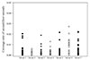
Background signals that interfere with interpretation of data from low-template or mixed DNA samples have to be excluded, but it is difficult to discriminate between these and signals possibly derived from minor contributors. In this study, we addressed the issue by comparing the percent of the second allele to the percent of the third+fourth alleles that might be background signals. If the second allele had a higher percent than those of the third+fourth alleles, it was filtered (Fig. 2). No remarkable difference in patterns of allele percentages among samples was found, similar to the results for heterozygote balance (Table 1). Because of the absence of reference data for mothers, it was only assumed that serum 6 and serum 7, which contained Y-SNPs, might contain fDNA. In serum 6 and serum 7, the highest ratio of an allele detected was 2.71% at rs4530059 and 2.23% at rs430046, respectively. No non-specific alleles were detected at these loci, which clearly showed only major and minor alleles. The alleles preserved after filtering out the possible error calls could be a potential clue for fetal allele detection in DNA extracted from maternal serum.
Discussion
Only limited information can be gained from DNA profiles of a mixed sample without reference genotypes, particularly regarding the genotype of the minor contributor, and so several indicators have been developed to best assess these samples [5]. Multiple studies have shown that heterozygote balance in a sample from a single heterozygous individual can range from 40%-60% [4578]; however, it is not possible to define exact limits for homozygotes and heterozygotes in 1:1 mixtures. This is because the balance varies depending on the relative amounts of DNA from each individual and on whether the minor contributor is the homozygote or the heterozygote [5]. However, imbalanced patterns in the heterozygote balance of mixed samples with 10% or less of the DNA from the minor contributor are routinely detected [5]. Changes in the observed level of heterozygosity in mixtures were also discussed. While a sample representing a single individual can be expected to present approximately 50% heterozygosity, a mixed sample will show increased heterozygosity. Lastly, for Y-SNP patterns in mixed DNA samples, it was reported that Y-SNP (haploid) coverage in a single male is half the coverage of A-SNPs (diploid). This implies that Y-SNPs are detected at low levels when the minor contributor is a male [5]. It has been recommended that data be analyzed using the somatic parameters for minimum threshold coverage if any of the indicators, e.g., increased heterozygosity or reduced Y-SNP coverage, for mixed samples are found, because the somatic analysis parameter for Y-SNPs can result in higher no-call rates [5]. According to the guidelines for the HID-Ion AmpliSeq™ Identity Panel provided by Thermo Fisher Scientific, 30× and 500× were suggested as minimum coverage thresholds for germline and somatic SNP detection, respectively [5]. Considering these guidelines, the detection of Y-SNPs in serum 6 and serum 7 could indicate that the maternal serum did contain male fDNA, although the minimum threshold coverage was not defined during this study in order to explore all features detectable in mixed DNA samples. It is also the case that low levels of Y-SNP coverage can indicate the presence of a minor male component in a mixed DNA sample when analyzing forensic samples of unknown origin [5].
In order to improve SNP genotyping of mixed DNA samples with MPS, analyses of known samples mixed in a variety of ratios should be performed. The current study could support the use of the HID-Ion AmpliSeq™ Identity Panel for diagnostic purposes (e.g., non-invasive prenatal testing) as well as for analysis of forensic serum samples [10].

 XML Download