Abstract
We report the case of a 42-year-old woman who died in hospital from severe respiratory failure, 10 days after the onset of symptoms. Autopsy and microscopic examination identified features of diffuse alveolar damage in both lungs including hyaline membranes and intra-alveolar exudate. Gomori's methenamine silver stain of pink frothy materials in these exudates revealed thin-walled and cup-shaped microorganisms and a diagnosis of Pneumocystis jirovecii pneumonia was made. There were small granulomas in the pulmonary interstitium and hepatic lobules representing an unusual inflammatory reaction against Pneumocystis jirovecii. Extrapulmonary involvement with pneumocystis infection is a rare event occurring in 1% to 2% of all pneumocystis cases. Screening and confirmatory tests for human immunodeficiency virus (HIV) detection were positive. There was no information available regarding the patient's medical history or the possibility of HIV infection prior to the autopsy, because the patient was a foreign worker who arrived in Korea 2 months before her death. Medical examiners often perform autopsies with limited information regarding the deceased person, even when person is a Korean national. Therefore, an awareness of protection protocols during autopsy, as well as of the atypical patterns of critical diseases, is crucial.
REFERENCES
1.Kim YJ., Woo JH., Kim MJ, et al. Opportunistic diseases among HIV-infected patients: a multicenter-nationwide Korean HIV/AIDS cohort study, 2006 to 2013. Korean J Intern Med. 2016 Apr 27. [Epub].http://dx.doi.org/10.3904/kjim.2014.322.

2.Centers for Disease Control and Prevention. 2014 Annual Report on the Notified HIV/AIDS in Korea [Internet]. Osong: Centers for Disease Control and Prevention;2015. [cited 2016 Jul 24]. Available from:. http://www.cdc.go.kr.
3.UNAIDS. AIDSinfo [Internet]. Geneva: UNAIDS;2016. [cited 2016 Jul 24]. Available from:. http://aidsinfo.unaids.org/.
4.Hartel PH., Shilo K., Klassen-Fischer M, et al. Granulomatous reaction to Pneumocystis jirovecii: clinicopathologic review of 20 cases. Am J Surg Pathol. 2010. 34:730–4.
5.Travis WD., Pittaluga S., Lipschik GY, et al. Atypical pathologic manifestations of Pneumocystis carinii pneumonia in the acquired immune deficiency syndrome: review of 123 lung biopsies from 76 patients with emphasis on cysts, vascular invasion, vasculitis, and granulomas. Am J Surg Pathol. 1990. 14:615–25.
7.Korea Immigration Service. Korea Immigration Service Statistics 2015 [Internet]. Gwacheon: Korea Immigration Service;2016. [cited 2016 Jul 24]. Available from:. http://www.immigration.go.kr/.
8.Nolte KB., Taylor DG., Richmond JY. Biosafety considerations for autopsy. Am J Forensic Med Pathol. 2002. 23:107–22.

9.Royal College of Pathologists. Guidelines on autopsy practice. London: Royal College of Pathologists;2002.
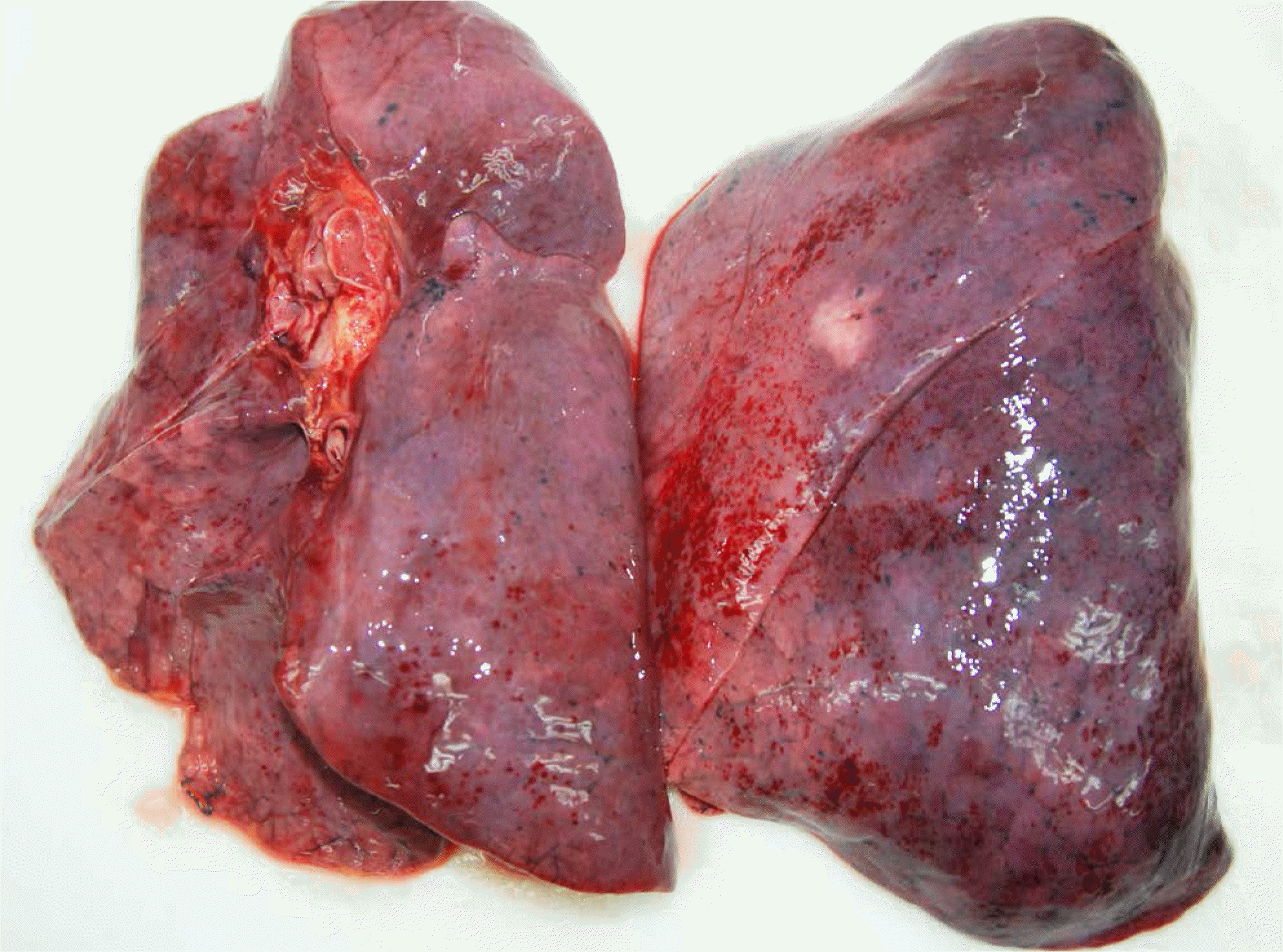
Fig. 1.
Pleural surface showed numerous petechial hemorrhages in both lobes. There were several focal whitish areas in right middle to lower lobe and left lobe. Among them, one in the left upper lobe is shown in this figure and its diameter was about 2 cm.

Fig. 2.
On microscopic examination of lung sections, there were hyaline membranes lining alveolar walls (A). Some alveolar spaces were filled with pink foamy amorphous materials (B), which were revealed as Pneumocystis jirovecii with cup shaped cysts in Gomori's methenamine silver (GMS) (C) and periodic acid-Schiff (PAS) (D) stains (A, H&E, ×40; B, H&E, ×100; C, GMS, ×400; D, PAS, ×400).

Fig. 3.
In grossly whitish areas of the lung, their interstitium were thickened with inflammatory infiltration, mainly lymphoplasmacytes. (A) Granulomatous reactions with multinucleated giant cells were also found in these areas, but central necrosis were not definite. (B) In the liver sections randomly selected, there were also small granulomas in hepatic lobules (A, H&E, ×200; B, H&E, ×100).


 PDF
PDF ePub
ePub Citation
 Print
Print


 XML Download