Abstract
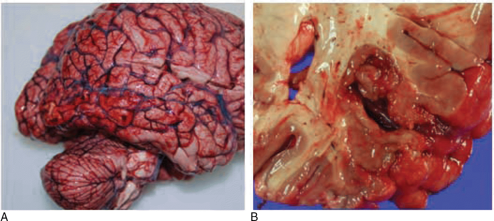
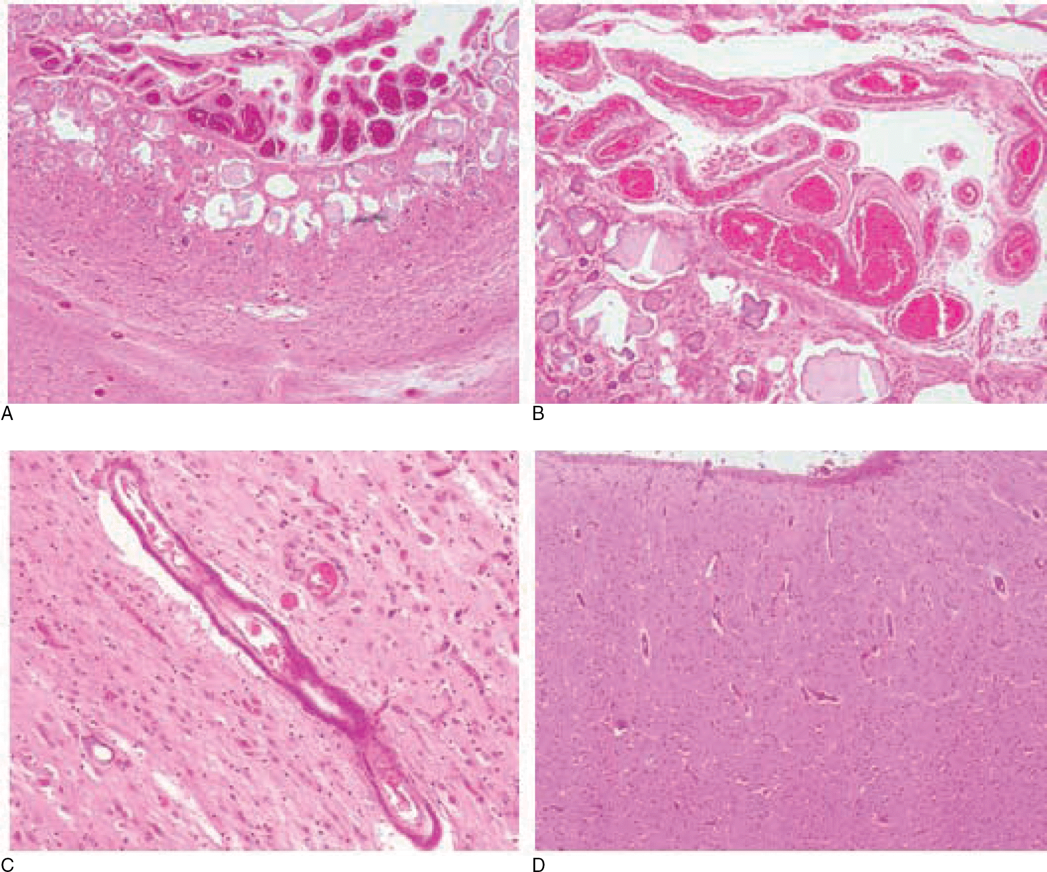
In some cases, it is difficult to determine a single cause of death even after conducting full autopsy and additional tests. A 49-year-old man, reportedly having diabetes mellitus, was found unconscious by his mother and revealed to be dead. He had several contusions all over his body, including the right periocular area, but they did not appear fatal. A focal area of polymicrogyria and cortical dysplasia was found on the right preoccipital notch, accompanied with dystrophic calcification and leptomeningeal angiomatosis. These findings were considered indicative of Sturge-Weber syndrome, a rare neurocutaneous disorder, of atypical type without facial lesions. Blood level of β -hydroxybutyrate was 859 μ g/mL, implying that he also had diabetic ketoacidosis. His ketoacidosis may not have been corrected appropriately because of status epilepticus in association with brain lesion, resulting in his death, but neither direct evidence nor statement was obtained. In cases with several apparent causes of death, the examiner's assumption should be based not on imagination but on evidence, and logic should not be overlooked. It is more helpful for the investigators or the bereaved to obtain more detailed information rather than come to a hasty conclusion.
REFERENCES
1. Maton B, Krsek P, Jayakar P, et al. Medically intractable epilepsy in Sturge-Weber syndrome is associated with cortical malformation: implications for surgical therapy. Epilepsia. 2010; 51:257–67.

2. Jagtap S, Srinivas G, Harsha KJ, et al. Sturge-Weber syndrome: clinical spectrum, disease course, and outcome of 30 patients. J Child Neurol. 2013; 28:725–31.
3. Geronemus RG, Ashinoff R. The medical necessity of evaluation and treatment of port-wine stains. J Dermatol Surg Oncol. 1991; 17:76–9.

4. Kitabchi AE, Umpierrez GE, Fisher JN, et al. Thirty years of personal experience in hyperglycemic crises: diabetic ketoacidosis and hyperglycemic hyperosmolar state. J Clin Endocrinol Metab. 2008; 93:1541–52.

5. Kanetake J, Kanawaku Y, Mimasaka S, et al. The relationship of a high level of serum beta-hydroxybutyrate to cause of death. Leg Med (Tokyo). 2005; 7:169–74.

6. Palmiere C, Bardy D, Mangin P, et al. Postmortem diagnosis of unsuspected diabetes mellitus. Forensic Sci Int. 2013; 226:160–7.

7. Palmiere C, Lesta Mdel M, Sabatasso S, et al. Unconsciousness and sedation as precipitating factors of diabetic ketoacidosis. J Forensic Leg Med. 2013; 20:830–5.

8. Park JP, Min BW, Na JY, et al. Sudden unexpected death of epileptic patient with dendriform pulmonary ossification. Korean J Leg Med. 2011; 35:53–6.
Fig. 1.
(A) Cerebral cortex of the right preoccipital area shows microgyria and cortical atrophy with mild congestion in leptomeninges. (B) Cut surface shows leptomeningial hypertrophy and cortical atrophy which is locally restricted.

Fig. 2.
Brain cortex shows severe dystrophic microcalcification combined with leptomeningial angiomatosis (A) which includes thickened vascular wall due to hyalinization (B). (C) Microcalcification was also found in the vascular wall of brain cortex parenchyme. (D) In the adjacent area of the lesion, there was no definite atrophy but cortical dysplasia was present, without normal development of horizontal six layers (A and D, H&E, ×40; B, H&E, ×100; C, H&E, ×200).


 PDF
PDF ePub
ePub Citation
 Print
Print


 XML Download