Abstract
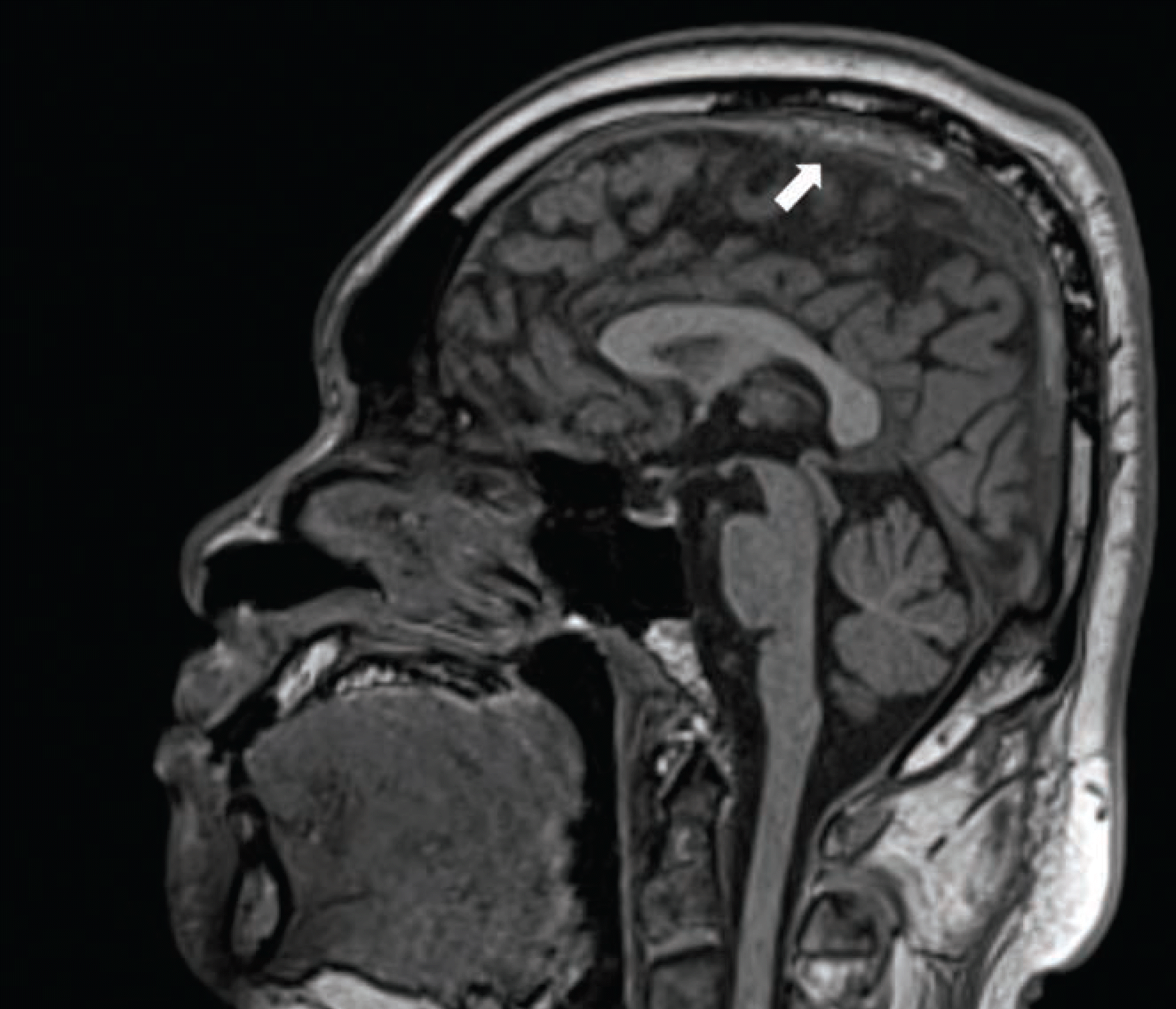
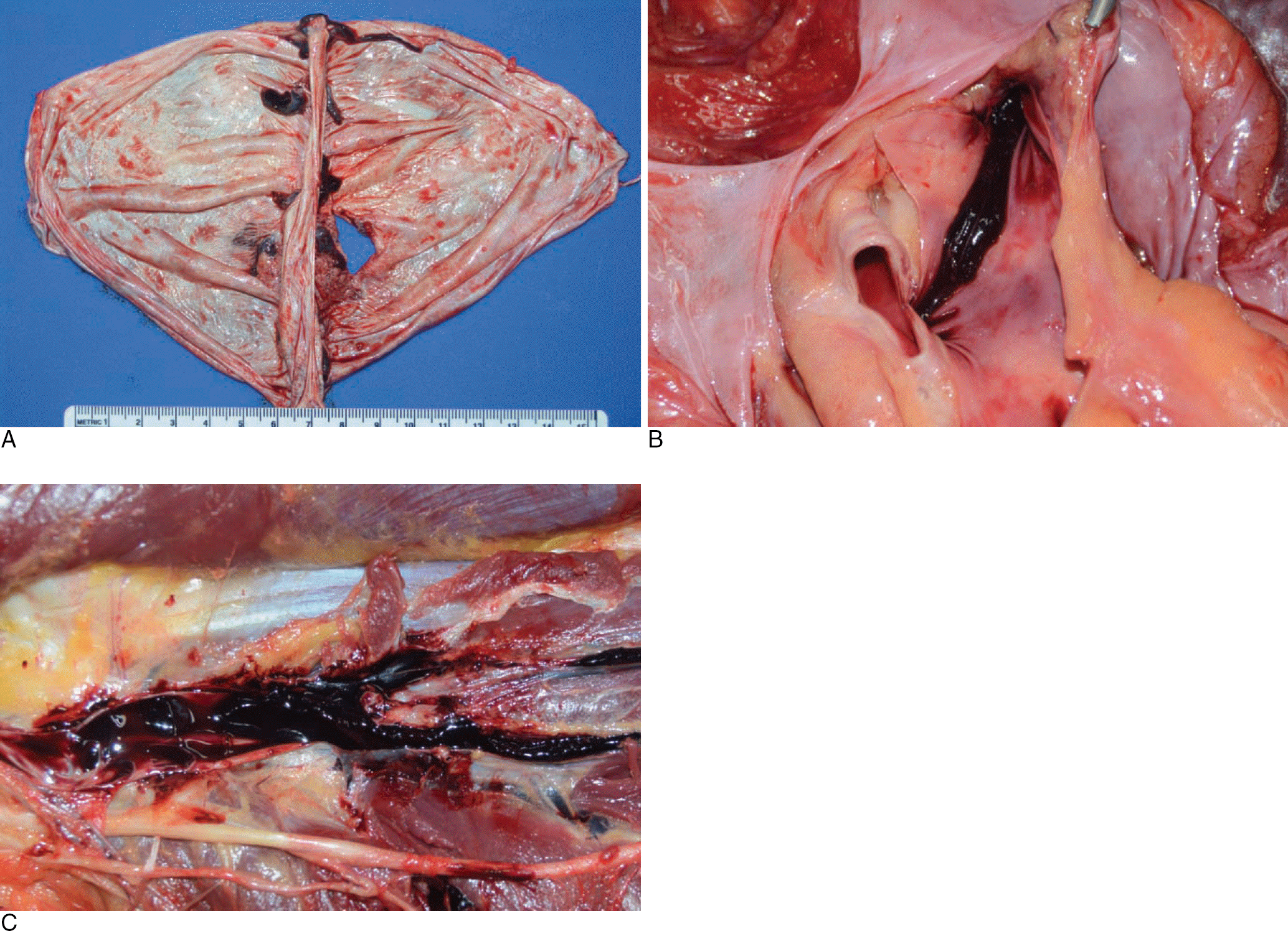
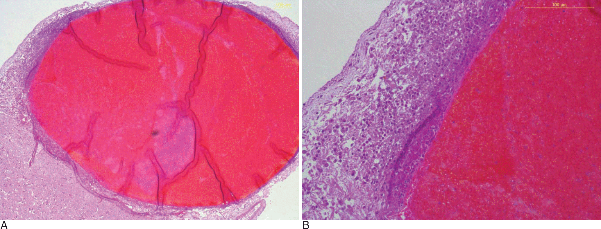
Cerebral venous thrombosis is an uncommon cause of cerebral infarction. A 31-year-old man complained of headache, weakness, and numbness of the left leg a day before being admitted to the hospital. After admission, brain computed tomography and brain magnetic resonance imaging revealed superior sagittal sinus thrombosis with cerebral infarction in the right hemisphere. He had no significant medical history. On the fourth hospital day, he suddenly collapsed and died. Medicolegal autopsy was performed 3 days later; medical malpractice was suspected. External examination revealed a few conjunctival petechiae. Internal examination revealed thrombi in the superior sagittal sinus and superficial cortical veins. Thrombi were noted in the pulmonary trunk and both pulmonary arteries. Upon dissection of the left leg, we found thrombi in the posterior tibial vein. A microscopic examination revealed vasculitis of the same cortical veins, and we therefore assumed that vasculitis of the cortical veins gave rise to thrombosis. In typical autopsy practice, an examination of the dura mater is often overlooked, but careful examination of this region should be performed in cases of cerebral infarction in young adults, such as this one.
REFERENCES
1. Saposnik G, Barinagarrementeria F, Brown RD Jr, et al. Diagnosis and management of cerebral venous thrombosis: a statement for healthcare professionals from the American Heart Association/American Stroke Association. Stroke. 2011; 42:1158–92.

3. Towbin A. The syndrome of latent cerebral venous thrombosis: its frequency and relation to age and congestive heart failure. Stroke. 1973; 4:419–30.

4. Ferro JM, Canhao P, Stam J, et al. Prognosis of cerebral vein and dural sinus thrombosis: results of the International Study on Cerebral Vein and Dural Sinus Thrombosis (ISCVT). Stroke. 2004; 35:664–70.
5. Biousse V, Ameri A, Bousser MG. Isolated intracranial hypertension as the only sign of cerebral venous thrombosis. Neurology. 1999; 53:1537–42.

6. Gulati D, Strbian D, Sundararajan S. Cerebral venous thrombosis: diagnosis and management. Stroke. 2014; 45:e16–8.
Fig. 1.
T1-weighted magnetic resonance image shows cord-like hyperintensity signals (arrow) representing a venous thrombosis of the superior sagittal sinus.


 PDF
PDF ePub
ePub Citation
 Print
Print


 XML Download