

 PDF
PDF ePub
ePub Citation
 Print
Print


INTRODUCTION
Ileostomy is associated with many complications, including prolapse, necrosis, stenosis, skin irritation, and infection, among others. Among them, prolapse accounts for 2% to 26% of stoma complications (1). Stoma prolapse is a full-thickness protrusion of intestine through the stoma. There are various causes of stoma prolapse, which are influenced by location of the bowel, stoma creation technique, and disease process. However, intussusception through an ileostomy is a rare cause of stoma prolapse (2). We found eight cases of intussusception through an ileostomy in the literature (3456789); however, there are no reports describing the radiological features. We report a rare case of intussusception of the small bowel through an ileostomy and describe the typical computed tomography (CT) features.
CASE REPORT
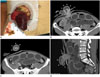
A 49-year-old man presented to the emergency department with a one-day history of pain at the stoma site created after loop ileostomy, which was performed four months previously to treat colonic pseudo-obstruction. On physical examination, the bowel had protruded through the proximal stoma, resulting in prolapse. The protruding bowel exhibited color change (Fig. 1A). Laboratory investigations revealed leukocytosis, indicating inflammation; however, other parameters were within normal limits. An abdominal CT scan was performed immediately to evaluate the extent of ischemic bowel. CT revealed an intussusception of the small bowel through the proximal stoma, which was the intussuscipiens. The small bowel segment protruding through the proximal stoma was intussuscepted with the inner and middle layers (Fig. 1B-D). CT imaging revealed no evidence of ischemic or necrotic change in the small bowel in the peritoneal cavity, or a visible lead point of intussusception. Diffuse, mild distention of the proximal small bowel in the peritoneal cavity was also noted on CT. The patient underwent emergency surgery. Intussusception through the proximal stoma, overlapping the small bowel for approximately 10 cm with ischemic changes, was observed. Consequently, the protruding bowel loop was resected, and a new ileostomy was created through the same site. Pathological examination of the resected portion of the small bowel revealed ischemic necrosis. The patient recovered well, with satisfactory function of the new ileostomy, and he was discharged on postoperative day 4.
DISCUSSION
Intussusception of the small bowel through an ileostomy is very rare. We found eight cases of intussusception through an ileostomy that have been reported in the literature, which are summarized in Table 1 (3456789). In these cases, intussusception occurred through an end ileostomy or a loop ileostomy. The patients underwent ileostomy due to ulcerative colitis, Crohn's disease, complicated diverticulitis, rectal perforation, or colorectal cancer. Antegrade intussusception is common due to the direction of peristalsis; however, retrograde intussusception can occur against the normal direction of peristalsis (56). In three cases (345), the patients were pregnant and increased intra-abdominal pressure during pregnancy was suspected to be an etiologic factor. Based on preoperative CT in our case, the patient may have experienced increased intra-abdominal pressure due to an ileus of the proximal small bowel loop. Nevertheless, risk factors for intussusception through an ileostomy remain unclear, and in all cases included in our review, there were no lead points, similar to our case.
In previously reported cases, bowel necrosis was observed in the intussusceptions and emergency surgery was performed. Radiological examinations were not performed before surgery (3456789). In our case, preoperative CT was performed to evaluate the extent of ischemic bowel. If necrotic changes involve the bowel in the peritoneal cavity, exploratory laparoscopy or laparotomy is mandatory. CT was able to clearly depict intussusception through a stoma in our case. Also, CT may reveal the lead point requiring surgery (10).
A prolapsed stoma can be treated conservatively. Before surgical repair, manual reduction can be attempted in patients without bowel necrosis. However, if reduction fails, surgical resection is required (5). Intussusception through a stoma may result in bowel strangulation and necrosis, which necessitates emergency surgery (8).
In summary, intussusception through an ileostomy should be treated promptly if bowel necrosis is detected. CT can clearly reveal intussusception at the ileostomy site.

 XML Download