INTRODUCTION
Mesenchymal tumors of the pancreas are rare and they account for only 1–2% of all primary pancreatic tumors (1). In order of decreasing frequency, the most commonly reported primary benign or intermediate (borderline) mesenchymal tumors of the pancreas are as follows: schwannoma, inflammatory myofibroblastic tumor, solid and cystic hamartoma, and solitary fibrous tumor (2). Owing to very few case reports describing primary mesenchymal tumors of the pancreas, no definitive protocol for the treatment of these lesions has been established (3).
Primary pancreatic angiomyolipoma (AML), a part of a family of mesenchymal tumors, is extremely rare; only a few cases have been reported (2345). Here, we describe the findings from multidetector computed tomography (MDCT) scan, endoscopic ultrasound (EUS), positron emission tomography (PET), magnetic resonance imaging, and provide a pathologic correlation.
CASE REPORT
A 59-year-old female patient was referred to our hospital for treatment of a pancreatic mass. The mass was incidentally noted on a MDCT scan for abdominal trauma work-up. Her physical examination was unremarkable, and laboratory findings including carcinoembryonic antigen and cancer antigen 19-9 levels were within normal limits. In addition to the abdominal MDCT scan, we performed EUS, PET, and MRI for further evaluation of the pancreatic tumor.
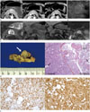
On a CT scan (Somatom Sensation-16; Siemens Medical Solutions, Erlangen, Germany and Brilliance 64; Philips Medical Systems, Cleveland, OH, USA), non-enhanced images demonstrated a 2-cm isodense mass with a mean attenuation value of 48 Hounsfield units (HU) and no evidence of fat components or calcifications. Enhanced CT scan images demonstrated peripheral enhancement during the arterial, parenchymal, and portal venous phases (Fig. 1A). The mass displayed a well-defined margin, no invasion of adjacent organs, and no signs of internal hemorrhage, necrosis, or duct dilatation. EUS revealed a 2-cm hyperechogenic mass in the body of the pancreas (Fig. 1B) without hypervascularity noted on Doppler ultrasound. On a PET scan, the mass exhibited a maximum standardized uptake value of 2.2, which suggested a low-grade malignant or benign tumor. On MRI (Avanto; Siemens Medical Solutions), the mass displayed a heterogeneous peripheral high signal intensity (SI) on half-Fourier acquisition single-shot turbo spin-echo T2-weighted imaging [repetition time (TR)/echo time (TE), 1000/154; flip angle, 160°; section thickness, 6 mm], and a homogenous low SI on T1-weighted images (TR/TE, 160/4.92; flip angle, 70°; section thickness, 6 mm). Chemical shift gradient-echo MR imaging showed no definite loss of SI. On diffusion weighted imaging (DWI; b-value of 1000), the mass revealed peripheral high SI (Fig. 1C). But on the apparent diffusion coefficient map, the periphery of the tumor did not show low SI. We suggested that high SI on DWI was due to the T2 shine-through artifact, not true diffusion restriction. Dynamic MRI also revealed strong peripheral enhancement and poor central enhancement during the arterial phase. As the arterial phase transitioned to the parenchymal and portal venous phases, the mass showed heterogeneous enhancement with peripheral isoenhancement and central subtle poor enhancement. Considering all the results, the final radiographic diagnosis indicated a neuroendocrine tumor in the body of the pancreas. Subsequently, a medial pancreatectomy was performed.
On gross pathological examination, the mass appeared as a well-defined, gray-white nodular tumor in the body of the pancreas measuring 2 × 2 cm. Hematoxylin and eosin staining revealed that the mass was composed of blood vessels, adipose tissue, and spindle cells. Additionally, the mass had a hypercellular central area and a hypocellular peripheral area with loose connective tissue (Fig. 1D). This histologic finding of the tumor was correlated with the enhancement pattern on the MDCT and MRI images. The immunohistochemistry analysis revealed that the mass had positive immunoreactivity to human melanoma black-45 (HMB-45) and smooth muscle actin (SMA) (Fig. 1E). Accordingly, the final pathological diagnosis of a primary pancreatic AML was determined. No signs of recurrence or metastasis have been identified on follow-up CT scans performed over 30 months.
DISCUSSION
The incidence and general radiographic features of primary pancreatic AML are not known; this is because this disease has previously been reported only twice in the literature (23). Pathologically, AML is composed of a mixture of smooth muscle, adipose tissue, and blood vessels. AMLs react with HMB-45 and SMA on immunohistochemical analysis (5). The mass in our case displayed the same pathological and immunohistochemical characteristics.
The imaging findings associated with the mass in our case differed from those of the first reported primary pancreatic AML, renal AML, and hepatic AML. In 2004, Heywood et al. (3) described the first known primary pancreatic AML. This mass was located in the uncinate process and was accompanied by hemorrhage, measuring 4.5 × 3 × 2.5 cm. Most of the pathological and immunohistochemical results in that report were similar to those in our case; however, other results such as imaging findings were quite different, which may be due to hemorrhage. The mass in that case appeared heterogeneous on US and as an irregular, thick-walled, cystic mass on a CT scan (3).
Renal AML is located in the renal cortex and exhibits diffusely high echogenicity on US (6). Most renal AMLs demonstrate fat density (less than -20 HU) on a non-enhanced CT scan, although lipid-poor AMLs show high attenuation. On an enhanced CT scan, they show homogeneous enhancement with a prolonged enhancement pattern (678). The MRI signals vary depending on the amount of intratumoral fat. However, with the exception of lipid-poor AMLs, most renal AMLs are easily detected using a fat suppression or chemical shift technique (78).
Hepatic AMLs appear as well-circumscribed, hyperechogenic lesions and may show relative hypervascularity on US (89). Unlike renal AMLs, 50% of hepatic AMLs lack an appreciable fat content. As a result, hepatic AMLs show two different imaging types on a CT scan. One type is a lipid-poor hepatic AML with a peripheral angiomyomatous component and soft tissue attenuation. The other type is a lipid-rich hepatic AML with an attenuation value less than -20 HU (9). In the early phase, hepatic AMLs demonstrate marked enhancement with or without visible large central vessels (10). On MR, hepatic AMLs have the same imaging characteristics as renal AMLs (78910). In conclusion, CT and MRI imaging findings of a primary pancreatic AML in our patient differ from those of the first reported primary pancreatic AML as well as renal and hepatic AMLs.
To date, this is the first known report describing CT, EUS, PET and MRI imaging findings of a primary pancreatic AML without hemorrhage. This report has limitations owing to the scarcity of documented cases of primary pancreatic AML. Further evaluation is recommended to establish the general imaging features, standard imaging modalities, and prognostic factors.


 PDF
PDF ePub
ePub Citation
 Print
Print


 XML Download