

 PDF
PDF ePub
ePub Citation
 Print
Print


INTRODUCTION
Hepatocellular carcinoma (HCC) is one of the most common cancers (1), and its incidence worldwide appears to be rising due to the increasing prevalence of hepatitis C virus infection (2). Spontaneous rupture of HCC is rare, and it has been found to be relatively more common in Asian countries than in Western countries (2). Because of the intraabdominal location of HCC, hemothorax resulting from ruptured HCC is extremely uncommon.
Hemothorax associated with ruptured HCC has been reported in only 18 cases in the English and Japanese literature (34). In one of these cases, the incidence occurred in Korea. In this case, hemothorax was caused by a ruptured metastatic mediastinal lymph node (3). In our study, we report a case of hemothorax caused by spontaneous rupture of primary HCC into the pleural cavity.
CASE REPORT
A 51-year-old male hepatitis B carrier was diagnosed with liver cirrhosis and HCC involving the right hepatic lobe in 2013. The patient was readmitted to the hospital due to abdominal pain in December 2015.
At the time of hospitalization, no abnormalities were identified through the physical examination, and the patient,s vital signs were normal. A chest radiograph showed a moderate amount of pleural effusion in the right hemithorax. In patients with liver cirrhosis, pleural effusion can occasionally be found, especially in the right side (5). Therefore, the patient was treated to relieve the abdominal pain, and nutritional support was provided.
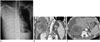
On the 14th day post admission, the patient experienced an abrupt onset of shortness of breath. His SpO2 level decreased to 89%, and his blood pressure fell to 80/50 mm Hg. In addition, his pulse rate (116/min) and respiration rate (40 breaths/min)were high. A chest radiograph showed complete white out of the right hemithorax with mediastinal shifting to the left (Fig. 1A). Bloody fluid was aspirated by thoracentesis. The red blood cell count in the aspirated fluid was 1650000/mcL. Chest tube thoracostomy was performed, and the bloody fluid was drained. The patient's hemoglobin (Hb) level decreased from 11.2 g/dL to 6.2 g/dL.
Contrast enhanced chest and abdominal computed tomography (CT) scans were obtained at this timepoint. On comparing these CT scans to the previous CT scans, obtained 2 months earlier, it was found that HCC had rapidly increased in size from 6 cm to 10 cm. Moreover, the tumor had invaded the right diaphragm, and it had also invaded the right pleural cavity (Fig. 1B). The tumor showed extensive necrosis, and the surface of the intrapleural portion of the tumor was irregular and ruptured (Fig. 1C). Metastatic nodules in the right lower lobe were surrounded by a collapsed lung, and intrathoracic lesions were excluded as the possible cause of hemothorax. These findings suggested that intrathoracic rupture of HCC resulted in hemothorax.
Transcatheter arterial embolization (TAE) was carried out immediately. Even though CT scans and angiography indicated no contrast extravasation, embolization of both the right anterior and middle hepatic arteries was undertaken using lipiodol and gelatin sponges.
Subsequently, the patient,s blood pressure rose to 105/60 mm Hg, and his Hb level increased to 9.3 g/dL. The amount of bloody fluid drained through a chest catheter gradually decreased, and the catheter was removed from the patient's chest on the 36th day post admission. Paracentesis was undertaken multiple times during admission, and a large amount of yellowish ascitic fluid was drained. Although the patient received intensive supportive care, the patient died of multiple organ failure on the 58th day post admission.
DISCUSSION
Spontaneous rupture of HCC is a rare but potentially life-threatening complication. The incidence of HCC rupture has been reported to be within the range of 2.3–26%. Since HCC is a hypervascular tumor and the pleural cavity pressure is negative, hemothorax caused by spontaneous rupture of HCC leads to high possibility of mortality due to uncontrollable bleeding.
Ruptured HCC usually presents with hemoperitoneum due to its intraabdominal location. Thus, hemothorax after HCC rupture is an extremely uncommon condition, with only 18 cases being reported in the English and Japanese literature (34). In four of the 18 cases, direct invasion of HCC into the right pleural cavity was the cause of hemothorax, as in our case. In 13 out of the 18 cases, instead of ruptured primary HCC, ruptured intrathoracic metastatic lesions resulted in hemothorax. In the remaining one case, it was reported that hemothorax was caused by blood flow from hemoperitoneum into the pleural cavity (4).
Previous reports have indicated that tumors with larger sizes and greater extrahepatic protrusion rupture more frequently (6). Besides the tumor size and the degree of extrahepatic protrusion, vascular dysfunction in the peritumoral region, where the blood supply to the tumor is the richest, may be related to spontaneous rupture of HCC (7). Portal vein thrombosis has also been reported to be a predictor of impending HCC rupture (8).
Multiplanar reformation images using multi-detector row CT are useful for detecting and defining the extent of extrahepatic protrusion of HCC, which is an indicator of the possibility of HCC rupture. To detect active bleeding from ruptured HCC, contrast enhanced CT is helpful in identifying extravasation of contrast material. If extravasation of contrast material is identified, this finding can be used to assist in the selection of appropriate vessels for TAE.
Hemostasis is the first step in the management of a ruptured HCC using TAE or surgery. TAE followed by liver resection seems to achieve the best treatment results (910). The prognosis of ruptured HCC is poor compared to that of non-ruptured HCC. Prognosis is worse in advanced stage HCC rupture than in early stage HCC rupture.
In conclusion, if pleural fluid accumulates or pleural effusion abruptly increases in amount in patients with a large-sized HCC and extrahepatic protrusion, the possibility of hemothorax due to intrathoracic rupture should be given due consideration, and in the actual event of intrathoracic rupture, immediate diagnosis and treatment will result in a better prognosis.
 XML Download