Journal List > J Korean Soc Radiol > v.77(1) > 1087881

문, 정, 이, 정, Moon, Jeong, Lee, and Jeong: 어깨충돌증후군에서 봉우리위팔뼈 불안정에 의한 어깨밑근 손상

Abstract

Purpose

To evaluate the association of subscapularis (SSC) injuries with acromiohumeral instability (AHI) in patients with shoulder impingement syndrome with supraspinatus (SSP) tears.

Materials and Methods

Pre-operative shoulder magnetic resonance images of 106 patients with subsequent arthroscopic confirmation of shoulder impingement syndrome were reviewed retrospectively. Patients with SSC injuries were divided into the following 3 groups: 1) those with no injury symbolized to the SSC[0] (n = 38), 2) those with partial injuries to the SSC[1] (n = 41), and 3) those with complete disruption of the SSC[2] (n = 27). AHI was categorized into 5 stages depending on the SSP lesions: partial tear or pretear tendinosis symbolized to the SSP[0] (n = 24) and according to retraction severity of the SSP tendon with complete tear such as SSP[1] (n = 19), SSP[2] (n = 27), SSP[3] (n = 29), and SSP[4] (n = 7). Also, AHI was divided into two groups such as the mild group summed with SSP plus SSP plus SSP and the severe group summed with SSP plus SSP, including 70 patients and 36 patients, respectively.

Results

Twenty-nine patients (63.0%) among 46 patients with SSC[0] and thirty-two patients (78.0%) among 41 patients with SSC[1] were related to mild AHI. Eighteen patients (66.7%) among 27 patients with SSC[2] were associated with severe AHI. The SSC injury groups were statistically significantly associated with AHI (estimate 0.207, standard error 0.057, p < 0.01).

Conclusion

SSC injuries could be related to AHI in patients with shoulder impingement syndrome.

Abstract

목적

가시위근의 파열을 초래하는 어깨충돌증후군에서 봉우리위팔뼈관절 불안정이 어깨밑근 손상에 미치는 영향을 평가하고자 한다.

대상과 방법

가시위근 손상을 가진 어깨충돌증후군 환자에서 관절경 수술 전 어깨 magnetic resonance images (이하 MRI)를 시행한 환자 106명에서 후향적으로 관절경 어깨밑근 소견과 관절경과 어깨 MRI로 봉우리윗팔뼈관절 불안정을 가시위근 손상 단계로 나타내 서로 비교하였다. 어깨밑근 소견은 손상이 없는 subscapularis (이하 SSC)[0] 46예, 부분손상 SSC[1] 41예, 그리고 완전 손상 SSC[2] 27예로 세 군을 나누고, 봉우리위팔뼈관절 불안정은 가시위근의 부분손상이나 힘줄염 소견을 supraspinatus (이하 SSP)[0] 24예, 큰결절에 국한된 완전손상 SSP[1] 19예, 찢어진 힘줄 뒤당김부분이 위팔뼈갈래 바깥쪽에 위치한 완전손상 SSP[2] 27예, 뒤당김부분이 위팔뼈갈래 안쪽에 위치한 완전손상 SSP[3] 29예, 그리고 뒤당김부분이 관절와(glenoid)에 위치한 완전손상 SSP[4] 7예, 5단계로 나누었다. 통계는 어깨밑근 손상 정도를 종속변수로 하고 가시위근 손상 정도를 설명변수로 하여 평균과 표준편차, 회귀분석, 그리고 대응분석으로 처리하였다.

결과

경한 어깨밑근 손상인 SSC[0] 63%와 SSC[1] 78%가 경한 봉우리위팔뼈관절 불안정인 SSP[0], SSP[1], 그리고 SSP[2]와 관계가 있었고, 심한 어깨밑근 손상인 SSC[2] 66.7%가 심한 봉우리위팔뼈관절 불안정인 SSP[3]과 SSP[4]와 관계가 있었다(estimate 0.207, standard error 0.057, p < 0.01).

결론

어깨충돌증후군에 있어서 어깨밑근 손상은 봉우리위팔뼈관절 불안정과 관련이 있을 것으로 생각된다.

서론

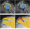
어깨봉우리뼈아래충돌증후군 환자에서 어깨밑근(subscapu-laris; 이하 SSC)의 손상은 관절경상 27%에서 43% 빈도로 보고하고 있으며, 가시위근(supraspinatus; 이하 SSP)의 완전파열 중 지방변화가 있는 만성인 경우 어깨밑근의 손상 빈도는 86%까지 높게 보고하고 있다(123). 최근 들어 1.5T magnetic reso-nance images (이하 MRI)로 어깨밑근의 부분손상은 79%, 완전손상은 89%로 그 진단적 정확성은 82%나 된다고 보고하였다(4). 어깨밑근의 손상 기전은 아직까지 정확하게 밝혀지지 않고 있다. 대부분 어깨밑근 손상은 가시위근의 파열손상이 있는 경우 발생하고 있으며 단독으로 오는 경우는 부리뼈아래충돌증후군이나 외상으로 인한 경우를 보고하고 있다(567). 이에 본 저자들은 어깨밑근 손상이 가시위근의 파열손상과 관련이 있다고 보고, 가시위근의 손상이 클수록 어깨봉우리뼈와 위팔뼈갈래 사이 간격은 좁아지고 이차적으로 어깨삼각근이 어깨관절 운동에 관여하여 위팔뼈갈래가 탈구되어 올라가고 따라서 위팔뼈갈래의 앞쪽 볼록한 면과 부리뼈(coracoid process)의 짧은갈래이두근(short head of biceps tendon) 사이에 어깨밑근이 끼여 압박을 받아 손상되는 것으로 가정하고, 어깨봉우리뼈아래충돌증후군에서 가시위근의 파열 정도에 따른 어깨밑근의 손상을 조사하므로 봉우리위팔뼈 불안정이 어깨관절 내장증을 초래하여 어깨밑근의 손상이 일어남을 확인하고자 하였다(Fig. 1).

대상과 방법

이 후향적 연구는 본 병원 임상시험심사위원회의 승인을 받았으며, 연구대상자에 대한 동의서는 면제되었다. 가시위근 손상을 가진 어깨충돌증후군 환자에서 관절경 수술 전 어깨 MRI를 시행한 환자 106명에서 후향적으로 관절경 어깨밑근 소견과 관절경과 어깨 MRI로 봉우리위팔뼈관절 불안정을 가시위근 손상 단계로 나타내 서로 비교하였다. 환자는 평균 연령이 61.9세(최고 83세, 최저 36세)였고 남자 39명, 여자 67명이었다. 자기공명영상은 Verio 3T (Siemens, Erlangen, Germany)와 Avanto 1.5T (Siemens, Erlangen, Germany) 두 기종이 사용되었으며, 전자는 3 mm 두께 T1 강조영상[repetition time (이하 TR)/echo time (이하 TE) = 600/20 ms]과 T2 강조영상(TR/TE = 4500/60 ms), 그리고 후자는 3 mm 두께로 T1 강조영상(TR/TE = 510~560/12 ms)과 T2 강조영상(TR/TE = 3000~ 4500/52 ms)으로 각각 축상, 빗관상 및 빗시상 단면영상을 얻었다. 관찰 범위는 가시위근의 전방, 큰결절의 앞쪽에서 작은결절 아래, 그리고 긴갈래이두근과 짧은갈래이두근 사이 어깨밑근을 포함하였다.
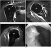
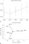
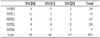
봉우리위팔뼈관절 불안정은, 설명변수로 자기공명영상에서 가시위근의 부분손상이나 힘줄염 소견을 SSP[0] 24예, 큰결절에 국한된 완전손상 SSP[1] 19예, 찢어진 힘줄 뒤당김부분이 위팔뼈갈래 바깥쪽에 위치한 완전손상 SSP[2] 27예, 뒤당김부분이 위팔뼈갈래 안쪽에 위치한 완전손상 SSP[3] 29예, 그리고 뒤당김부분이 관절와(glenoid)에 위치한 완전손상 SSP[4] 7예, 5단계로 나누었다. 종속변수로 어깨밑근 소견은 수술 후 의무기록지를 참조하여, 손상이 없는 SSC[0] 46예, 부분손상 SSC[1] 41예, 그리고 완전 손상 SSC[2] 27예, 세 군으로 나누었다(Table 1, Fig. 2). 통계는 어깨밑근 손상 정도를 종속변수로 하고 가시위근 손상 정도를 설명변수로 하여 두 가지 척도 모두 숫자로 간주하여 평균과 표준편차를 나타내고, 회귀분석(regression anal-ysis), 그리고 대응분석(correspondence analysis)으로 처리하였다(Fig. 3).

결과

SSP[0] 24예 중 SSC[1]이 13예(54.2%), SSP[1] 19예 중 SSC[0]이 12예(63.2%), SSP[2] 27예 중 SSC[1]이 14예(51.9%), SSP[3] 29예 중 SSC[2]가 13예(44.8%), 그리고 SSP[4] 7예 중 SSC[2]가 5예(71.4%)로 각각 가장 빈도가 높았다. SSC[0] 38예 중 SSP[1]이 12예(31.6%), SSC[1] 41예 중 SSP[2]가 14예(34.1%), 그리고 SSC[2] 27예 중 SSP[3]이 13예(48.1%)로 각각 가장 빈도가 높았다. 경한 어깨밑근 손상인 SSC[0] 63%와 SSC[1] 78%가 경한 봉우리위팔뼈관절 불안정인 SSP[0], SSP[1], 그리고 SSP[2]와 관계가 있었고, 심한 어깨밑근 손상인 SSC[2] 66.7%가 심한 봉우리위팔뼈관절 불안정인 SSP[3]과 SSP[4]와 관계가 있었다(estimate 0.207, standard error 0.057, p < 0.01) (Table 2, Fig. 3).

고찰

봉우리뼈아래 충돌증후군(subacromial impingement syn-drome)이란 봉우리뼈아래 뼈돌기나 부리봉우리인대 비대(cor-acoacromial ligament hypertrophy) 등으로 인하여 가시위근의 손상을 초래하여 근육둘레띠(rotator cuff)의 기능을 상실하게 하는 질환군을 말한다(8). 하지만 가시위근이 손상되어 그 기능을 상실하게 되면 이차적으로 어깨삼각근(deltoid muscle)이 팔을 거상시키는 역할을 하므로 봉우리위팔뼈 불안정을 초래하고 관절내장증(internal derangement)이 일어나 어깨밑근이나 긴두갈래근힘줄(long head of biceps tendon), 그리고 주변 구조물의 손상을 초래하게 된다(910). 위팔어깨관절 불안정(gleno-humeral instability)은 임상적으로 그 원인에 따라 전방 탈구와 전방 관절순 손상을 초래하는 전방불안정(anterior instability), 후방 탈구와 후방 관절순 손상을 초래하는 후방불안정(posterior instability), 그리고 관절낭의 느슨함(laxity)으로 인한 뭇방향불안정(multidirectional instability)이 보고되어 있다(11). 하지만 본 저자의 경우처럼 가시위근의 손상과 어깨삼각근의 수축으로 인한 상방불안전성(superior instability)에 대한 문헌 기록이 없는 관계로 이를 봉우리뼈위팔불안정(acromiohumeral instability)이라고 명명하였다.
어깨 운동에 관여하는 위팔어깨관절 근육으로는 어깨 근육둘레띠로 알려져 있는 가시위근, 어깨밑근, 가시아래근, 그리고 작은원근(teres minor)이 있고, 위팔어깨관절 근육둘레띠 바깥쪽으로 어깨세모근, 큰가슴근(pectoralis major), 위팔세갈래근중 안갈래(medial head of triceps brachii), 부리위팔근(coraco-brachialis), 위팔두갈래근(biceps brachii), 그리고 큰원근(teres major)이 어깨 운동에 관여하고 있다. 그중 어깨세모근은 위팔어깨관절 근육 중 가장 크며 어깨 빗장뼈(clavicle) 바깥쪽, 봉우리뼈 바깥쪽 후방, 그리고 어깨뼈가시(scapular spine) 아래쪽에서 기시하여 위팔뼈(humerus) 상부 바깥쪽에 삽입되어 어깨 벌림(abduction), 굽힘(flexion), 폄(extension), 그리고 돌림(rota-tion) 운동에 관여한다. 어깨 운동은 주로 근육둘레띠에 의해 일어나지만 가시위근이 손상되는 경우 어깨세모근이 어깨 운동에 주 역할을 하게 된다(10). 어깨밑근 손상은 그렇게 드물지 않은 질병 중 하나이지만 임상적으로 봉우리뼈아래충돌증후군에서 가시위근의 손상으로 인해 대부분 간과되어 왔던 어깨 근육둘레띠 손상 중 하나다(2). 가시위근의 손상이 있는 경우 어깨위팔뼈관절은 불안정해지고 근육둘레띠의 대부분이 본래의 위치에서 이탈하여 긴장성 이완수축을 하게 되므로 어깨관절 내장증을 일으키게 된다. 이는 가시위근의 손상이 만성화될수록 어깨밑근의 손상 빈도가 높아진다는 그 이유를 설명하고 있다(3). 어깨밑근은 어깨위팔뼈관절의 앞쪽 안쪽에 위치하며 어깨밑근의 작은결절 삽입부 상부는 부리위팔뼈인대(coracohumeral ligament)와 접하고 있다. 해부학적으로 가시위근이 심하게 손상되면 위팔뼈갈래(humeral head)가 위로 올라가게 되고 위팔뼈갈래의 앞쪽 안쪽면이 부리위팔뼈인대와 가까워져 물리적으로 어깨밑근이 손상될 뿐만 아니라 어깨밑근의 상부가 왜곡되고 당겨져 긴장성 손상까지 일어나게 된다(Fig. 1). 이를 부리아래충돌증후군(subcoracoid impingement)이라고 하고 자기공명영상 코호트조사에서 어깨밑근 손상이 없는 환자군(n = 15)의 부리돌기와 위팔뼈 사이 평균간격은 10.48 mm였고 어깨밑근 손상이 있는 환자군(n = 9)의 경우 평균간격은 5.33 mm였다(9). 어깨밑근의 손상은 부분파열과 완전파열로 나눌 수 있으며, 부분파열은 주로 삽입부 상부 1/3에서 발생하고 내시경 소견으로 세로째짐(vertical tear), 판분리(laminar separation), 가로째짐(horizontal tear) 등이 있으나 현존하는 자기공명영상으로 이들 병소를 구별하기에는 해상력에 한계가 있다. 어깨밑근의 완전파열은 자기공명영상에서 어깨밑근의 힘줄섬유단절(tendon fiber disruption), 미만부기(diffuse swelling), 누덕신호부위(patchy signal) 소견 등을 관찰할 수는 있으나 진단적 예민성이 낮은 편이다(12). 어깨밑근 손상은 단독으로 발생하는 경우는 드물고 대부분 심한 가시위근의 파열을 동반하고 있으며 봉우리뼈위팔뼈관절 불안정 또한 동반하고 있어 가시위근 파열을 치료하면서 관절 간격을 유지하는 시술을 하므로 더 이상의 어깨밑근 손상이 일어나지 않도록 예방하거나 비치료 치유를 기대할 수 있다. 본 저자들의 연구에 의하면 가시위근의 완전파열 정도가 심하면 심할수록 어깨밑근의 손상 또한 심해진다는 것을 알 수 있었고, 이는 가시위근의 손상으로 어깨세모근이 어깨윗팔운동에 관여하게 되고 윗팔뼈가 탈구되어 봉우리뼈 방향으로 거상하게 되면 수리의 부리처럼 어깨뼈(scapula)에서 전방으로 돌출한 부리돌기(coracoid process)와 위팔뼈갈래의 불룩 나온 면이 근접하게 되어 그 사이에 있는 어깨밑근이 끼이게 되어 부리돌기하 충돌(subcoracoid impingement)을 일으키고 어깨밑근이 손상을 받게 된다. 또한 긴갈래이두근힘줄이 안쪽으로 부분탈구가 일어나 작은거친면(lesser tuberosity)에 삽입되어 있는 어깨밑근이 손상을 입게 된다. 결론적으로 봉우리뼈위팔뼈관절의 불안정이 어깨관절 내장증을 악화시키기 때문에 술전에 어깨 MRI를 검사하여 가시위근의 완전파열 정도와 지방변성의 만성화를 평가하므로 어깨밑근의 손상을 예측하고 어깨밑근 손상의 치료계획을 세우는 한편 봉우리뼈위팔뼈관절 불안정을 동시에 교정하는 시술이 필요하리라 생각된다.

Figures and Tables

Fig. 1

Schematic images explaning mechanical SSC tendon injuries in patients with subacromial impingement syndrome.

A. The oblique sagittal image at the level of the coracoid process illustrates a normal anatomical structure of the SSP and SSC (small arrows) in the absence of subacromial impingement.
B. In subacromial impingement with acromiohumeral instability, the oblique sagittal image demonstrates acromiohumeral joint space narrowing and elevated position of the humeral head resulting in SSC tendon injury (large arrows) caused by dynamic force of subcoracoid impingement between the coracoid process and the anterior convexity of the humeral head.
C. The coronal image with a center of the coracoid process illustrates the normal anatomical position of the SSP and SSC (small arrows).
D. In subacromial impingement with acromiohumeral instability, the coronal image reveals acromiohumeral joint space narrowing and elevated position of the humeral head (large arrows) resulting in SSC tendon tears caused by shearing force of the coracoid process. Also, medial subluxation of the LHBT could result in injury to the LT insertion portion of the SSC.
A = acromion, GT = greater tuberosity, ISP = infraspinatus, LHBT = long head of biceps tendon, LT = lesser tuberosity, SSC = subscapularis, SSP = supraspinatus
jksr-77-47-g001
Fig. 2

A 54-year-old female patient was arthroscopically confirmed to have complete tears of the SSP[3] and the SSC[2].

A. The T2 fat saturation (FS) coronal MR image demonstrates stage complete tear of the SSP with retracted portion on the medial side of the humeral head (white arrows).
B. The T2 FS axial MR image depicts complete tear of the proximal portion of the SSC (black arrows).
C. The T2 FS oblique-sagittal MR image illustrates marked narrowing of the acromiohumeral space due to full-thickness tear of the SSP (white arrow) and absence of the SSC on the medial side of the lesser tuberosity suggestive of full-thickness tear of the SSC (black arrows).
D. Arthroscopy shows SSC stump due to its complete disruption (black arrows).
A = acromion, GL = glenoid, H = humeral head, SSC = subscapularis, SSP = supraspinatus
jksr-77-47-g002
Fig. 3

Regression analysis (A) and correspondence analysis (B) related between 5 stages of SSP lesions and 3 groups of SSC injuries (SSC = 0.529 + 0.207 SSP, regression coefficient estimate 0.207). SSC = subscapularis, SSP = supraspinatus

jksr-77-47-g003
Table 1

The Definitions of 5 Stages of SSP Lesions on MR Images and 3 Groups of SSC Injuries on Arthroscopy

jksr-77-47-i001
Stages/Groups Definitions
SSP[0] Partial tear or pretear tendinosis of SSP
SSP[1] Complete tear of SSP with torn cuff edge adjacent to greater tuberosity
SSP[2] Complete tear of SSP with tendon retraction to lateral humeral head
SSP[3] Complete tear of SSP with tendon retraction to medial humeral head
SSP[4] Complete tear of SSP with tendon retraction to glenoid
SSC[0] No injury of SSC
SSC[1] Partial injuries of SSC
SSC[2] Complete disruption of SSC

SSC = subscapularis, SSP = supraspinatus

Table 2

The Numbers of SSC Injuries According to SSP Tear Staging in Subacromial Impingement

jksr-77-47-i002
SSC[0] SSC[1] SSC[2] Total
SSP[0] 9 13 2 24
SSP[1] 12 5 2 19
SSP[2] 8 14 5 27
SSP[3] 8 8 13 29
SSP[4] 1 1 5 7
Total 38 41 27 106

SSC = subscapularis, SSP = supraspinatus

Acknowledgments

이 논문은 부산대학교 기본연구지원사업(2년)에 의하여 연구되었음.

References

1. Bennett WF. Arthroscopic repair of anterosuperior (supraspinatus/subscapularis) rotator cuff tears: a prospective cohort with 2- to 4-year follow-up. Classification of biceps subluxation/instability. Arthroscopy. 2003; 19:21–33.
2. Adams CR, Schoolfield JD, Burkhart SS. The results of arthroscopic subscapularis tendon repairs. Arthroscopy. 2008; 24:1381–1389.
3. Bergin D, Parker L, Zoga A, Morrison W. Abnormalities on MRI of the subscapularis tendon in the presence of a full-thickness supraspinatus tendon tear. AJR Am J Roentgenol. 2006; 186:454–459.
4. Malavolta EA, Assunc¸ão JH, Guglielmetti CL, de Souza FF, Gracitelli ME, Bordalo-Rodrigues M, et al. Accuracy of preoperative MRI in the diagnosis of subscapularis tears. Arch Orthop Trauma Surg. 2016; 136:1425–1430.
5. Giaroli EL, Major NM, Lemley DE, Lee J. Coracohumeral interval imaging in subcoracoid impingement syndrome on MRI. AJR Am J Roentgenol. 2006; 186:242–246.
6. Tung GA, Yoo DC, Levine SM, Brody JM, Green A. Subscapularis tendon tear: primary and associated signs on MRI. J Comput Assist Tomogr. 2001; 25:417–424.
7. Pfirrmann CW, Zanetti M, Weishaupt D, Gerber C, Hodler J. Subscapularis tendon tears: detection and grading at MR arthrography. Radiology. 1999; 213:709–714.
8. Shinohara H, Urabe Y, Maeda N, Xie D, Sasadai J, Fujii E. Does shoulder impingement syndrome affect the shoulder kinematics and associated muscle activity in archers? J Sports Med Phys Fitness. 2014; 54:772–779.
9. Nair AV, Rao SN, Kumaran CK, Kochukunju BV. Clinico-radiological correlation of subcoracoid impingement with reduced coracohumeral interval and its relation to subscapularis tears in Indian patients. J Clin Diagn Res. 2016; 10:RC17–RC20.
10. Sakoma Y, Sano H, Shinozaki N, Itoigawa Y, Yamamoto N, Ozaki T, et al. Anatomical and functional segments of the deltoid muscle. J Anat. 2011; 218:185–190.
11. Watson L, Warby S, Balster S, Lenssen R, Pizzari T. The treatment of multidirectional instability of the shoulder with a rehabilitation program: part 1. Shoulder Elbow. 2016; 8:271–278.
12. Moon TY, Lee IS, Song YS, Jang JY, Ahn S, Park JH. MR imaging of subscapularis injuries in subacromial impingement. J Pusan Natl Univ Hosp. 2013; 34:1–12.
TOOLS
Similar articles