

 PDF
PDF ePub
ePub Citation
 Print
Print


INTRODUCTION
Osteoma is a benign bone-forming tumor composed of compact or mature trabecular bone and is limited almost exclusively to craniofacial bones, especially those of paranasal sinuses (1). This tumor occurs more often in females (12). It is a slow growing lesion that is usually asymptomatic, unless it enlarges and causes symptoms (123). Its radiological appearance depends on its location. Central osteomas are circumscribed sclerotic lesions with smooth borders, whereas peripheral or juxtacortical osteomas are radiopaque lesions with expansive borders that can be sessile or pedunculated (24). Most osteomas are solitary; when osteomas are present at multiple sites, Gardner's syndrome should be considered (1-3). In cases of juxtacortical osteoma, it is crucial to differentiate between this type of tumor and parosteal osteosarcoma, sessile osteochondroma, and matured juxtacortical focus of myositis ossificans (235). We describe a case of an osteoma arising from the second metatarsal bone, a rare anatomical location for osteoma. To our knowledge, this is the first reported case of osteoma of the metatarsal bone. The tumor was removed surgically and had lamellar bone, suggesting osteoma.
CASE REPORT
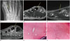
A 61-year-old woman was presented to our hospital complaining of right foot pain. This symptom had started 1 month prior and was worse with ambulation. She did not report history of trauma and had no external wounds. On physical examination, she had tenderness on the dorsal aspect of the second metatarsal area. Laboratory findings were normal. Plain radiograph revealed a small, well-demarcated, exophytic sessile osseous lesion arising from the shaft of the right second metatarsal bone (Fig. 1A). MRI revealed a mass lesion arising from the mid-shaft of the second metatarsal bone, showing homogeneously low signal intensity on T1- and T2-weighted images compared to that of the foot muscle (Fig. 1B, C). This mass revealed the same signal intensity as the bony cortex, had clear cortical margins, and no evidence of a cartilaginous cap. After intravenous administration of gadolinium, the mass did not reveal enhancement (Fig. 1D). Based on imaging findings, our tentative diagnosis was benign osteochondroma, and differential diagnosis included bizarre parosteal osteochondromatous proliferation, parosteal osteoma, and parosteal osteosarcoma. The patient underwent surgical excision of the lesion after interdepartmental discussion. Gross specimen was a 1.5 × 0.8 × 0.5-cm-sized solid mass with grayish-white firm bony tissue and without cartilage formation on the surface of the cut. Histological examination revealed mature bone tissue organized in wide lamellar patterns, with multiple osteocytes inside the lacunas (Fig. 1E, F). The excised mass was diagnosed as an osteoma.
DISCUSSION
Osteoma of bone other than the skull or facial bones is extremely rare (267). Small lesions are usually asymptomatic and are identified incidentally (23). Juxtacortical osteoma is a distinct entity and is diagnosed based on clinical, radiographic, and histological features (2). Usual presentation of the lesion is as an asymptomatic and radiographically long-standing uniform dense sclerotic lesion attached to the surface of the diaphysis or metadiaphysis of long bones in adults (2). In some reports, osteoma arising from craniofacial bones occasionally caused symptoms such as pain from mass effect from its large size (123). Foot pain has not been reported previously in literature, as juxtacortical osteoma from the foot bone is rare. To our knowledge, our patient is the first case to be diagnosed with an osteomafrom metatarsal bone. Because of its anatomical position, the osteoma may have caused pain from recurring stress on ambulation.
Basic imaging technique used to diagnose osteoma is conventional radiography, and the tumor typically appears as a homogeneous osseous mass with well-delineated smooth margins (58). CT is helpful in evaluating presence of cortical invasion. A heavily ossified mass attached to the cortex with no areas of lucency or cortical invasion is a typical CT finding of osteoma (9). Low signal intensity of the mass seen in T1 and T2 sequences of MRI is suitable for assessment of cortical bone lesions (5).
Differential diagnosis of potential symptoms related to osteoma includes sessile osteochondroma, matured juxtacortical focus of myositis ossificans, and parosteal osteosarcoma, and it is particularly crucial to differentiating benign osteoma tumors from osteosarcoma (235). Osteochondromas can be distinguished from osteomas on MRI because osteochondromas have a cartilaginous cap that reveals high signal intensity on T2-weighted images and a medullary space that is continuous with the parent bone (9). Myositis ossificans occur after repeated trauma and progressively ossify from the periphery with time (3). Juxtacortical or surface osteosarcomas account for approximately 4% of osteosarcomas (3). Radiographically, parosteal osteosarcoma usually has an irregular margin; unlike osteoma, scattered areas of radiolucency or a heterogeneous appearance are often detected (2). Parosteal osteosarcomas incorporate surrounding soft tissues as they grow and therefore typically have an indistinct interface between surrounding soft tissue and the leading edge of the tumor on CT or MR imaging (2). Parosteal osteosarcoma may cause extrinsic erosion of thickened underlying diaphyseal cortex and perpendicular periosteal reaction extending into the soft-tissue component (10). On MRI, reactive marrow changes are commonly observed (10).
Since osteoma is a benign disease, invasive surgical treatment is not recommended. Recurrence is rare, and no malignant transformation has been reported (2). Therefore, close follow-up with conventional radiography or marginal resection is likely to be sufficient for osteoma.
We described a rare case of juxtacortical osteoma of the metatarsal bone. Because it was hard to differentiate between osteoma and other bone tumors radiographically, open biopsy was required to rule out malignant bone tumor.
 XML Download