Abstract
Purpose
There is a relatively higher risk of peri-procedural stroke following elective stenting of patients with basilar artery (BA) occlusion compared with stenting of in- tracranial arteries. We sought to diagnose stroke risks in patients with BA steno-oc-clusive disease by describing their clinicoradiological features and by demonstrating that appropriate treatment would lead to favorable outcomes.
Materials and Methods
A total of 92 patients who were treated from 2004 to 2016 for severe stenosis or occlusion of BA based on MR or CT angiography were en-rolled in the study. We assessed clinical features, radiologic findings, and other clinical outcomes such as the degree of disability as determined by the modified Rankin Scale.
Results
A total of 49 of the 92 patients (53.3%) had no relevant symptoms. The risk of a recurrent or new infarct in the relevant area was 4.59%/year. Following treat-ment, more than 50% of the 92 patients had favorable outcomes. A recurrent or new infarct was found in 9 (20.5%) of the 44 patients who had a poor prognosis. There was no significant difference between the two groups with respect to compromised circulation; however, the initial infarct (16 of 48 and 29 of 44, p = 0.002) was statis-tically significant between the two groups.
Go to : 
REFERENCES
1.Mohammadian R., Pashapour A., Sharifipour E., Mansouriza-deh R., Mohammadian F., Taher Aghdam AA, et al. A compari-son of stent implant versus medical treatment for severe symptomatic intracranial stenosis: a controlled clinical trial. Cerebrovasc Dis Extra. 2012. 2:108–120.

2.Gorelick PB., Wong KS., Bae HJ., Pandey DK. Large artery in-tracranial occlusive disease: a large worldwide burden but a relatively neglected frontier. Stroke. 2008. 39:2396–2399.
4.Derdeyn CP., Chimowitz MI., Lynn MJ., Fiorella D., Turan TN., Janis LS, et al. Aggressive medical treatment with or with-out stenting in high-risk patients with intracranial artery ste-nosis (SAMMPRIS): the final results of a randomised trial. Lancet. 2014. 383:333–341.
5.Derdeyn CP., Fiorella D., Lynn MJ., Rumboldt Z., Cloft HJ., Gib-son D, et al. Mechanisms of stroke after intracranial angio-plasty and stenting in the SAMMPRIS trial. Neurosurgery. 2013. 72:777–795. discussion 795.
6.Jiang WJ., Du B., Hon SF., Jin M., Xu XT., Ma N, et al. Do patients with basilar or vertebral artery stenosis have a higher stroke incidence poststenting? J Neurointerv Surg. 2010. 2:50–54.

7.Nahab F., Lynn MJ., Kasner SE., Alexander MJ., Klucznik R., Zai-dat OO, et al. Risk factors associated with major cerebrovas-cular complications after intracranial stenting. Neurology. 2009. 72:2014–2019.

8.Levy EI., Hanel RA., Boulos AS., Bendok BR., Kim SH., Gibbons KJ, et al. Comparison of periprocedure complications re-sulting from direct stent placement compared with those due to conventional and staged stent placement in the basi-lar artery. J Neurosurg. 2003. 99:653–660.

9.Datar S., Lanzino G., Rabinstein AA. An unusually benign course of extensive posterior circulation occlusion. J Stroke Cerebrovasc Dis. 2015. 24:e165–e168.

10.Woolfenden AR., Tong DC., Norbash AM., Ali AO., Marks MP., O'Brien MW, et al. Basilar artery stenosis: clinical and neu-roradiographic features. J Stroke Cerebrovasc Dis. 2000. 9:57–63.

11.Caplan LR. Occlusion of the vertebral or basilar artery. Follow up analysis of some patients with benign outcome. Stroke. 1979. 10:277–282.

12.Caplan LR., Rosenbaum AE. Role of cerebral angiography in vertebrobasilar occlusive disease. J Neurol Neurosurg Psy-chiatry. 1975. 38:601–612.

13.Caplan L. Posterior circulation ischemia: then, now, and to-morrow. The Thomas Willis Lecture-2000. Stroke. 2000. 31:2011–2023.
14.Devuyst G., Bogousslavsky J., Meuli R., Moncayo J., de Freitas G., van Melle G. Stroke or transient ischemic attacks with basilar artery stenosis or occlusion: clinical patterns and outcome. Arch Neurol. 2002. 59:567–573.
15.Mattle HP., Arnold M., Lindsberg PJ., Schonewille WJ., Schroth G. Basilar artery occlusion. Lancet Neurol. 2011. 10:1002–1014.

16.Abuzinadah AR., Alanazy MH., Almekhlafi MA., Duan Y., Zhu H., Mazighi M, et al. Stroke recurrence rates among patients with symptomatic intracranial vertebrobasilar stenoses: sys-tematic review and meta-analysis. J Neurointerv Surg. 2016. 8:112–116.

17.Chimowitz MI., Kokkinos J., Strong J., Brown MB., Levine SR., Silliman S, et al. The warfarin-aspirin symptomatic intracra-nial disease study. Neurology. 1995. 45:1488–1493.

18.Poletti PA., Pereira VM., Lovblad KO., Canel L., Sztajzel R., Beck-er M, et al. Basilar artery occlusion: prognostic signs of se-verity on computed tomography. Eur J Radiol. 2015. 84:1345–1349.

19.Lee WJ., Jung KH., Ryu YJ., Lee KJ., Lee ST., Chu K, et al. Acute symptomatic basilar artery stenosis: MR imaging predic-tors of early neurologic deterioration and long-term out-comes. Radiology. 2016. 280:193–201.

Go to : 
 | Fig. 1.Basilar artery stenosis due to atherosclerosis. An MR angiogram (A) shows a short stenosis at the middle basilar artery (short arrow). A proton density-weighted high-resolution MR image (B) shows eccentric wall thickening (arrow) at the middle basilar artery, which may be indic-ative of an atherosclerotic plaque. In an MR angiogram (A), the stenosis at the fourth segment of vertebral artery (long arrow), left is also noted. |
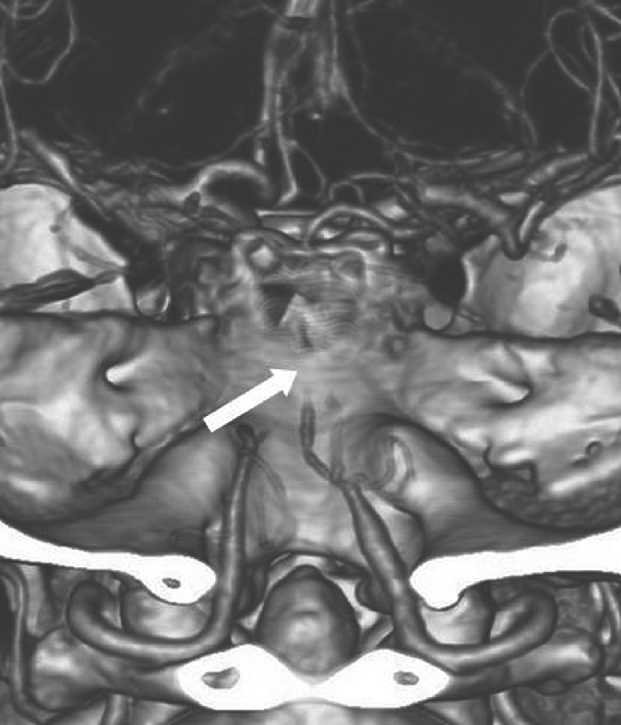
 | Fig. 2.Basilar artery occlusion. An CT angiogram reconstruction scan shows the entire length of the basilar artery occlusion (arrow), along with underlying vertebrobasilar hypoplasia. |
Table 1.
Clinicoradiological Characteristics of Patients with Basilar Artery Steno-Occlusion
Table 2.
Comparison of New Infarct and Clinical Outcome between Symptomatic and Asymptomatic Patients with Atherosclerotic Basilar Artery Steno-Occlusion
| All (n = 92) | Symptomatic (n = 43) | Asymptomatic (n = 49) | p-Value | |
|---|---|---|---|---|
| Recurrent or new infarct, (%) | 11 (12.0) | 5 (11.6) | 6 (12.2) | 0.927 |
| Overall poor outcome, (%) | 22 (23.9) | 8 (18.6) | 14 (28.6) | 0.263 |
| Relevant poor outcome, (%) | 15 (16.3) | 8 (18.6) | 7 (14.3) | 0.576 |
| Inflow compromise† | ||||
| Yes | 61 (8/11)* | 27 (3/5)* | 34 (5/6)* | 0.504 |
| No | 31 (3/4)* | 16 (2/3)* | 15 (1/1)* |
Table 3.
Radiological Characteristics of Patients with Atherosclerotic Basilar Artery Steno-Occlusion with Poor and Favorable Outcomes
| Radiological Characteristics | All (n = 92) | Favorable Outcome (n = 48) | Poor Outcome | |||
|---|---|---|---|---|---|---|
| Overall (n = 44) | p-Value | p-Value | Relevant (n = 23) | |||
| Inflow compromise, (%) | ||||||
| Undetectable PCom | 35 (38.0) | 21 (43.8) | 14 (31.8) | 0.239 | 10 (43.5) | 0.535 |
| VA steno-occlusion | 45 (48.9) | 23 (47.9) | 22 (50.0) | 0.842 | 11 (47.8) | 0.904 |
| VA hypoplasia | 20 (21.7) | 8 (16.7) | 12 (27.3) | 0.218 | 3 (13.0) | 0.355 |
| Infarct*, (%) | 45 (48.9) | 16 (33.3) | 29 (65.9) | 0.002 | 17 (73.9) | 0.006 |
| Recurrent/new infarct†, (%) | 11 (7/4) (12.0) | 2 (0/2) (4.2) | 9 (7/2) (20.5) | 0.016 | 9 (7/2) (39.1) | < 0.001 |


 PDF
PDF ePub
ePub Citation
 Print
Print


 XML Download