

 PDF
PDF ePub
ePub Citation
 Print
Print


Abstract
A gadolinium-based contrast is the preferred agent when differentiating acute neu-rological diseases. Since the renal route is the main pathway for excretion of gadolini-um chelates, prolonged extracellular distribution of gadolinium has previously been reported in dialysis-dependent patients. Hence, gadolinium-based contrast agents are used cautiously in patients with known renal disease. Retention of gadolinium manifests as increased fluid-attenuated inversion recovery (FLAIR) signal intensity in the subarachnoid space, leading to diagnostic errors. Here, we describe a patient who presented to our emergency room with an acute cerebral infarction. Enhanced brain magnetic resonance imaging performed 2 days later revealed high signal inten-sity in the cerebrospinal fluid spaces on follow-up FLAIR images.
REFERENCES
1.Ong EM., Yeh IB. High signal in the cerebrospinal fluid following prior gadolinium administration in a patient with renal impairment. Singapore Med J. 2007. 48:e296–e298.
2.Rai AT., Hogg JP. Persistence of gadolinium in CSF: a diagnos-tic pitfall in patients with end-stage renal disease. AJNR Am J Neuroradiol. 2001. 22:1357–1361.
3.Lev MH., Schaefer PW. Subarachnoid gadolinium enhance-ment mimicking subarachnoid hemorrhage on FLAIR MR im-ages. Fluid-attenuated inversion recovery. AJR Am J Roent-genol. 1999. 173:1414–1415.

4.Tombach B., Bremer C., Reimer P., Schaefer RM., Ebert W., Geens V, et al. Pharmacokinetics of 1M gadobutrol in patients with chronic renal failure. Invest Radiol. 2000. 35:35–40.
5.Maramattom BV., Manno EM., Wijdicks EF., Lindell EP. Gado-linium encephalopathy in a patient with renal failure. Neu-rology. 2005. 64:1276–1278.
6.Stuckey SL., Goh TD., Heffernan T., Rowan D. Hyperintensity in the subarachnoid space on FLAIR MRI. AJR Am J Roentgen-ol. 2007. 189:913–921.
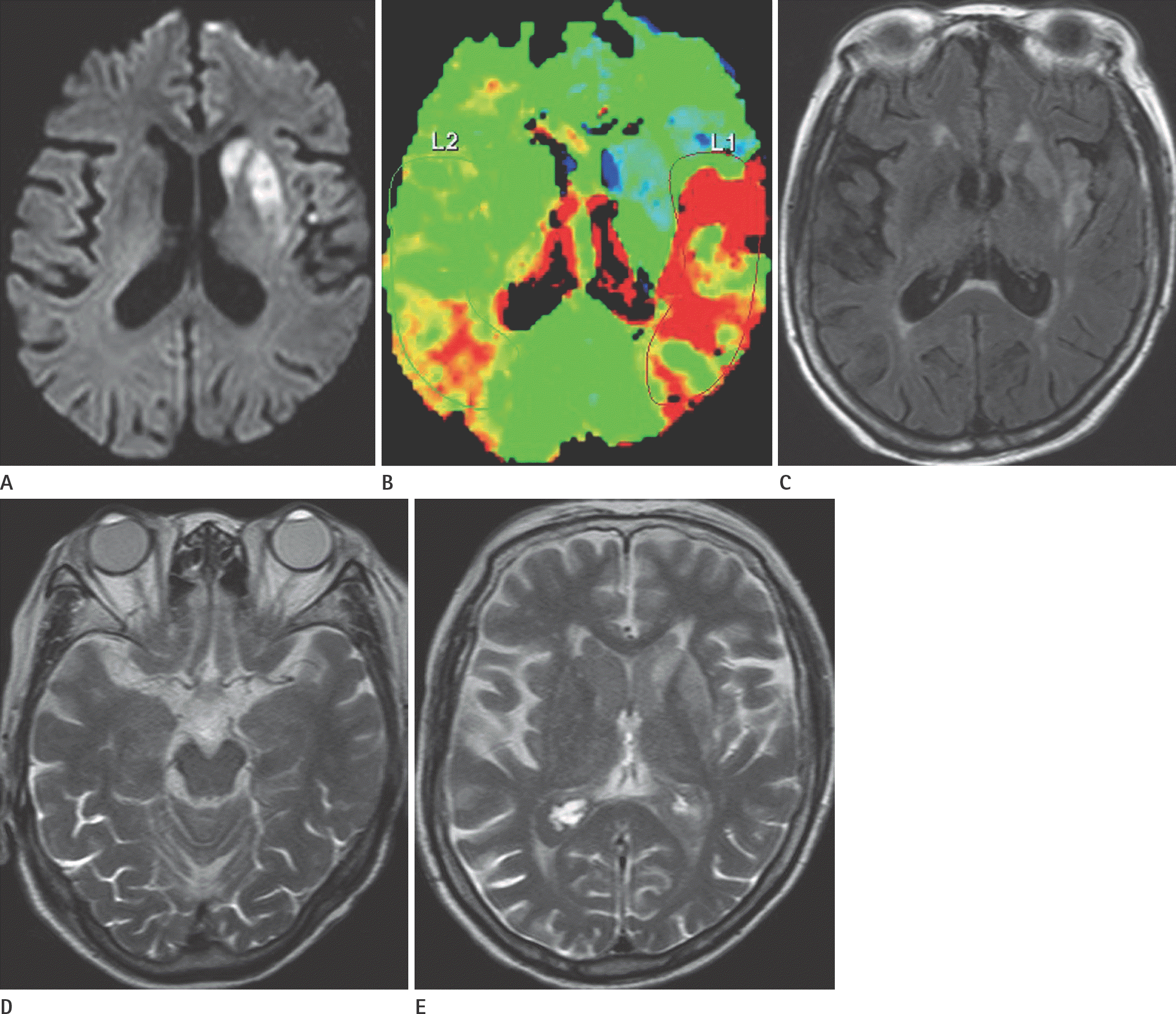
Fig. 1.
An 81-year-old woman with acute cerebral infarction. A. A diffusion-weighted image shows lesions with diffusion restriction in the left basal ganglia. B. Perfusion–weighted magnetic resonance imaging shows increased time to peak in left middle cerebral artery territory, suggesting acute infarction with perfusion-diffusion mismatch. C. Fluid-attenuated inversion recovery image shows multifocal hyperintense lesions in the left basal ganglia and insular cortex, but normal signal intensity in the cerebrospinal fluid spaces. D, E. Two-day follow-up axial fluid-attenuated inversion recovery images (D, E) show high signal intensity in the bilateral eye globes, subarach-noid spaces, and ventricular system.
 XML Download