

 PDF
PDF ePub
ePub Citation
 Print
Print


INTRODUCTION
Gestational trophoblastic disease (GTD) comprises a broad spectrum of disorders that arise from uncontrolled growth of placental trophoblastic tissue, and hydatidiform mole is a pre-malignant lesion of GTD originating from aberrant fertilization (1). GTD refers to a type of tumor which is, characteristically, highly vascularized and can cause heavy or life-threatening bleeding (23). The development of uterine arteriovenous malformations (AVMs) can be found in patients with GTD because of abnormal trophoblastic proliferation and increased angiogenesis caused by high production of human chorionic gonadotropin (hCG) (4).
The primary site of metastasis in GTD is the lung, with an incidence of 76–87% (1). However, the formation of AVMs within pulmonary metastatic lesions is rare in patients with GTD. We are reporting on a case of AVMs which developed in the areas of pulmonary metastases before treatment in a patient with hydatidiform mole. This report was approved by the Institutional Review Board of our institution.
CASE REPORT
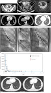
A nineteen-year-old woman visited our emergency room due to a febrile sensation and lower abdominal pain in June of 2014. She reported a four month history of vaginal bleeding. She was suspected of hydatidiform mole on pelvic sonography. On admission, the serum hCG level was 62200.02 mIU/mL. The findings on abdominal computed tomography (CT) and pelvic magnetic resonance imaging (MRI) revealed an enlarged uterus with abnormal heterogeneous enhancement, typical of molar pregnancy (Fig. 1A, B). At that time, chest radiography and contrast-enhanced chest CT scanning revealed multiple metastatic nodules in both lungs, and there were curvi-linear structures that were connected to some metastatic nodules in both lower lobes and the inferior lingular segment of the left upper lobe. Around the metastatic nodule, there were patchy ground-glass opacity lesions in the inferior lingular segment of the left upper lobe, consistent with pulmonary hemorrhage. The findings on chest CT consisted of multiple pulmonary metastases with AVMs, complicated by recent hemorrhage (Fig. 1C). No other metastasis was found in the brain or abdomen on abdominal CT or brain MRI. During admission, she was in septic shock (body temperature; 38.5℃, blood pressure; 80/60 mm Hg, heart rate; 120 beats per minute). After uterine embolization, dilatation and curettage was performed twice. Pulmonary arteriography confirmed the presence of two left AVMs and pulmonary AVMs were successfully embolized with a 2 × 5 mm coil one week later, as shown in Fig. 1D. Then, she was given three cycles of Methotrexate weekly and she attained a normal hCG level (Fig. 1E). She was regularly followed up and the findings on follow-up chest CT showed that multiple metastatic nodules with residual AVMs in both lungs had markedly decreased in size in January 2015 (Fig. 1F).
DISCUSSION
GTD is a rare entity, and it manifests as an hCG-producing hypervascular tumor (2). The key regulatory role of hCG in angiogenesis and vascular function is well known (4). AVMs in the uterus are a relatively frequent event, and the relationship between GTD and uterine AVMs is well-documented by abnormal trophoblastic proliferation and angiogenesis induced by high hCG production (245).
Pulmonary metastases of GTD are seen most often and are thought to be most common. Angiographic studies have shown that pulmonary metastases of GTD are hyper-vascular tumors, and are supplied by the pulmonary artery (6). Pulmonary AVMs are abnormally dilated vessels which provide an anatomic right-to-left shunt between the pulmonary artery and the pulmonary vein without an intervening pulmonary capillary system (7). Most pulmonary AVMs are congenital in etiology, and are associated with hereditary hemorrhagic telangiectasia. Acquired pulmonary AVMs can occur in the context of a variety of physical injuries or disease processes such as liver cirrhosis, chest trauma, infection, metastatic carcinoma, mitral stenosis or systemic amyloidosis (78). Pulmonary metastases of GTD have the potential to form pulmonary AVMs, which can cause life-threatening hemorrhage or hemoptysis (2). A few studies have shown that pulmonary AVMs were secondary to GTD at the metastatic site during or after chemotherapy (26910). Our patient showed multiple pulmonary metastases and AVMs within these lesions accompanied by pulmonary hemorrhage even before the onset of chemotherapy.
Uterine AVMs in patients with GTD have the potential risk of heavy vaginal bleeding or intraperitoneal hemorrhage resulting from disorganized vascular structures and high blood flow (5). Uterine or pulmonary AVMs may be spontaneously resolved with successful chemotherapy, and these patients were treated for persistent or life-threatening bleeding with embolization (35). However, AVMs might persist after successful completion of chemotherapy (2910). In our case, embolization was performed to reduce the bleeding risk, because our patient had pulmonary hemorrhage associated with pulmonary AVMs. In addition to metastatic nodules, residual pulmonary AVMs had disappeared or were markedly decreased with successful chemotherapy.
In conclusion, AVMs associated with pulmonary metastatic lesions are extremely rare. When the early diagnosis of pulmonary AVM using CT scanning is established, appropriate embolization and chemotherapy could be considered in an effort to reduce the risk of bleeding and to improve the outcome.
 XML Download