Abstract
Immunoglobulin G4 (IgG4)-related periaortitis and periarteritis are rare systemic inflammatory and fibrosclerosing diseases, usually involving the aorta and its main branches. We report a pathologically confirmed case of IgG4-related periaortitis involving the thoracoabdominal aorta, which can be confused with intramural hematoma or periaortic lymphoma.
Immunoglobulin G4 (IgG4)-related systemic disease is a recently recognized systemic inflammatory and sclerosing condition. Histopathologically, it is characterized by diffuse infiltration of inflammatory cells containing numerous IgG4-expressing plasma cells and associated fibrosis (1). This disease affects various systemic organs; it was first identified in the pancreas as autoimmune pancreatitis, and may involve extrapancreatic organs including the biliary system, kidney, lung, retroperitoneum, mesentery, lacrimal glands, and blood vessels (2). Although IgG4-related vascular lesion is a rare condition, it is important to understand its radiological and clinical characteristics since this disease is treatable with corticosteroids, unlike cases of neoplasm, aortic wall hemorrhage, or idiopathic aortitis (345). Here, we present a case of IgG4-related periaortitis and periarteritis, involving the junction of the thoracoabdominal aorta and right proximal common carotid artery (CCA) on computed tomography aortography (CTA).
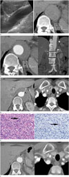
A 63-year-old woman, having a nonspecific past history, had left flank discomfort for two weeks. Her blood pressure was 141/64 mm Hg, her respiratory rate was 20 breaths per min, and body mass index was 22.92 kg/m2. Initial laboratory investigations found a white blood cell count of 6.6 K/mcL (normal range: 3.5–10.0 K/mcL), erythrocyte sedimentation rate of 83 mm/h (normal range: 0–20 mm/h) and C-reactive protein level of 1.9 mg/dL (normal range: 0–0.5 mg/dL), thereby suggesting an inflammatory reaction. Abdominal ultrasonography revealed localized eccentric hypoechoic aortic wall thickening (about 1.1 cm thickness) at the upper abdominal aorta (Fig. 1A). The lesion seemed to be a relatively preserved intima layer, with thickening mainly in the adventitia and only partly in the media layer of the aorta. For further evaluation, she underwent CTA on the same day. On pre-enhanced computed tomography (CT) images, a semi-circular eccentric, high-attenuated soft tissue lesion around the lower thoracic to upper abdominal aorta was detected (Fig. 1B). The maximal wall thickness and longitudinal length of the lesion were 11 and 105 mm, respectively, and the attenuation of the internal area was 40–50 Hounsfield units. The post-enhanced CT images of this lesion demonstrated some homogenous enhancement with a smooth border, with thickening mainly in the adventitia and only partly in the media layer of the aorta (Fig. 1C). The proximal celiac artery was also affected by aortic wall thickening, without any luminal narrowing. No atherosclerotic changes were seen in the affected segment of the aorta, and no streaky infiltration was observed around the thickened aortic wall. We assumed that the possibility of an inflammatory or infectious lesion (including aortitis) was low in this case. Based on the following observations, the differential diagnosis was assumed as intramural hematoma (IMH): high attenuation with little enhancement, smooth semi-circular eccentric aortic wall thickening without luminal narrowing, longitudinal long segment involvement, no surrounding streaky infiltration around the thickened aortic wall, and high blood pressure. She was admitted for blood pressure control with symptomatic management, and discharged a week later after symptoms improved. On follow-up CTA taken two months after initial CTA, the thickness of the aortic wall at the same location of the thoracoabdominal junction area was markedly increased from 11 to 18 mm, and remained highly attenuated on pre-enhanced CT images. The proximal celiac artery enclosed within the lesion showed diffuse luminal narrowing, which was not seen on the last follow-up CTA (Fig. 1D, left). Additionally, prominent circumferential soft tissue thickening around the right proximal CCA was detected (Fig. 1D, right). Given the rapid aggravation with the same characteristics as periaortic and periarterial lesions, but without distinct surrounding inflammatory changes, we decided that periaortic lymphoma could be ruled out for this lesion. She underwent surgical biopsy of the periaortic lesion at the upper abdominal aorta, four months after her first visit. The surgery report indicated it was mainly located around the aorta, manifesting as a firm mass. The histopathological finding of the resected periaortic lesion was negative for malignancy; however, diffuse lymphoplasmacytic cell infiltration and some eosinophils were observed, suggesting a chronic fibrosclerosing disorder. Immunochemical staining of IgG4 confirmed elevated IgG4 positive plasma cells in the tissue. The pathology report was consistent with IgG4-related periaortitis (Fig. 1E). On laboratory examination, serum IgG4 level was within the normal range [138 mg/L (range, 30–2010 mg/L)]. By considering the lesion as IgG4-related periaortitis, the patient was treated with steroid pulse therapy. The follow-up aorta CT three months after steroid treatment revealed decreased thickness of the aortic wall, from 18 to 12 mm, at the junction of the thoracoabdominal aorta, and circumferential soft tissue thickening around the right CCA also decreased (Fig. 1F).
We report here a case of IgG4-related periaortitis and periarteritis in hyper-arterial phase of CTA involving the thoracoabdominal aorta and right proximal CCA, which were misinterpreted as IMH or confused with periaortic lymphoma on early or hyper-arterial phase scan of CTA.
IgG4-related periaortitis and periarteritis are rare inflammatory and sclerosing conditions, which are a group of IgG4-related systemic diseases characterized by diffuse infiltration of inflammatory cells containing numerous IgG4-expressing plasma cells and associated fibrosis (1). IgG4-related perivascular lesions predominantly occur in the aorta and its main branches, are two or three times more likely in adult males, and have elevated serum IgG4 level (67). There is an overlap between the definition of IgG4-related periaortitis and retroperitoneal fibrosis. Retroperitoneal fibrosis can be classified as IgG4-related and nonrelated. Periaortitis is defined as predominant periaortic involvement; however, predominant retroperitoneal lesions (such as periureteral lesion) should be termed as retroperitoneal fibrosis (2).
Radiologically, IgG4-related perivascular lesions usually appear relatively well-circumscribed, with arterial wall thickening, homogenous enhancement on regular contrast-enhanced abdominal CT, and possible association with luminal changes (such as aneurysmal dilatation and focally exaggerated atherosclerotic change), occasionally involving other organs (67). The arterial wall thickening observed on CT images corresponds to the adventitial thickening with sclerosing inflammation (18). However, in this case, the high-attenuated aortic wall thickening on pre-contrast images, which has not been previously reported, could be confused with IMH. It might be attributed to high lymphoplasmacytic proliferation in the aortic wall as well as fibrosis. On post-enhanced CT images, this lesion demonstrated little homogenous enhancement. Unlike routine abdominal CT, CTA had a much earlier arterial phase scan, making it difficult to identify the correct enhancement. Also, the patient was a 63- year-old woman with slightly high blood pressure. Therefore, IMH had to be the differential initial diagnosis. Another unique finding in our case was that the proximal celiac artery enclosed with lymphoplasmacytic proliferation in the aortic wall, showed diffuse stenosis on follow-up CTA. According to previous reports, it may be associated with luminal changes such as aneurysmal dilatation, focally exaggerated atherosclerotic change, and penetration of small vessels in the lesions; however, vessel stenosis in IgG4-related periaortitis or periarteritis is rarely observed (69). Furthermore, the follow-up CTA revealed an increase in the thickness of the aortic wall at the thoracoabdominal junction area, and another skip lesion was observed around the proximal CCA.
Generally, a skip lesion indicates a systemic disease; thus, periaortic lymphoma, large vessel vasculitis, sarcoidosis-induced aortitis, and other autoimmune diseases were considered as differential diagnosis (10). However, despite the radiological aggravation of the disease course in our case, there were no definite inflammatory changes adjacent to the aortic wall changes, which made differentiation from periaortic lymphoma challenging. Several studies (235) have stated malignant lymphoma is the most important radiologic differential diagnosis of IgG4 related periaortitis and periarteritis, and the presence or absence of IgG4-related disease at other organs (including autoimmune pancreatitis, renal lesions, and pulmonary lesions) seems to be most helpful for this discrimination. However, in our case, no evidence of IgG4-related disease was found in other organs, even with normal serum IgG4 levels. Takayasu aortitis was considered another differential diagnosis. It is characterized by a highly attenuated ring in the aortic wall on pre-enhanced CT, and an inner lower and outer highly-enhanced ring (double ring) on post-enhanced (venous phase) routine abdomen CT. However, in the case of the Takayasu aortitis, predominant pathological changes occur as medial inflammation associated with luminal narrowing or stenosis, which could be ruled out from differential diagnosis. In this case, the arterial wall thickening on CT images seemed to be adventitial thickening with relatively preserved intima and media, features which could be a differential point between IgG4-related periaortitis and other types of classical aortitis, including Takayasu aortitis (1910).
Serum IgG4 levels are helpful in the diagnosis of IgG4-related systemic disease, including periaortitis; nevertheless, a confirmed case of IgG4-related periaortitis, such as in this case, can also show normal serum IgG4 levels. Therefore, histopathological confirmation, characterized by infiltration of IgG4-positive plasma cells and associated fibrosis, is needed in many cases (12).
IgG4-related periaortitis usually shows a favorable response to corticosteroid therapy within a few weeks (3). Hence, accurate diagnosis of IgG4-related periaortitis is essential. In our case, the follow-up aorta CT performed three months after steroid treatment demonstrated a good response to corticosteroid therapy. Immediate CT diagnosis of IgG4-related periaortitis based on CT findings may also be helpful to avoid the invasive process of surgical biopsy, although this potential role should be elucidated through further studies.
In summary, we present a rare case of IgG4-related periaortitis and periarteritis involving the thoracoabdominal aorta and right proximal CCA, which were confused with IMH and periaortic lymphoma. Although this disease is rare, it involves several unique radiological findings, including relative adventitial high-attenuated aortic wall thickening on pre-enhanced CT images and homogenous enhancement with a smooth border on post-enhanced CT images, which had a relatively preserved intima layer without distinct luminal or periaortic inflammatory change.
Figures and Tables
Fig. 1
A 63-year-old woman with IgG4-related periaortitis involving the thoracoabdominal aorta.
A. Abdominal ultrasonography revealed localized eccentric hypoechoic aortic wall thickening (arrows, 11 mm in maximal thickness) at the upper abdominal aorta. The lesion seems to be a relatively preserved intima layer, and the thickening mainly occurs in the adventitia and only partly in the media layer of the aorta.
B. On initial CTA, semi-circular eccentric, high-attenuated soft tissue lesion at the thoracoabdominal junction of the aorta (arrows) is seen on pre-enhanced axial image. The attenuation of the internal area is 42 HU.
C. On initial post-enhanced axial and coronal CTA images, little homogenous enhancement (47 HU) with a smooth border at the same location (arrows), with thickening mainly in the adventitia and only partly in the media layer of the aorta is seen. The maximal wall thickness and longitudinal length of the lesion are about 11 and 105 mm, respectively. Notably, there is no distinct atherosclerotic change in the affected segment of the aorta, and no streaky infiltration around this lesion.
CTA = computed tomography aortography, IgG4 = immunoglobulin G4, HU = Hounsfield unit, SD = standard deviation
D. After two months, the aortic wall thickness at the thoracoabdominal junction area markedly increased from 11 to 18 mm. The proximal celiac artery enclosed within the lesion shows diffuse luminal narrowing (left arrow). Another prominent circumferential soft tissue lesion around the right proximal common carotid artery is noted (right arrow).
E. Histologic specimens reveal diffuse lymphoplasmacytic cell infiltration and some eosinophils, suggesting chronic fibrosclerosing disorder (left arrow, hematoxylin and eosin stain, × 400). Immunochemical staining of IgG4 revealed elevated IgG4-positive plasma cells in the tissue, confirming IgG4-related periaortitis (right arrow, IgG4 immunohistochemical stain, × 400).
IgG4 = immunoglobulin G4

References
1. Lipton S, Warren G, Pollock J, Schwab P. IgG4-related disease manifesting as pachymeningitis and aortitis. J Rheumatol. 2013; 40:1236–1238.
2. Stone JH. L45. Aortitis, retroperitoneal fibrosis, and IgG4-related disease. Presse Med. 2013; 42(4 Pt 2):622–625.
3. Mizushima I, Inoue D, Yamamoto M, Yamada K, Saeki T, Ubara Y, et al. Clinical course after corticosteroid therapy in IgG4-related aortitis/periaortitis and periarteritis: a retrospective multicenter study. Arthritis Res Ther. 2014; 16:R156.
4. Babur Güler G, Cantürk E, Güler E, Oran G, Demir GG, Akçevin A, et al. IgG4-related aortitis mimicking intramural hematoma. Anatol J Cardiol. 2016; 16:728–729.
5. Inoue D, Zen Y, Abo H, Gabata T, Demachi H, Yoshikawa J, et al. Immunoglobulin G4-related periaortitis and periarteritis: CT findings in 17 patients. Radiology. 2011; 261:625–633.
6. Siddiquee Z, Smith RN, Stone JR. An elevated IgG4 response in chronic infectious aortitis is associated with aortic atherosclerosis. Mod Pathol. 2015; 28:1428–1434.
7. Nishimura S, Amano M, Izumi C, Kuroda M, Yoshikawa Y, Takahashi Y, et al. Multiple coronary artery aneurysms and thoracic aortitis associated with IgG4-related disease. Intern Med. 2016; 55:1605–1609.
8. Zambetti BR, Garrett E Jr. Plasmacytic aortitis with occlusion of the right coronary artery. Am J Case Rep. 2016; 17:549–552.
9. Tran MN, Langguth D, Hart G, Heiner M, Rafter A, Fleming SJ, et al. IgG4-related systemic disease with coronary arteritis and aortitis, causing recurring critical coronary ischemia. Int J Cardiol. 2015; 201:33–34.
10. Rousselin C, Pontana F, Puech P, Lambert M. [Differential diagnosis of aortitis]. Rev Med Interne. 2016; 37:256–263.


 PDF
PDF ePub
ePub Citation
 Print
Print


 XML Download