

 PDF
PDF ePub
ePub Citation
 Print
Print


Abstract
Purpose
To evaluate which brain MR images obtained with a clinical 3T MR system using surface coils less than 15.4 cm in diameter are best in rabbit and rat models, and to assess the feasibility of the clinical 3T MR machine in the study of morphologic brain in a preclinical study using medium- and small-sized animal models.
Materials and Methods
Brain T2-weighted image (T2WI), T1-weighted image (T1WI), diffusion-weighted image (DWI), and susceptibility-weighted image (SWI) were obtained, and MR angiography was performed with a clinical 3T MR system using a rat, a cat, and a knee coil (5, 12, and 15.4 cm in diameter, respectively) in normal rabbits (n = 3) and using a rat and a cat coil in normal rats (n = 3). MR images were assessed qualitatively by consensus of two neuroradiologists and quantitatively using signal-to-noise ratio (SNR) and statistical analysis (using analysis of variance or t-test) in terms of which images obtained with different coils were the best. Brain T2WI, DWI, SWI, and Gd-T1WI MR images were obtained 2 hours after embolization with triolein emulsion infused into the carotid artery in rabbits (n = 3) and rats (n = 3) using the coil which showed highest SNR in the above study, and the images were assessed in terms of abnormal findings and image quality.
Results
Brain MR images obtained with the rat coil revealed better image quality and higher SNR compared with those obtained with other coils, and they showed statisti-cal significance (p < 0.05) in rabbits. In rats, brain MR images obtained with the rat coil were better than those obtained with the cat coil in qualitative analysis; however, they revealed no statistical significance except for DWI in quantitative analysis. MR images obtained after triolein emulsion showed T2 hyperintensity and lesional contrast enhancement on Gd-T1WI without evidence of infarction or hemorrhage.
Go to : 
REFERENCES
1.Liang CC., Liu HL., Chang SD., Chen SH., Lee TH. The protective effect of human umbilical cord blood CD34+ cells and es-tradiol against focal cerebral ischemia in female ovariecto-mized rat: cerebral MR imaging and immunohistochemical study. PLoS One. 2016. 11:e0147133.

2.Bearer EL., Zhang X., Janvelyan D., Boulat B., Jacobs RE. Reward circuitry is perturbed in the absence of the serotonin trans-porter. Neuroimage. 2009. 46:1091–1104.
3.Kim HJ., Lee CH., Kim HG., Lee SD., Son SM., Kim YW, et al. Re-versible MR changes in the cat brain after cerebral fat em-bolism induced by triolein emulsion. AJNR Am J Neuroradiol. 2004. 25:958–963.
4.Kim HJ., Lee CH., Lee SH., Cho BM., Kim HK., Park BR, et al. Early development of vasogenic edema in experimental cerebral fat embolism in cats: correlation with MRI and electron microscopic findings. Invest Radiol. 2001. 36:460–469.
5.Kim HJ., Lee CH., Lee SH., Moon TY. Magnetic resonance im-aging and histologic findings of experimental cerebral fat embolism. Invest Radiol. 2003. 38:625–634.
6.Kim HJ., Lee JH., Lee CH., Lee SH., Moon TY., Cho BM, et al. Ex-perimental cerebral fat embolism: embolic effects of triole-in and oleic acid depicted by MR imaging and electron mi-croscopy. AJNR Am J Neuroradiol. 2002. 23:1516–1523.
7.Kim HJ., Pyeun YS., Kim YW., Cho BM., Lee TH., Moon TY, et al. A model for research on the blood-brain barrier disruption induced by unsaturated fatty acid emulsion. Invest Radiol. 2005. 40:270–276.
8.Kim HJ., Kim YW., Choi SH., Cho BM., Bandu R., Ahn HS, et al. Triolein emulsion infusion into the carotid artery increases brain permeability to anticancer agents. Neurosurgery. 2016. 78:726–733.
9.Lee IS., Lee JE., Kim HJ., Song JW., Choi SH. Immediate break-down of blood retinal barrier by infusion of triolein emulsion observed by fluorescein angiography. Curr Eye Res. 2011. 36:358–363.
10.Lee JE., Jea SY., Oum BS., Kim HJ., Ohn YH. Effect of fat embo-lism with triolein emulsion on blood-retinal barrier. Oph-thalmic Res. 2009. 41:14–20.
11.Lee JY., Eun CK., Kim YW., Kim HJ., Jung YJ., Jae SY, et al. The steroid effect on the blood-ocular barrier change induced by triolein emulsion as seen on contrast-enhanced MR im-ages. Korean J Radiol. 2008. 9:205–211.
12.Kim YW., Kim HJ., Cho BM., Moon TY., Eun CK. The study of ce-rebral hemodynamics in the hyperacute stage of fat embo-lism induced by triolein emulsion. AJNR Am J Neuroradiol. 2006. 27:398–401.
13.Kim YW., Park YM., Yoon S., Kim HJ., Park DY., Cho BM, et al. Effect of intra-arterial infusion with triolein emulsion on rabbit liver. World J Gastroenterol. 2014. 20:14442–14449.
14.Haenold R., Herrmann KH., Schmidt S., Reichenbach JR., Schmidt KF., Löwel S, et al. Magnetic resonance imaging of the mouse visual pathway for in vivo studies of degeneration and re-generation in the CNS. Neuroimage. 2012. 59:363–376.
15.Chang NK., Jeong YY., Park JS., Jeong HS., Jang S., Jang MJ, et al. Tracking of neural stem cells in rats with intracerebral hemorrhage by the use of 3T MRI. Korean J Radiol. 2008. 9:196–204.
16.Wu B., Wang C., Pang Y., Zhang X. Comparison of SNR calcu-lation methods for in vivo imaging. Available at:. http://cds.ismrm.org/protected/10MProceedings/files/3153_1012.pdf. Accessed Mar 4, 2017.
17.Pfeuffer J., Merkle H., Beyerlein M., Steudel T., Logothetis NK. Anatomical and functional MR imaging in the macaque mon-key using a vertical large-bore 7 Tesla setup. Magn Reson Imaging. 2004. 22:1343–1359.
18.Mezer A., Yeatman JD., Stikov N., Kay KN., Cho NJ., Dougherty RF, et al. Quantifying the local tissue volume and composition in individual brains with magnetic resonance imaging. Nat Med. 2013. 19:1667–1672.
Go to :
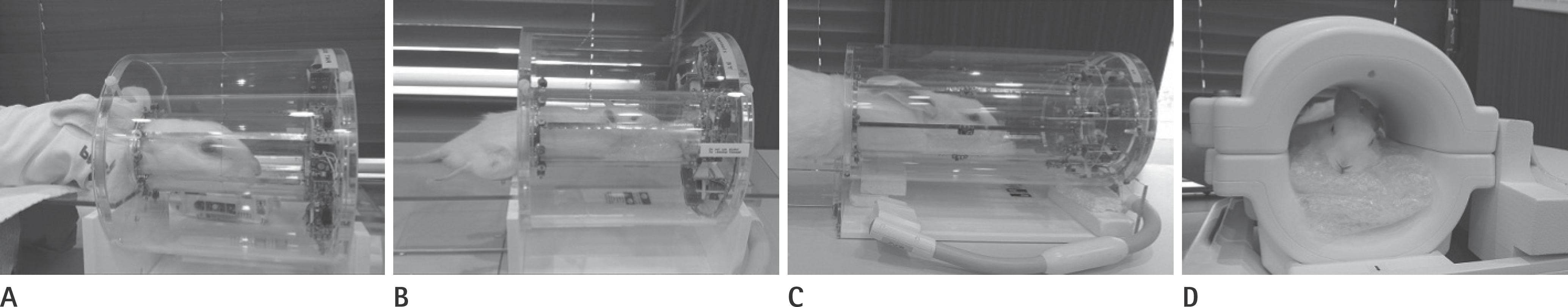 | Fig. 1.Surface coils used in the present study. Rat coil (5 cm in diameter) with a rabbit (A) and a rat (B) within the coil. Cat coil (C, 12 cm in di-ameter) and knee coil (D, 15.4 cm in diameter) with a rabbit within the coils. |
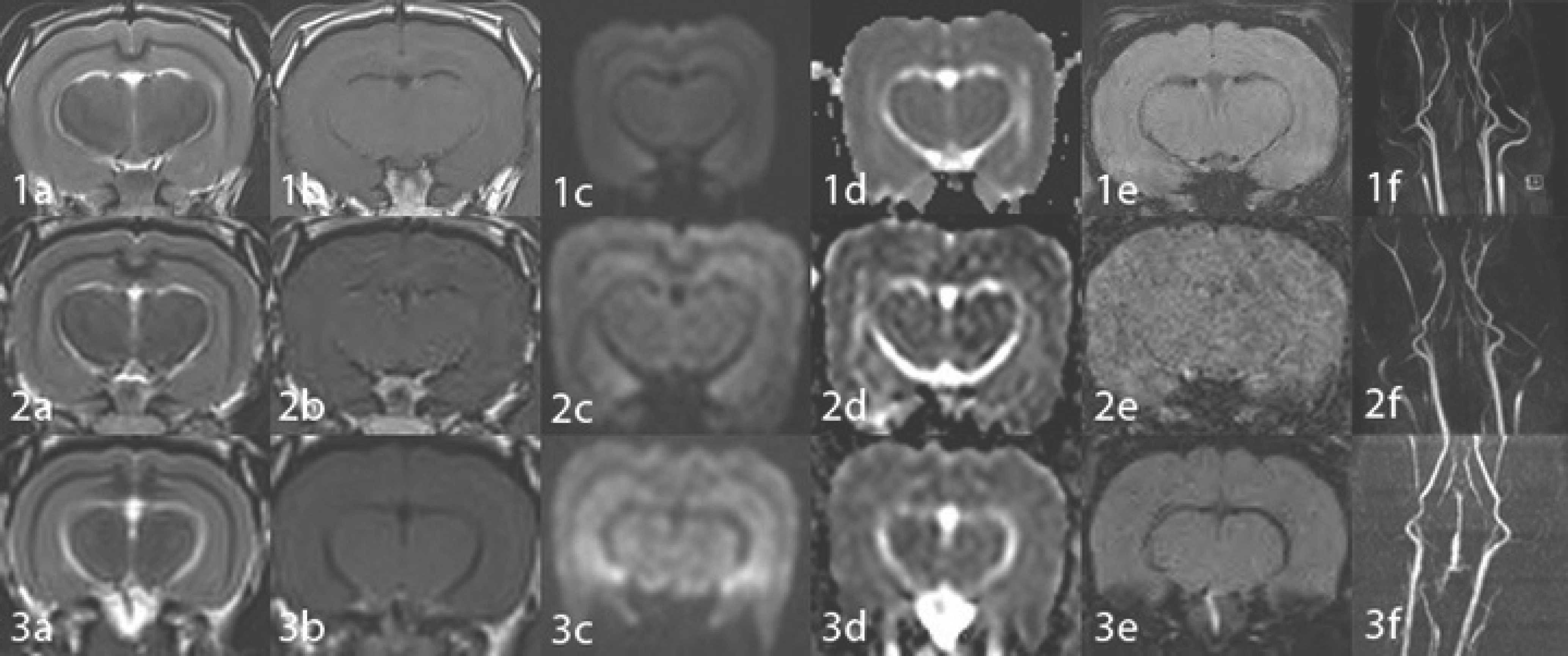 | Fig. 2.Brain MR images of a normal rabbit obtained with a rat coil (1), a cat coil (2) and a knee coil (3). T2WI (a), T1WI (b), DWI (c), ADC map image (d), SWI (e) and MRA (f). All MR images obtained with the rat coil reveal good differentiation of the gray/white matter and smoothness of the parenchyma. However, images obtained with the rat coil (1) show better quality compared to those obtained with the cat (2) or knee coil (3). The sharpness of MRA is best on the image obtained with the rat coil (1f) compared to the image obtained with the cat (2f) or knee coil (3f). ADC = apparent diffusion coefficient, DWI = diffusion-weighted image, MRA = MR angiography image, SWI = susceptibility-weighted image, T1WI = T1-weighted image, T2WI = T2-weighted image |
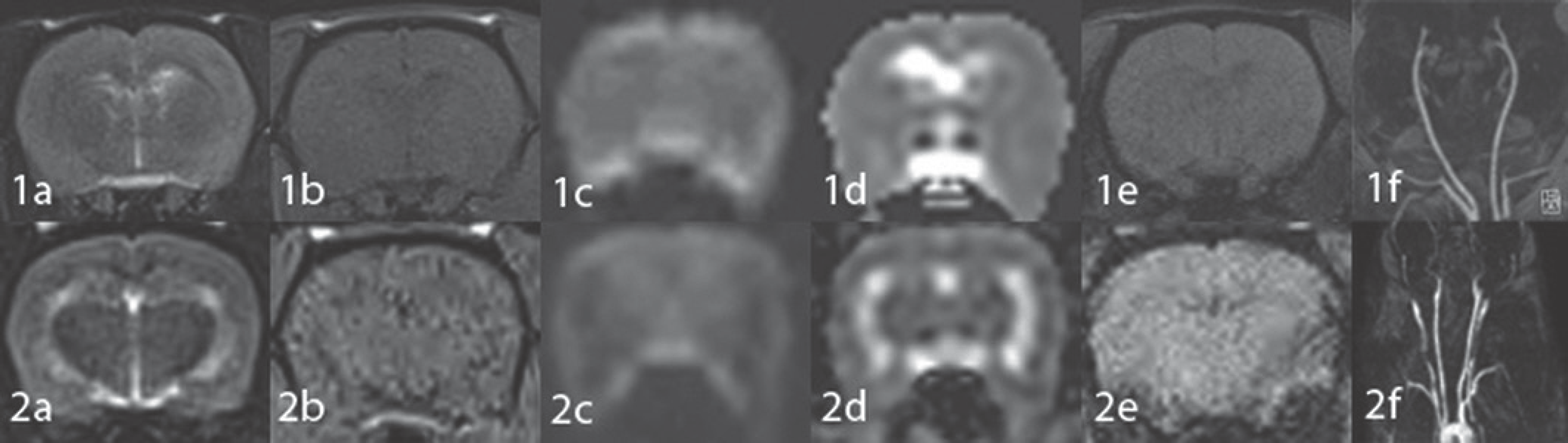 | Fig. 3.Brain MR images of a normal rat obtained with the rat coil (1) and with the cat coil (2). T2WI (a), T1WI (b), DWI (c), ADC map image (d), SWI (e) and MRA (f). MR images obtained with the rat coil (1) reveal better quality in terms of gray/white matter differentiation and smooth-ness compared to those obtained with the cat coil (2). MRA image obtained with the rat coil (1f) shows better sharpness compared to that ob-tained with the cat coil (2f). ADC = apparent diffusion coefficient, DWI = diffusion-weighted image, MRA = MR angiography image, SWI = susceptibility-weighted image, T1WI = T1-weighted image, T2WI = T2-weighted image |
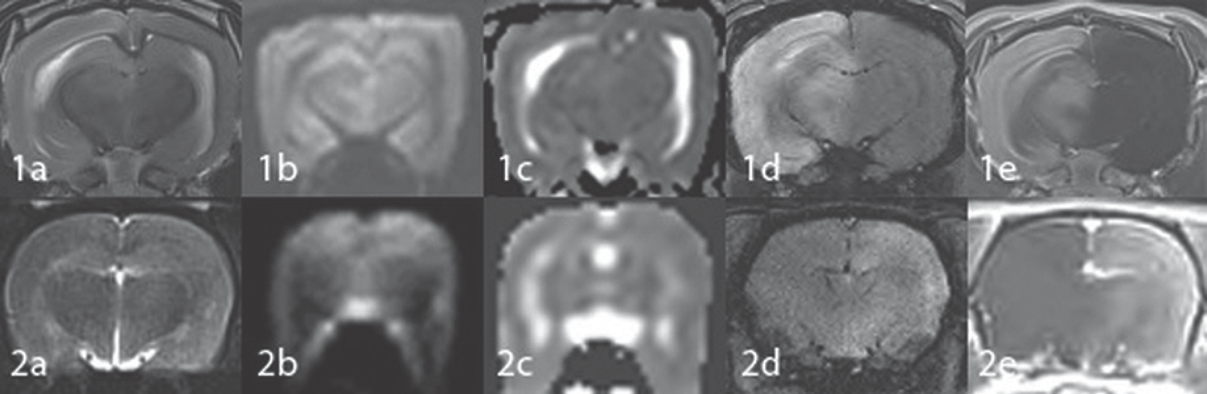 | Fig. 4.MR images of a rabbit (1) and a rat (2) obtained with the rat coil 2 hours after embolization of triolein emulsion into the carotid artery. Embolized hemispheres show mild hyperintensity on T2WI (a), no evidence of diffusion restriction (b, c), no hemorrhage on SWI (d) and diffuse contrast enhancement on Gd-T1WI (e). Lesion conspicuity is better on images obtained from the rabbit (1) compared with those obtained from the rat (2). SWI = susceptibility-weighted image, T1WI = T1-weighted image, T2WI = T2-weighted image |
Table 1.
Parameters of MR Sequences Used in Rabbit MR Brain
bV = b value, DWI = diffusion-weighted image, ET = echo train length, FA = flip angle, FOV = field of view, Mat = matrix number, NEX = number of excitation, ST = slice thickness, SWI = susceptibility-weighted image, TE = echo time, TR = repetition time, T1WI = T1-weighted image, T2WI = T2-weighted image
Table 2.
Parameters of MR Sequences Used in Rat MR Brain
bV = b value, DWI = diffusion-weighted image, ET = echo train length, FA = flip angle, FOV = field of view, Mat = matrix number, NEX = number of excitation, ST = slice thickness, SWI = susceptibility-weighted image, TE = echo time, TR = repetition time, T1WI = T1-weighted image, T2WI = T2-weighted image
Table 3.
Signal-To-Noise Ratios of Brain MR Images
 XML Download