

 PDF
PDF ePub
ePub Citation
 Print
Print


INTRODUCTION
Major vascular injuries present a complex challenge to trauma surgeons and interventional radiologists. Trauma to the portal vein (PV) and the superior mesenteric vein (SMV) is uncommon, and few surgeons have significant experience in treating these injuries (1). A great majority of these patients have an associated vascular injury and the overall mortality rate is high. Due to the technical difficulty in isolating injured vessels, associated abdominal injuries and massive bleeding are the most common causes of death (2). We present a case of blunt abdominal trauma in a patient who sustained an atypical pancreaticoduodenal vein (PDV) injury with hemodynamic instability and was successfully treated with percutaneous transhepatic embolization of the PDV.
CASE REPORT
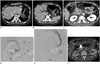
A 33-year-old male who sustained injury in a motorcycle accident was transferred to our trauma center in a tertiary care hospital due to unstable vital signs and right upper quadrant abdominal pain. His systolic blood pressure was 70 mm Hg on initial physical examination and tachycardia exceeded 120 bpm. Laboratory findings revealed a hemoglobin level of 11.2 g/dL, hematocrit of 31.9%, and platelet count of 110000/µL. Initial focused assessment with sonography in trauma revealed a hemoperitoneum. Contrast enhanced computed tomography (CT) of the abdomen and pelvis also revealed a hemoperitoneum (Fig. 1A, B), and portal venous phase images showed a pseudoaneurysm in the anterior pancreatic head, but arterial phase images showed only increased strands within fatty tissue (Fig. 1C). There were no other abnormalities in the abdominal solid organs or a hollow viscus on CT.
We immediately performed angiography. Celiac angiography and superior mesenteric angiography did not reveal an arterial bleeding focus with contrast extravasation or pseudoaneurysm formation, and no definite lesion was seen on indirect portal venography. Therefore, we decided to perform direct portal venography in consideration of the presence of a pseudoaneurysm on portal venous CT.
A 21-G Chiba needle (Cook, Bloomington, IN, USA) was used to puncture the peripheral PV (liver segment 5) under ultrasonography and fluoroscopy guidance, and a 0.018-inch guide wire was placed in the PV. A 5 Fr introducer sheath was inserted, and the SMV was selected with a 0.035-inch guide wire. A 5 Fr Cobra catheter (Cook) was placed in the SMV and direct portal venography was performed. However, venous rupture or pseudoaneurysm was not observed. Therefore, venography was performed by selecting the branches of the SMV. Selection of the ruptured vein was based on indirect venography using SMA and CT images, and the location of the PDV was determined. On selective venography, a ruptured venous pseudoaneurysm of the PDV was identified (Fig. 1D). Then, a 2.2 Fr Progreat microcatheter (Terumo, Tokyo, Japan) was used to engage the PDV and the microcatheter was negotiated through the venous pseudoaneurysm into the normal segment just distal to the pseudoaneurysm. Seventeen Tornado pushable microcoils (Cook) with diameters ranging from 3 to 4 mm were delivered through the microcatheter to embolize the pseudoaneurysm, using the sandwich technique (proximal and distal embolization) to avoid distal reconstitution of the injured vein. Reflux venography was repeated to confirm that the distal flow was blocked. Postembolization venography showed complete exclusion of the pseudoaneurysm and successful filling of the SMV (Fig. 1E). The transhepatic tract was embolized using an N-butyl cyanoacrylate (NBCA; Histoacryl, B Braun, Melsungen, Germany) plus lipiodol (Lipiodol Ultrafluid, Laboratoire Guerbet, Paris, France) mixture at a ratio of 1:1. After the procedure, the patient's systolic blood pressure was restored to 110 mm Hg and the vital signs stabilized. No additional contrast extravasation or hemoperitoneum was observed on follow-up CT after 3 days (Fig. 1F). The patient subsequently underwent surgery for a femoral fracture and was discharged on the 24th day of hospitalization. He is currently doing well, without any evidence or symptoms of traumatic sequelae or postembolization complications, after a follow-up period of 7 months.
DISCUSSION
SMV injuries are rare and incur high mortality. Given their low incidence, little data exist delineating treatment methods. The controversy about repair versus ligation for SMV injuries remains unresolved (34).
Since the development of angiography and transcatheter techniques, interventional radiology has played an important role in the management of trauma patients. The ability to treat life-threatening hemorrhage with transcatheter embolization has reduced the morbidity associated with surgery, and mortality has been reduced by quick access to a bleeding focus with use of angiography techniques (5).
Venous bleeding is considered relatively insignificant compared to arterial bleeding and it often stops spontaneously because of low pressure. However, large vein injuries due to trauma can cause massive bleeding and SMV injuries are highly lethal (3). In this case, the injured vessel was located in the retroperitoneal space, but it resulted in a hemoperitoneum. It is possible that both the vessels and the peritoneum of the mesenteric root were damaged simultaneously. Therefore, active and rapid intervention is thought to have prevented death and reduced morbidity.
Percutaneous transhepatic catheterization of the PV has been used to safely and accurately evaluate the portal venous system since its introduction in the 1970s (6). The technique is also used in various interventions related to portal hypertension and liver transplantation (78). Additionally, the potential bleeding complications of access tracts can be safely managed through NBCA embolization (9).
An isolated injury to the PDV is extremely rare, and this is the first reported case in which percutaneous transhepatic venous embolization was used to treat a PDV injury. Although there are several treatment options for PDV injury, surgical treatments are accompanied by complications such as pancreatic fistula and intraabdominal abscess (10). In cases of severe pancreatic injury or duodenal rupture, surgical treatment should be given priority; however, if there is only vascular damage, as in this case, percutaneous transhepatic embolization may be a useful alternative minimally invasive treatment.
 XML Download