Abstract
Adenomyoepithelioma of the breast is a rare tumor. A myoepithelial carcinoma arising within an adenomyoepithelioma is even more unusual. There are a limited number of reports discussing myoepithelial carcinoma; most of them describe pathological findings, but not imaging findings. We present a case of a 55-year-old woman who had a screen-detected myoepithelial carcinoma arising within an adenomyoepithelioma in her right breast. Upon the completion of a mammography and sonography an oval shaped mass with an indistinct margin in the upper portion of the right breast had been seen. It as appeared to be a spiculated, irregular-shaped, peripheral-enhancing mass on an MRI. On sonography-guided biopsy, an epithelial-myothelial tumor was confirmed, and the possibility of myoepithelial carcinoma was suggested. Breast-conserving surgery with a sentinel lymph node dissection was performed, and a pathological examination revealed a myoepithelial carcinoma arising within an adenomyoepithelioma.
Epithelial-myoepithelial lesions of the breast, also known as adenomyoepithelial lesions, consist of a heterogeneous group of entities that exhibit a wide spectrum of benign lesions to malignant ones (1). Adenomyoepithelioma is a biphasic neoplasm composed of ductal and myoepithelial cells, and exhibits potential for malignant progression (1). When malignant progression occurs, the malignant component can be epithelial, myoepithelial, or both and can exhibit a wide histological grade spectrum ranging from low-to-high (1). Due to the rare occurrence of these tumors, there is very limited literature describing them, especially their radiological appearance. In the present study a case of myoepithelial carcinoma arising within an adenomyoepithelioma of the breast is being reported. This study also discusses the imaging findings of the case obtained by a mammography, sonography, and magnetic resonance imaging (MRI).
A 55-year-old woman visited our hospital for breast cancer screening. She had no significant medical history, and no history of personal or familial breast cancer. Physical examination of the right breast revealed a painless small mass in the right upper breast. There was no skin retraction, nipple discharge, or palpable axillary-lymph-node seen during the physical examination.
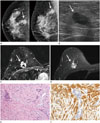
Mammography was performed using Lorad Selenia (Hologic Inc., Bedford, MA, USA). On the screening mammography (Fig. 1A), an oval-shaped, isodense mass with indistinct margins and a long-diameter of 2 cm was seen in the 12 o1clock direction of right breast. There was no associated calcification or axillary-lymphadenopathy observed. Breast sonography was performed using a LOGIQ 9 (GE Healthcare, Milwaukee, WI, USA) with a broad-bandwidth 14–5 MHz linear probe. On breast sonography (Fig. 1B), the lesion presented as an oval, hypoechoic mass with indistinct margins in the half past 12 o'clock direction of the right breast, 8 cm from the nipple. The legion was classified as a category 4 per the American College of Radiology Breast Imaging-Reporting and Data System (ACR BI-RADS) criteria (2). A breast MRI was performed using a 3.0-T Scanner (Achieva 3.0T TX; Philips Healthcare, Best, the Netherlands) with a breast coil (MRI Devices; InVivo Research, Orlando, FL, USA), with the patient maintained in the prone position. Images were acquired in the axial plane. The interpretation of the degree and patterns of enhancement were performed using CAD stream TM (Merge Health Care, Chicago, IL, USA). An irregular mass with spiculated margins and rim enhancement was seen in the right upper mid-to-outer portion (Fig. 1C). It was hyperintense on the fat-saturated T2-weighted image (Fig. 1D) and isointense on the T1-weighted image. Upon a kinetic curve assessment, it initially showed fast-enhancement with a delayed plateau pattern. It also showed diffusion restriction on a diffusion-weighted image and apparent diffusion coefficient map. There were not any significantly enlarged lymph nodes in either axillae.
Sonography-guided core needle biopsy was done for the right breast mass. It was confirmed to be an epithelial-myothelial tumor with myoepithelial overgrowth, moderate degree of atypism and frequent mitoses. The possibility of myoepithelial carcinoma was suggested and a complete excision was recommended. Therefore, breast-conserving surgery was performed. On gross examination, the cut surface of the specimen showed a well-demarcated, ovoid, grayish mass with multifocal hemorrhagic changes. Microscopic examination (Fig. 1E, F) revealed a myoepithelial carcinoma in an adenomyoepithelioma. The tumor was 1.8 × 1.4 cm in size, and was composed of a ductal and spindle cell component. The ductal component was small, tubular and cystically dilated, with two cell components, epithelial and myoepithelial cells. The spindle cells were epithelioid, vaguely clustered, and fibroblastic or smooth muscle-like. On immunostaining, the spindle cell components were positive for smooth muscle markers, epithelial markers (CK, HMW-CK), and a myoepithelial cell marker (p63). The stromal cells showed frequent mitoses (10/10 HPF), necrosis, and 30–40% Ki-67 labeling. The surgical margin was free of carcinoma. An 18F-fludeoxyglucose positron emission tomography-CT scan was performed as a part of the metastatic work up; no distant metastasis was found.
Myoepithelial cells are the normal components of breast lobules and ducts (34). These cells have the characteristics of both epithelial and smooth muscle cells. Thus, tumors that arise from these cells show characteristics of epithelial and smooth muscle cells (4). They can arise in the salivary glands, skin, soft tissue, retroperitoneum, breast, and lungs (4). Epithelial-myoepithelial lesions of the breast are also known as adenomyoepithelial lesions (1). These are a heterogeneous group of lesions, exhibiting a wide spectrum from benign to malignant (1).
Adenomyoepithelioma is an extremely rare neoplasm of the breast, and occurs almost exclusively in women (5). It is generally benign and has a low potential for malignancy and local recurrence (67). However, malignant progression can occur, and be associated with distant metastasis, mainly via a hematogenous spread (56). In more than 150 case reports studied, about 40 lesions were found to be malignant or potentially malignant, according to Tavassoli (8). Malignant degeneration of adenomyoepithelioma typically presents as a sudden, rapid growth of a long-standing, often-large breast mass (1). Adenomyoepithelioma is common after the fifth decade of life, and malignant degeneration may occur in older age (1). However, the 55-year-old patient presented in this study had no symptoms because of the relatively small size of her breast mass which measured less than 2 cm.
There are several reports of adenomyoepithelioma and myoepithelial carcinoma of the breast, but the imaging findings in these reports are not well-described. According to the results of previous studies, these imaging only states that the features may mimic radiological findings of breast cancer (57910), but other details were not provided. The most common mammographic finding is an irregular, non-calcified mass with variable margins (57910). Calcification and parenchymal distortions are uncommon findings, and are usually associated with malignancy (37). Upon sonography, a hypoechoic, irregular or oval mass with microlobulated or indistinct margins is the most common finding (5710). Lesions may also show posterior acoustic enhancement (37). Although there are a very limited number of reports that describe MRI findings, the most common are of an enhancing mass with an irregular or spiculated margin, and delayed washout or plateau kinetics (57). In the present case, on the basis of mammographic and sonographic findings, the mass was classified as being suspiciously abnormal (BI-RADS 4) per the ACR classification. The MRI features were consistent with a diagnosis of malignancy. Overall, the imaging features of the mass in our present case were consistent with those seen in previous studies.
Adenomyoepitheliomas have more prominent proliferative image features than do other common benign lesions (10) that adenomyoepitheliomas considered to be almost benign were categorized in BI-RADS classifications as 4 or higher in previous studies (57910). Since there are no definite differences in imaging features between benign and malignant adenomyoepithelial tumors, a sonography-guided core needle biopsy may be required for diagnosis and future treatment planning (5). Irrespective of whether a lesion is benign or malignant, a wide local surgical excision is recommended. If the lesion is confirmed to be malignant a lymph node dissection may be done in addition to the aforementioned wide excision (45). Additional adjuvant chemotherapy or radiotherapy can be considered, even though the precise role of both these in the management of adenomyoepithelial tumors is unknown (14). In this case, the patient underwent breast-conserving surgery with a sentinel lymph node biopsy. After surgery, she was treated with adjuvant chemotherapy and radiotherapy to reduce the possibility of tumor recurrence.
Myoepithelial carcinoma arising within an adenomyoepithelioma of the breast is extremely rare. There are few reports about the radiological features of this tumor, especially describing MRI findings. Understanding the radiological findings of this rare entity can be helpful in making an accurate diagnosis, appropriate treatment planning and follow-up.
Figures and Tables
Fig. 1
A 55-year-old woman with myoepithelial carcinoma arising within adenomyoepithelioma.
A. Screening mammography shows a 2 cm-sized, oval-shaped, isodense mass with indistinct margins (arrows) in the 12 o'clock direction of the right breast.
B. Breast sonography shows an indistinct, oval, hypoechoic mass (arrow) seen in the right upper breast. There is no observed abnormal axillary lymphadenopathy.
C. On MRI, axial fat-saturated T1-weighted subtraction image with post-contrast gadolinium injection shows an irregular mass with spiculated margins and rim enhancement (arrow) in the right upper mid-to-outer portion.
D. On axial fat-saturated T2-weighted image, the mass was hyperintense (arrow). On dynamic study (not shown), this mass shows initial fast-enhancement and delayed plateau pattern.
E. On histological examination (hematoxylin and eosin stain, × 100), the tumor shows a relatively uniform admixture of scattered, glandular, epithelial-lined spaces, and surrounding spindle and epithelioid myoepithelial-cell proliferation.
F. On immunostaining (SMA, × 200), the myoepithelial cells are immunoreactive for SMA.
SMA = smooth muscle actin

References
1. Ali RH, Hayes MM. Combined epithelial-myoepithelial lesions of the breast. Surg Pathol Clin. 2012; 5:661–699.
2. American College of Radiology. ACR BI-RADS atlas: breast imaging reporting and data system. Reston, VA: American College of Radiology;2013.
3. Howlett DC, Mason CH, Biswas S, Sangle PD, Rubin G, Allan SM. Adenomyoepithelioma of the breast: spectrum of disease with associated imaging and pathology. AJR Am J Roentgenol. 2003; 180:799–803.
4. Endo Y, Sugiura H, Yamashita H, Takahashi S, Yoshimoto N, Iwasa M, et al. Myoepithelial carcinoma of the breast treated with surgery and chemotherapy. Case Rep Oncol Med. 2013; 2013:164761.
5. Ruiz-Delgado ML, López-Ruiz JA, Eizaguirre B, Saiz A, Astigarraga E, Fernández-Temprano Z. Benign adenomyoepithelioma of the breast: imaging findings mimicking malignancy and histopathological features. Acta Radiol. 2007; 48:27–29.
6. Petrozza V, Pasciuti G, Pacchiarotti A, Tomao F, Zoratto F, Rossi L, et al. Breast adenomyoepithelioma: a case report with malignant proliferation of epithelial and myoepithelial elements. World J Surg Oncol. 2013; 11:285.
7. Adejolu M, Wu Y, Santiago L, Yang WT. Adenomyoepithelial tumors of the breast: imaging findings with histopathologic correlation. AJR Am J Roentgenol. 2011; 197:W184–W190.
8. Tavassoli FA. Myoepithelial lesions of the breast. Myoepitheliosis, adenomyoepithelioma, and myoepithelial carcinoma. Am J Surg Pathol. 1991; 15:554–568.
9. Moritz AW, Wiedenhoefer JF, Profit AP, Jagirdar J. Breast adenomyoepithelioma and adenomyoepithelioma with carcinoma (malignant adenomyoepithelioma) with associated breast malignancies: a case series emphasizing histologic, radiologic, and clinical correlation. Breast. 2016; 29:132–139.
10. Park YM, Park JS, Jung HS, Yoon HK, Yang WT. Imaging features of benign adenomyoepithelioma of the breast. J Clin Ultrasound. 2013; 41:218–223.


 PDF
PDF ePub
ePub Citation
 Print
Print


 XML Download