

 PDF
PDF ePub
ePub Citation
 Print
Print


INTRODUCTION
Although neuroblastoma is the most common solid tumor in childhood, primary cervical neuroblastoma is infrequent, accounting for between 2 and 6% of all neuroblastomas (1). Retropharyngeal neuroblastomas in infant were also documented in only small case reports (123). We report a case of primary retropharyngeal neuroblastoma with an ipsilateral lymph node metastasis manifesting as a growing neck mass in the retropharyngeal space in an 11-month old infant.
CASE REPORT
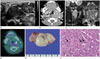
An 11-month-old boy presented with a left palpable neck mass at our hospital. He was born normally by spontaneous vaginal delivery at 40 ± 1 weeks, and weighed 3.1 kg. The left neck mass grew sufficiently to become palpable. The initial ultrasound (US) scan revealed an approximately 2.0-cm, low echoic mass at the inferior lateral aspect of the left lobe of the thyroid, and a 1.6-cm heterogeneous mixed echoic mass in the superficial area of the left neck (Fig. 1A). As a differential diagnosis, we initially suggested cervical lymph node enlargement by infection or inflammation. However, head and neck malignancy such as lymphoma, rhabdomyosarcoma, and neuroblastoma, could not be excluded. To further characterize the lesion, the patient underwent a neck computed tomography (CT) scan with contrast enhancement. The post-contrast CT scan showed an approximately 1.0 × 1.5 × 2.5-cm soft-tissue mass with mild homogeneous enhancement in the left retropharyngeal space, and enlarged lymph nodes with similar enhancement pattern in the left neck (level II) (Fig. 1B). There was no evidence of calcification or internal hemorrhage in the lesion. There were also multiple small enhancing lymph nodes at the lower level of the lateral neck, supraclavicular fossa, and left axilla. The CT findings favored a tumorous condition such as lymphoma, neuroblastoma, Langerhans cell histiocytosis, or rhabdomyosarcoma, rather than infection or inflammation. To obtain pathological confirmation of the lesion and enlarged lymph nodes, we performed a 18 G ultrasound-guided biopsy (performed using a biopsy gun). The results indicated a small round cell tumor, suggestive of neuroblastoma. The patient underwent neck magnetic resonance imaging (MRI) for further work up. The mass in the retropharyngeal space demonstrated moderately high signal intensity on T2-weighted image, and low signal intensity on T1-weighted image with homogenous enhancement. However, there was no evidence of neural foraminal extension of cervical spine (Fig. 1C). Next, an 18F-fludeoxyglucose positron emission tomography-computed tomography (18F-FDG PET/CT) was performed, to evaluate any systemic involvement. On 18F-FDG PET/CT, the dumbbell-shaped mass present in the left retropharyngeal space showed heterogeneous FDG uptake, which is a typical finding of malignant tumors rather than inflammatory lymphadenopathy (Fig. 1D). However, there was no evidence of systemic metastasis. Upon I-123 Metaiodobenzylguanidine (MIBG) scintigraphy, increased tracer uptake was noted in the left lateral neck. A bone scan showed no evidence of skeletal metastasis. The patient underwent surgical excision of the neck mass and selective dissection of the lymph nodes. Grossly, the mass had a size of approximately 4.8 × 2.5 × 1.7 cm, and was a well-encapsulated whitish gray mass (Fig. 1E). The diagnosis of neuroblastoma was confirmed histopathologically. On microscopic evaluation, the tissue had a mitosis-karyorrhexis index of less than 0.1%, and no vascular or lymphatic invasion. The mass primarily consisted of small round tumor cells with hyperchromatic nuclei and multiple immature ganglion cells (Fig. 1F). However, of the 93 dissected lymph nodes of the neck on both sides, there was only a single metastatic lymph node in the left neck (level II). The urinary levels of vanillylmandelic acid and homovanillic acid, which reflect the levels of secreted catecholamines by the tumor, were negative in the initial study. The laboratory findings also demonstrated a continuously low level of vanillylmandelic acid and homovanillic acid. According to the international neuroblastoma staging system (INSS), the staging of this tumor was 2B: a unilateral tumor with ipsilateral non-adherent lymph node metastasis. There has been no evidence of tumor recurrence for the 2 years follow up.
DISCUSSION
Primary cervical neuroblastoma accounts for less than 5% of all cases of neuroblastoma (2). According to the report of Okazaki et al. (1), about 44% of the neonatal cervical neuroblastoma were reported as retropharyngeal neuroblastoma. Retropharyngeal neuroblastoma in infants was also documented in small case reports (123). The initial symptoms of the patient were dyspnea, stridor, feeding difficulties, Horner syndrome, and a palpable mass (1).
According to previous case reports (123), a neuroblastoma patient of stage 4 with systemic metastasis has a poorer prognosis than a lower stage patient. Primary cervical neuroblastoma in infants under 1 year old showed a relatively good prognosis when the disease was stage 1 or 2. When only a single site in the cervical sympathetic chain is involved (stage 1), excision can eradicate the disease (4). The most important predictors of outcome are age at diagnosis and the INSS disease stage (1). The clinical course of neuroblastoma in infants has a better outcome and survival rate than in older patients, due to greater spontaneous regression, particularly if the child is under one-year, with localized disease, and the tumor cells are found to have favorable biologic features (45). In our case, the child was less than 1 year at the time of obtaining the initial ultrasound scan, and was diagnosed with neuroblastoma with INSS stage 2B. The survival rate for such a case is relatively good.
Similar to our patient, two case reports regarding the location of the mass and lymph node metastasis have been reported by Smith et al. (4). The cases were reported in 1985 and 1979, and both had no remarkable image presentation. Although after 1990s there have been several case reports that have presented the image of patient, there are no case reports involving the use of US, CT, MRI and PET. Okazaki et al. (1) reported a retropharyngeal mass with calcification on CT, and Abramson et al. (2) reported a case where MRI of the neck showed a solid mass with or without calcifications, which displaced the major vessels of the neck and encased vessels. On ultrasound scans, in addition to the displacement of surrounding structures, fetal cervical neuroblastomas have also shown hyperechoic, heterogeneous echogenicity of the solid lateral neck with small echoic foci, suggesting calcifications and airway deviation (6). As a differential diagnosis based on imaging findings, we considered inflammatory lymphadenopathy, and head and neck malignancies as other possibilities. Reactive lymph nodes may result from viral, bacterial, tuberculous infection or Bacille Calmette-Guerin lymphadenitis. Such nodes are typically seen bilateral with mildly enlarged cervical lymph node, with or without periadenitis. Especially in case of tuberculous lymphadenitis, the lymph node not only shows enlargement, but are also characterized by suppurative nodes or intranodal abscess, which were different from our case. Calcification, necrosis, and hemorrhage are uncommon with lymphoma and therefore, lymphoma appears more homogeneous at imaging, as compared to neuroblastoma. The cut surface of specimens may vary from tan to hemorrhagic. Necrosis, hemorrhage, and calcification may be detected. However, this was not indicated in the gross image obtained in our study; this could be because approximately 80–90% of neuroblastoma cases demonstrate calcification on CT (7). This was also concurrent with our CT findings. Microscopic characteristics of neuroblastoma generally involve immature neuroblasts and undifferentiated sympathetic cells that are small and round in shape with little cytoplasm, small, indistinct nucleoli, and stroma surrounding the neuroblasts or ganglion cells. According to the histopathologic grading system of Shimada (8), a mitosis-karyorrhexis index of less than 0.1% favors a better prognosis.
Even though radiation exposure for pediatric patients is very high, 18F-FDG PET/CT was performed before 131I-MIBG scintigraphy, to evaluate systemic involvement of neuroblastoma. This is because 18F-FDG PET/CT shows high accuracy in pediatric patients with neuroblastoma, and is superior to 131I-MIBG scintigraphy, especially in the detection of lymph nodal invasion or bone marrow involvement (9).
As there are limited case reports of retropharyngeal neuroblastoma, especially those including analysis by US, CT, MRI, PET, and fewer reports with histological characteristics, our case report provides further information about retropharyngeal neuroblastoma associated with the imaging findings.
In conclusion, we describe here a rare case of neuroblastoma in the retropharyngeal space with lymph node metastasis. Multi-modality inspection and histological correlation can be useful to diagnose and treat primary neuroblastomas in the retropharyngeal space.
 XML Download