

 PDF
PDF ePub
ePub Citation
 Print
Print


Abstract
A herniation pit is a benign bone pit characterized by its femoral location and sur-rounding sclerotic margin. Few reports have been issued on the fluorodeoxyglucose (FDG) positron emission tomography findings of herniation pit. Here, we report the unique case of a thyroid cancer patient, having a herniation pit showing transient FDG uptake, which mimicked bone metastasis.
REFERENCES
1.Kim JA., Park JS., Jin W., Ryu K. Herniation pits in the femo-ral neck: a radiographic indicator of femoroacetabular im-pingement? Skeletal Radiol. 2011. 40:167–172.

2.Sopov V., Fuchs D., Bar-Meir E., Gorenberg M., Groshar D. Clinical spectrum of asymptomatic femoral neck abnormal uptake on bone scintigraphy. J Nucl Med. 2002. 43:484–486.
3.Gao ZH., Yin JQ., Ma L., Wang J., Meng QF. Clinical imaging characteristics of herniation pits of the femoral neck. Or-thop Surg. 2009. 1:189–195.
4.Yoo SW., Song HC., Oh JR., Kim J., Kang SR., Chong A, et al. Herniation pit mimicking osseous metastasis on 18F-FDG PET/CT in patient with lung cancer. Clin Nucl Med. 2012. 37:682–683.
5.Pitt MJ., Graham AR., Shipman JH., Birkby W. Herniation pit of the femoral neck. AJR Am J Roentgenol. 1982. 138:1115–1121.
6.Panzer S., Esch U., Abdulazim AN., Augat P. Herniation pits and cystic-appearing lesions at the anterior femoral neck: an anatomical study by MSCT and microCT. Skeletal Radiol. 2010. 39:645–654.
7.Kim SH., Yoo HJ., Kang Y., Choi JY., Hong SH. MRI findings of new uptake in the femoral head detected on follow-up bone scans. AJR Am J Roentgenol. 2015. 204:608–614.
8.Borody C. Symptomatic herniation pit of the femoral neck: a case report. J Manipulative Physiol Ther. 2005. 28:449–451.
9.Min JW., Um SW., Yim JJ., Yoo CG., Han SK., Shim YS, et al. The role of whole-body FDG PET/CT, Tc 99m MDP bone scintig-raphy, and serum alkaline phosphatase in detecting bone metastasis in patients with newly diagnosed lung cancer. J Korean Med Sci. 2009. 24:275–280.
10.Gibiezaite S., Ozdemir S., Shuja S., McCook B., Plazarte M., Sheikh-Ali M. Unexpected bone metastases from thyroid cancer. Case Rep Endocrinol. 2015. 2015:434732.
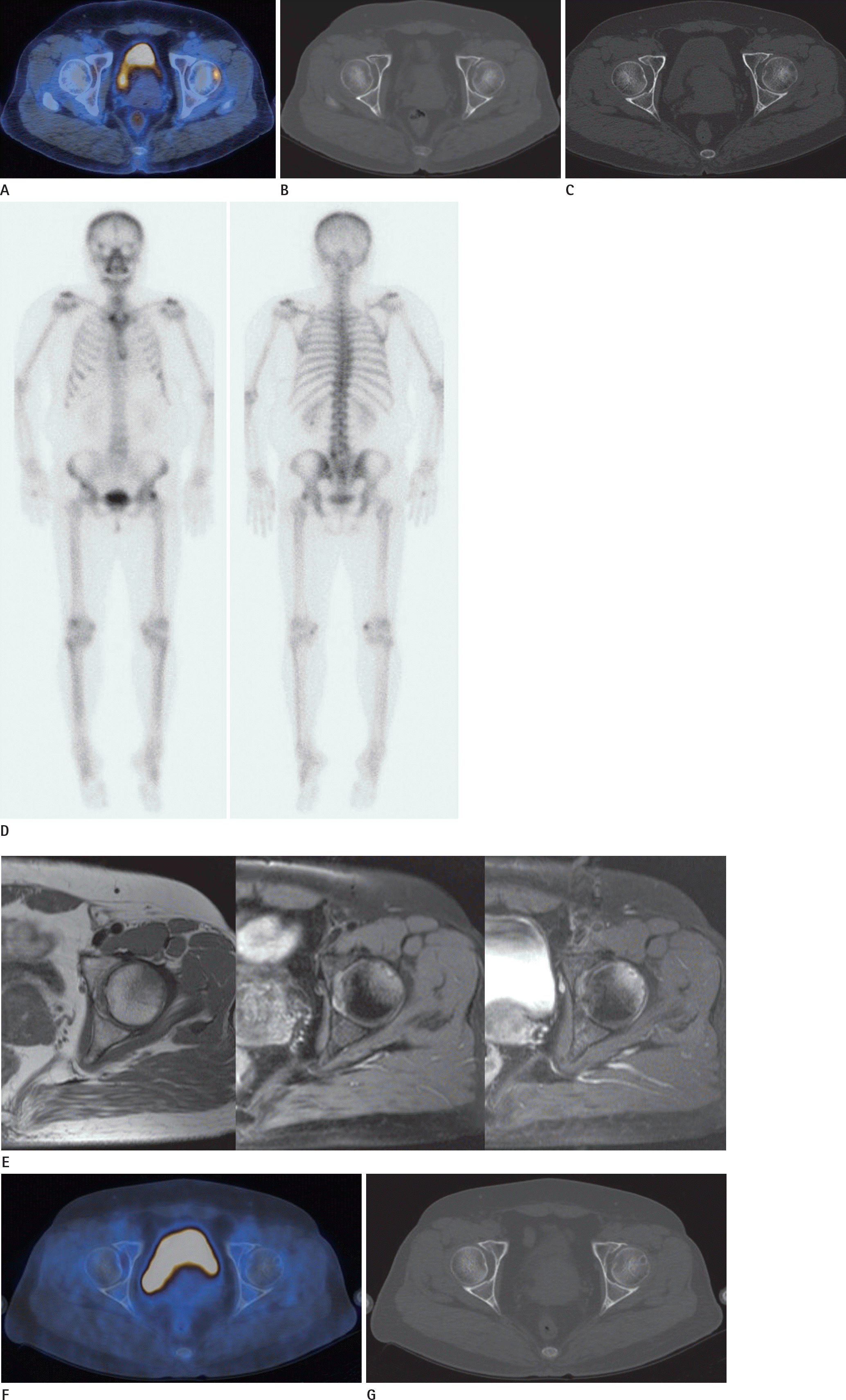
Fig. 1.
Transient 18 F-FDG activity of herniation pit, in a 53-year-old woman with thyroid cancer. A. PET/CT fusion image shows a focal hypermetabolism in the left anterolateral femoral head. B. CT bone setting finding, which corresponds to the location of the hypermetabolic lesion, shows no definite abnormality. C. After 3 weeks, axial CT of pelvic bone shows a small hypodense lesion without a sclerotic rim, in the left anterolateral femoral head. D. Bone scan shows focal increased uptake in the left lateral femoral head. CT = computed tomography, FDG = fluorodeoxyglucose, PET = positron emission tomography. Transient 18 F-FDG activity of herniation pit, in a 53-year-old woman with thyroid cancer. E. MR T1-weighted (left), T2-weighted (middle), contrast enhanced T1-weight (right) images. T1 weighted image shows ill defined low SI lesion in the anterosuperior femoral head/neck junction. T2-weighted image shows high SI with adjacent marrow edema. Contrast enhanced T1-weighted image shows focal rim enhancing lesion with mild diffuse enhancement of marrow. F, G. Follow up PET/CT images after 9 months show no FDG uptake (F), and a hypodense lesion with a sclerotic border in the left anterolateral femo-ral head, characteristic of a herniation pit (G). CT = computed tomography, FDG = fluorodeoxyglucose, PET = positron emission tomography, SI = signal intensity
 XML Download