

 PDF
PDF ePub
ePub Citation
 Print
Print


Abstract
Tumor implantation of lung cancer at the wire localization site has not been reported previously while there have been several reported cases of chest wall implantation after percutaneous core needle biopsy and chest tube insertion for malignant pleural effusion. We report the case of a 64-year-old-man with lung cancer (squamous cell carcinoma). The patient had lung cancer implantation in the chest wall 8 months after computed tomography-guided wire localization and subsequent anterior segmentectomy of right upper lobe with mediastinal lymph node dissection.
Figures and Tables
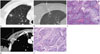
Fig. 1
Imaging and histology findings of a chest wall implantation from primary lung cancer following CT-guided wire localization in a 64-year-old man.
A. CT scan shows a 0.9 cm cavitary nodule in the subpleural region of the RUL. The distance between the nodule and visceral pleura is approximately 0.5 cm.
B. CT scan demonstrates that the wire tip is located at the nodule through the anterior part of the right second intercostal space.
C. Photomicrograph (hematoxylin and eosin stain, × 200) of the primary lung cancer in the RUL reveals tumor cells with intracellular bridges. This finding indicated the nodule was squamous cell carcinoma.
D. Enhanced CT scan reveals a well-defined 3-cm enhancing mass (arrows) in the right second intercostal space. The mass invades the right pectoralis minor and the second intercostal muscles.
E. Photomicrograph (hematoxylin and eosin stain, × 200) of the chest wall mass shows tumor cells with intracellular bridges. This histological characteristic was identical to that of primary lung cancer.
CT = computed tomography, RUL = right upper lobe
References
1. Kang DK, Min HK, Jun HJ, Hwang YH, Kang MK. Single-port video-assisted thoracic surgery for lung cancer. Korean J Thorac Cardiovasc Surg. 2013; 46:299–301.
2. Partik BL, Leung AN, Müller MR, Breitenseher M, Eckersberger F, Dekan G, et al. Using a dedicated lung-marker system for localization of pulmonary nodules before thoracoscopic surgery. AJR Am J Roentgenol. 2003; 180:805–809.
3. Mayo JR, Clifton JC, Powell TI, English JC, Evans KG, Yee J, et al. Lung nodules: CT-guided placement of microcoils to direct video-assisted thoracoscopic surgical resection. Radiology. 2009; 250:576–585.
4. Sugi K, Kaneda Y, Hirasawa K, Kunitani N. Radioisotope marking under CT guidance and localization using a handheld gamma probe for small or indistinct pulmonary lesions. Chest. 2003; 124:155–158.
5. Mullan BF, Stanford W, Barnhart W, Galvin JR. Lung nodules: improved wire for CT-guided localization. Radiology. 1999; 211:561–565.
6. Chen YR, Yeow KM, Lee JY, Su IH, Chu SY, Lee CH, et al. CT-guided hook wire localization of subpleural lung lesions for video-assisted thoracoscopic surgery (VATS). J Formos Med Assoc. 2007; 106:911–918.
7. Robertson EG, Baxter G. Tumour seeding following percutaneous needle biopsy: the real story! Clin Radiol. 2011; 66:1007–1014.
8. Yoshikawa T, Yoshida J, Nishimura M, Yokose T, Nishiwaki Y, Nagai K. Lung cancer implantation in the chest wall following percutaneous fine needle aspiration biopsy. Jpn J Clin Oncol. 2000; 30:450–452.
9. Jung SM, Won TG, Kim TH, Hwang HG, Kim MY, Jeong WJ, et al. Case report: implantation metastasis of lung cancer to chest wall after percutaneous fine-needle aspiration biopsy. Tuberc Respir Dis. 2001; 50:718–725.
10. Seo JM, Lee HY, Kim HK, Choi YS, Kim J, Shim YM, et al. Factors determining successful computed tomography-guided localization of lung nodules. J Thorac Cardiovasc Surg. 2012; 143:809–814.
 XML Download