Abstract
Purpose
To evaluate radiology residents’ performance in interpretation and compre-hension of breast ultrasonographic descriptors in the Breast Imaging Reporting and Data System (BI-RADS) to suggest the adequate duration of training in breast ultraso-nography.
Materials and Methods
A total of 102 radiology residents working in the Depart-ment of Radiology were included in this study. They were asked to answer 16 questions about the ultrasonographic lexicon and 11 questions about the BI-RADS category. We analyzed the proportion of correct answers according to the radiology residents’ year of training and duration of breast imaging training.
Results
With respect to the duration of breast imaging training, the proportion of correct answers for lexicon descriptors ranged from 77.2% to 81.3% (p = 0.368) and the proportion of correct answers for the BI-RADS category was highest after three-four months of training compared with after one month of training (p = 0.033). The proportion of correct answers for lexicon descriptors and BI-RADS category did not differ significantly according to the year of residency training.
Conclusion
Radiology residents’ comprehension of the BI-RADS category on breast ultrasonography was not associated with their year of residency training. Based on our findings, radiology residents’ assessment of the BI-RADS category was significantly im-proved with three-four months of training compared with one month of training.
Go to : 
REFERENCES
1.Mandelson MT., Oestreicher N., Porter PL., White D., Finder CA., Taplin SH, et al. Breast density as a predictor of mammo-graphic detection: comparison of interval- and screen-de-tected cancers. J Natl Cancer Inst. 2000. 92:1081–1087.

2.Kolb TM., Lichy J., Newhouse JH. Comparison of the perfor-mance of screening mammography, physical examination, and breast US and evaluation of factors that influence them: an analysis of 27,825 patient evaluations. Radiology. 2002. 225:165–175.

3.Berg WA., Gutierrez L., NessAiver MS., Carter WB., Bhargavan M., Lewis RS, et al. Diagnostic accuracy of mammography, clinical examination, US, and MR imaging in preoperative assessment of breast cancer. Radiology. 2004. 233:830–849.

4.Leong LC., Gogna A., Pant R., Ng FC., Sim LS. Supplementary breast ultrasound screening in Asian women with negative but dense mammograms-a pilot study. Ann Acad Med Singapore. 2012. 41:432–439.
5.Kelly KM., Dean J., Comulada WS., Lee SJ. Breast cancer de-tection using automated whole breast ultrasound and mammography in radiographically dense breasts. Eur Radiol. 2010. 20:734–742.

6.Parris T., Wakefield D., Frimmer H. Real world performance of screening breast ultrasound following enactment of Con-necticut Bill 458. Breast J. 2013. 19:64–70.

7.Shen SJ., Sun Q., Xu YL., Zhou YD., Guan JH., Mao F, et al. [Comparative analysis of early diagnostic tools for breast cancer]. Zhonghua Zhong Liu Za Zhi. 2012. 34:877–880.
8.Feig SA., Hall FM., Ikeda DM., Mendelson EB., Rubin EC., Segel MC, et al. Society of Breast Imaging residency and fellow-ship training curriculum. Radiol Clin North Am. 2000. 38:915–920. xi.

9.Bassett LW., Monsees BS., Smith RA., Wang L., Hooshi P., Far-ria DM, et al. Survey of radiology residents: breast imaging training and attitudes. Radiology. 2003. 227:862–869.

10.Burhenne LJ., Smith RA., Tabar L., Dean PB., Perry N., Sickles EA. Mammographic screening: international perspective. Semin Roentgenol. 2001. 36:187–194.
11.American College of Radiology. ACR BI-RADS Atlas, Breast Imaging Reporting and Data System. 5th ed.Reston, VA: American College of Radiology;2013.
12.American College of Radiology. ACR BI-RADS Breast Im-aging and Reporting Data System: Breast Imaging Atlas. 4th ed.Reston, VA: American College of Radiology;2003.
13.Kim EK., Ko KH., Oh KK., Kwak JY., You JK., Kim MJ, et al. Clini-cal application of the BI-RADS final assessment to breast sonography in conjunction with mammography. AJR Am J Roentgenol. 2008. 190:1209–1215.

14.Lazarus E., Mainiero MB., Schepps B., Koelliker SL., Livingston LS. BI-RADS lexicon for US and mammography: interob-server variability and positive predictive value. Radiology. 2006. 239:385–391.

15.Hamy AS., Giacchetti S., Albiter M., de Bazelaire C., Cuvier C., Perret F, et al. BI-RADS categorisation of 2,708 consecutive nonpalpable breast lesions in patients referred to a dedicated breast care unit. Eur Radiol. 2012. 22:9–17.

16.Park CS., Lee JH., Yim HW., Kang BJ., Kim HS., Jung JI, et al. Observer agreement using the ACR Breast Imaging Re-porting and Data System (BI-RADS)-ultrasound, first edition (2003). Korean J Radiol. 2007. 8:397–402.

17.Lee HJ., Kim EK., Kim MJ., Youk JH., Lee JY., Kang DR, et al. Ob-server variability of Breast Imaging Reporting and Data Sys-tem (BI-RADS) for breast ultrasound. Eur J Radiol. 2008. 65:293–298.

18.Lee EH., Lyou CY. Radiology residents' performance in screening mammography interpretation. J Korean Soc Radiol. 2013. 68:333–341.

19.U.S. Food and Drug Administration. Mammography facilities. Web site.http://www.accessdata.fda.gov/scripts/cdrh/cfdocs/cfMQSA/mqsa.cfm. Accessed Sep 2, 2015.
Go to : 
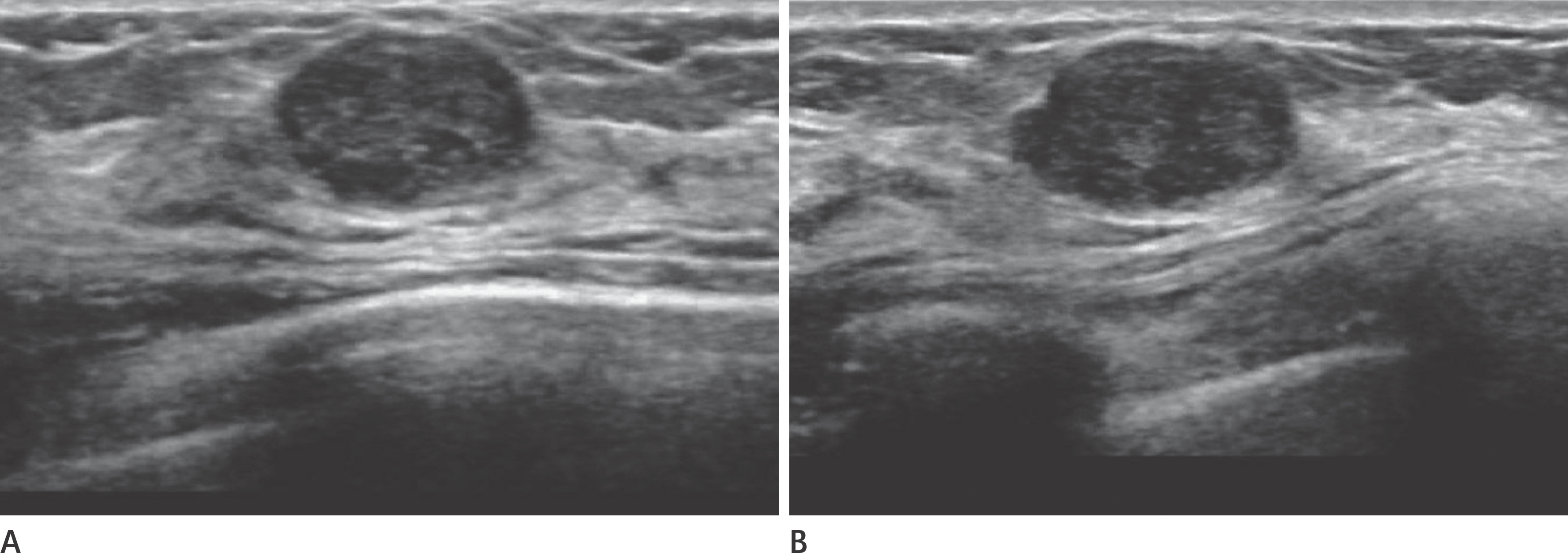
 | Fig. 1.Transverse (A) and longitudinal (B) ultrasonographic images showing an irregular hypoechoic mass with spiculated margins. The proportion of correct answers for margin was 74.1% (60/81). The mass was confirmed as a complex sclerosing lesion. Complex sclerosing adenosis can be seen as a spiculated margin. |
 | Fig. 2.Transverse (A) and longitudinal (B) ultrasonographic images of an oval circumscribed hypoechoic mass showing BI-RADS category 3. The proportion of correct answers for this BI-RADS category was 38.2% (26/68). The mass was confirmed to be a fibroadenoma. BI-RADS = Breast Imaging Reporting and Data System |
Table 1.
The Proportion of Correct Answers According to Ultrasono-graphic BI-RADS Lexicon
| Variable | Proportion of Correct Answers | p-Value |
|---|---|---|
| Orientation | 93.1% (161/173) | < 0.001 |
| Shape | 91.3% (232/254) | |
| Margin | 66.4% (263/396) | |
| Echo pattern | 68.1% (233/342) | |
| Posterior features | 94.3% (164/174) |
Table 2.
The Proportion of Correct Answers for Ultrasonographic BI-RADS Lexicon According to Year in Resident Training and the Duration of Training for Breast Imaging
Table 3.
The Proportion of Correct Answers According to BI-RADS Category
Table 4.
The Proportion of Correct Answers for BI-RADS Category According to Year in Resident Training
| Number of Residents | Proportion of Correct Answers | p-Value | |
|---|---|---|---|
| Year in resident training | 0.186 | ||
| 1st | 16 | 30.4% (28/92) | |
| 2nd | 16 | 37.8% (59/156) | |
| 3rd | 16 | 39.6% (55/139) | |
| 4th | 33 | 42.9% (130/303) |
Table 5.
The Proportion of Correct Answers for BI-RADS Category According to the Duration of Training for Breast Imaging
| Duration of Training (Months) | Number of Residents | Proportion of Correct Answers |
p-Value |
||||
|---|---|---|---|---|---|---|---|
| Among 3 Group | Test for Trend | vs. 1 | vs. 2 | vs. 3-4 | |||
| 1 | 28 | 35.4% (76/215) | 0.033 | 0.018 | 0.032 | 0.015 | |
| 0.096* | 0.044* | ||||||
| 2 | 24 | 37.0% (87/235) | 0.032 | 0.712 | |||
| 0.096* | > 0.999* | ||||||
| 3–4 | 19 | 47.8% (77/161) | 0.015 | 0.712 | |||
| 0.044* | > 0.999* | ||||||


 PDF
PDF ePub
ePub Citation
 Print
Print


 XML Download