Abstract
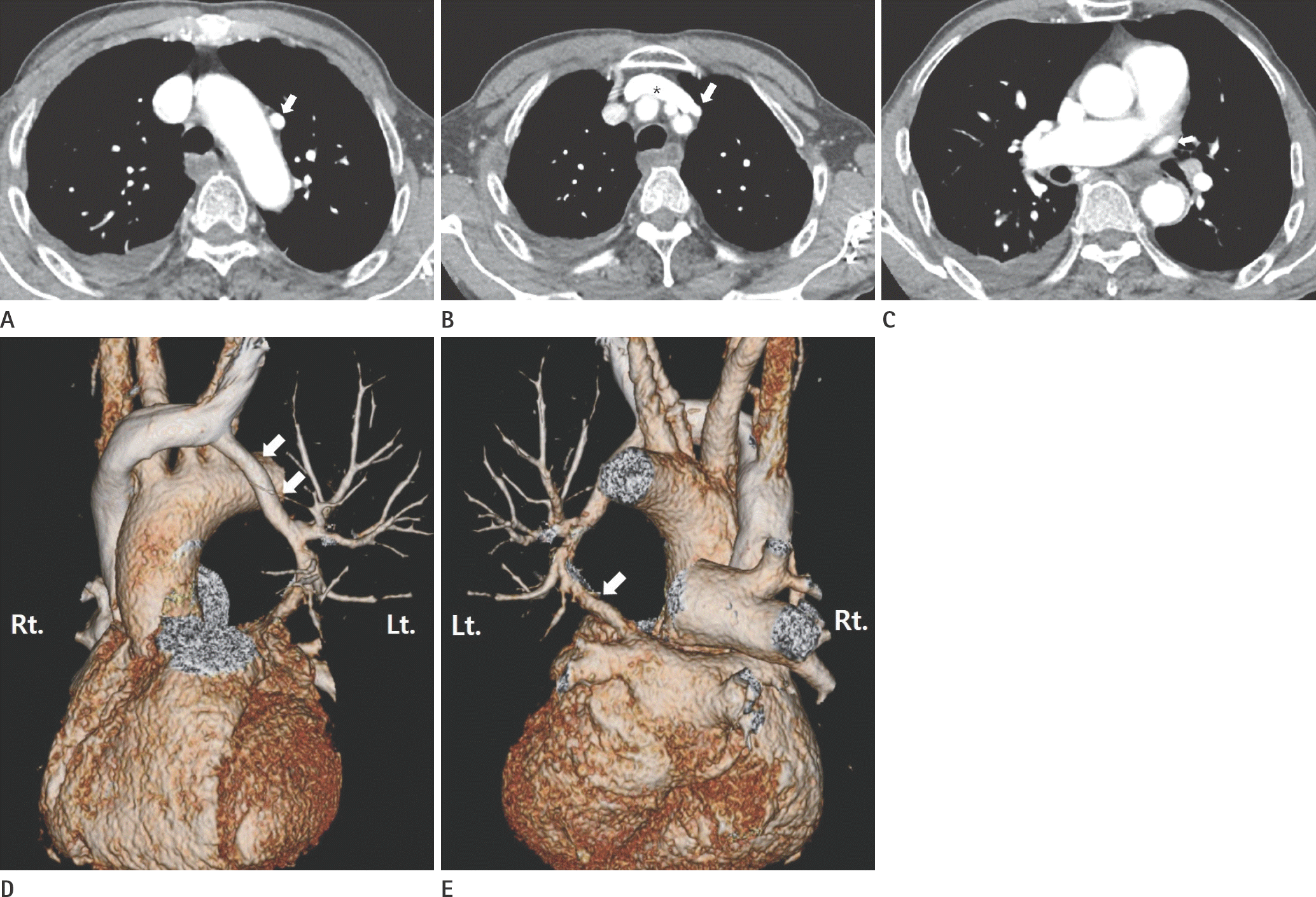
Partial anomalous pulmonary venous return is a rare congenital pulmonary venous anomaly, in which some of the pulmonary veins drain into the systemic circulation rather than the left atrium. Many variants of partial anomalous pulmonary venous return have been reported. We present a rare type of partial anomalous pulmonary venous return in which the anomalous left upper lobe pulmonary vein drained into the left innominate vein via the vertical vein, accompanying the left upper lobe pulmonary vein in the normal location.
References
1. Dillman JR, Yarram SG, Hernandez RJ. Imaging of pulmonary venous developmental anomalies. AJR Am J Roentgenol. 2009; 192:1272–1285.

2. Katre R, Burns SK, Murillo H, Lane MJ, Restrepo CS. Anomalous pulmonary venous connections. Semin Ultrasound CT MR. 2012; 33:485–499.

3. Park GM, Kang MJ, Lee HB, Bae KE, Lee J, Kim JH, et al. Anomalous pulmonary venous return: a case report. J Korean Soc Radiol. 2013; 69:279–282.

4. Zylak CJ, Eyler WR, Spizarny DL, Stone CH. Developmental lung anomalies in the adult: radiologic-pathologic correlation. Radiographics. 2002; 22:S25–S43.

5. Sears EH, Aliotta JM, Klinger JR. Partial anomalous pulmonary venous return presenting with adult-onset pulmonary hypertension. Pulm Circ. 2012; 2:250–255.

6. Moore JW, Kirby WC, Rogers WM, Poth MA. Partial anomalous pulmonary venous drainage associated with 45,X Turner's syndrome. Pediatrics. 1990; 86:273–276.

7. Haramati LB, Moche IE, Rivera VT, Patel PV, Heyneman L, McAdams HP, et al. Computed tomography of partial anomalous pulmonary venous connection in adults. J Comput Assist Tomogr. 2003; 27:743–749.

Fig. 1.
A 75-year-old man with partial anomalous pulmonary venous return accompanied by a normal left superior pulmonary vein. A, B. Contrast-enhanced chest CT scan shows a vertical vein (arrow) which courses through the paraaortic area and drains into the left innominate vein (asterisk). Note the right small pleural effusion. C. Contrast-enhanced chest CT scan shows a normal left superior pulmonary vein (arrow) anterior to the left main bronchus. Note the right small pleural effusion. D, E. Three-dimensional volume-rendering images show the vertical vein (arrow in D) draining into the left innominate vein and the normal left superior pulmonary vein (arrow in E) draining into the left atrium.


 PDF
PDF ePub
ePub Citation
 Print
Print


 XML Download