

 PDF
PDF ePub
ePub Citation
 Print
Print


INTRODUCTION
Stent implantation for stenosis of the iliac arteries is an established procedure for claudication. In particular, balloon-expandable stents (BES) have been preferred in heavily calcified lesions because of their strong radial force and expanded condition that can persist against the elastic recoil of arteries (1). Therefore, we have used the BES for a relatively short segmental stenosis of the iliac arteries with a severely calcified plaque over a period of approximately 10 years. During the follow-up period, there has been one case of a collapsed BES. We herein report a case of a collapsed BES due to minor external pressure in a lean patient.
CASE REPORT
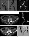
A 60-year-old man underwent aorto-iliac angiography three months following claudication. His initial ankle-brachial pressure index (ABI) was 0.78. The angiogram revealed focal severe stenosis with a heavily calcified lesion in the right common iliac artery (CIA). We performed primary stenting using the Express LD BES (9.0 × 40 mm) which was deployed, and the post-stenting angiogram showed a good angiographic result (Fig. 1A). His poststenting ABI was 1.1 and claudication had resolved after endovascular treatment. His ABI was maintained within the range of 1.02 to 1.1 during 18 months on a regular follow-up every 2 months. According to the Cardiovascular and Interventional Radiological Society of Europe Guidelines, follow up computed tomography angiography (CTA) was not considered because the patient did not complain of any symptoms and his ABI was maintained at normal levels. However, he suddenly developed claudication and coldness in his right leg after 19 months. His right leg ABI was 0.77. CTA showed that the stent in the right CIA was normally deployed on the anteroposterior view (Fig. 1B), but it was compressed (about 70% luminal narrowing) in the anteroposterior plane on the axial scan and the lateral plane (Fig. 1C, D). The patient explained that before returning to the hospital, his grandson had played with him and jumped once on his stomach, and that was the time when his symptoms relapsed. His grandson was 2 years old, and his weight was about 17 kg. We extrapolated that his grandson's jump had caused compression of the stent against the lumbar spine. The patient was thin and his body mass index (BMI) was below 19. We recommended an additional interventional treatment that he deferred. One year later, his symptoms deteriorated and CTA revealed that the right CIA was totally occluded due to a collapsed stent and thrombosis (Fig. 1E). We performed balloon angioplasty and additional self-expandable stent (SES) deployment across the site of occlusion. The post-stenting angiogram showed satisfactory patency and the patient's pain was alleviated in addition to the increase in ABI to 1.1 (Fig. 1F). After SES deployment, he has had an uneventful follow-up to date.
DISCUSSION
In atherosclerotic aorto-iliac steno-occlusive lesions, the BES is a useful option due to its accurate placement, straightforward delivery, satisfactory radial force in heavily calcified lesions and minimal foreshortening (12). In general, we know that the BES is more rigid and provides a higher radial force compared to the SES (3). Although this rigidity provides resistance against elastic recoil, it also makes the stent susceptible to deformity from external pressure (14). However, the BES can be fractured or collapsed when deployed in a superficial vessel or at places where external forces can lead to compression. Thus, the number of available target vessels is limited to the CIA, the subclavian artery, and the renal artery (5).
The SES has a multi-segmented design that allows each segment to extend the vessels and to adapt to different diameters along the vessel treated. The SES is commonly used in tortuous and longer segments (6). The BES is not suitable for placement in arteries traversing or in close proximity to a joint that could cause kinking and overall failure. Therefore, the target lesion site of the BES is defined as a deep-lying vessel or a heavily calcified, short stenosis/occlusion, especially in the CIA (7). However, we suggest that the CIA in a lean person could be susceptible to external compressive forces, as observed in this case.
Complications associated with the BES may be technical, related to the procedure, or biological, a result of the presence of the stent within the vascular lumen (8). These complications generally manifest as sudden vessel thrombosis or vessel dissection in an acute situation. In the chronic course, these complications present as restenosis, stent fracture, or delayed thrombosis.
Other than the possibility of incomplete expansion due to subintimal stenting, no acute complications associated with the stent could be demonstrated either during the placement or on the immediate follow-up arteriogram. No other abnormalities were noted. Stent compression was only observed 19 months later when repeat CTA was performed for recurrent symptoms of claudication. Unfortunately, no abdominal or pelvic radiographs had been obtained. Instead, the stent was compressed in precisely front and rear directions on follow-up CTA. We speculate that the orientation of the collapsed stent suggests sudden collapse rather than gradual stent occlusion.
Collapse or fracture of stents, especially of the BES, was usually seen in the superficial femoral arteries, but a collapsed BES in the iliac arteries has been observed very infrequently (59). Only a few reported cases indicate that cyclic axial force from repeated abdominal massage or vigorous exercise can predispose the stents to fracture and compression. Ichihashi et al. (10) described compression fracture of the BES in a 76-year-old man who had a lower abdominal massage and Sawhney et al. (5) reported a case of kissing balloon-expandable iliac stents complicated by stent fracture in a man who performed strong daily stretching and calisthenics. They assumed that his vigorous exercise program induced fracture of the stents. They did not mention the patients' BMI in the literature, but we speculate that they were lean individuals based on the figures and contents in these case reports. Both cases are similar to our case.
In conclusion, stenting in patients with lower BMI requires careful selection of the target vessel and the type of the stent. Thus, BES implantation for CIA stenosis/occlusion should be carefully considered in a lean person.
 XML Download