

 PDF
PDF ePub
ePub Citation
 Print
Print


INTRODUCTION
Ectopic liver is an unusual condition because gross abnormalities of the liver are exceptional, despite its complex developmental process. The gallbladder (GB) is the most common site of ectopic liver development, but the incidence of ectopic liver is low, at 0.24–0.47% (12). Although the GB is the most frequent site of ectopic liver development, due to the lack of experience with ectopic liver, it is rarely considered as a differential diagnosis when a soft tissue mass arising from the GB wall is encountered. Most ectopic livers are small, and most of them are detected during surgery or autopsy rather than by imaging studies (3). Few recent reports have focused on the radiologic features of ectopic liver (34). Moreover, there are no previous case reviews of ectopic livers that exhibit a tendency to grow during follow up. Here, we present a case of ectopic liver with normal liver parenchyma that was attached to the serosal surface of the GB and grew slowly, which developed in a patient with liver cirrhosis.
CASE REPORT
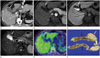
A 53-year-old man who was diagnosed with liver cirrhosis due to drug-induced toxic hepatitis in June 2013 received routine surveillance with computed tomography (CT) examination for liver cirrhosis. He was asymptomatic at the time of CT examination that was performed in July 2014. This CT examination revealed a well-circumscribed, oval mass in the GB fossa. The lesion was isodense to the liver parenchyma, but it was separated from the liver and it abutted the GB. A review of an abdominopelvic CT performed in 2009 also revealed a mass at the same location. It had gradually increased in size from 0.6 × 1.8 cm to 1.5 × 2.5 cm without any changes in its characteristics (Fig. 1A). However, the mass was not mentioned in the radiology report of the CT examination performed in 2009. For further evaluation, magnetic resonance imaging (MRI) using gadoxetic acid (Primovist; Bayer HealthCare, Berlin, Germany) and positron emission tomography (PET)-CT were also performed due to the possibility of tumorous condition of the GB. MRI showed findings similar to CT. The mass abutted the GB, exhibited peripheral enhancement during dynamic T1-weighted images (T1WI) obtained after contrast injection (Fig. 1B), and then it was isointense with respect to the liver in the 20-minute hepatobiliary phase (HBP) (Fig. 1C), T2-weighted images (T2WI) (Fig. 1D), and diffusion-weighted images (DWI) without diffusion restriction. On whole-body PET-CT performed at that time, the mass exhibited minimal fluorodeoxyglucose (FDG) uptake that was similar to that in the liver (Fig. 1E). The α-fetoprotein level was 4.38 ng/mL (normal: < 13.4 ng/mL), and liver function tests revealed no abnormalities (aspartate transaminase: 22 IU/L; alanine aminotransferase: 32 IU/L). The mass seemed unlikely to be malignant, but surgical resection was performed because the mass showed interval growth. The patient underwent elective laparoscopic cholecystectomy. During the procedure, a smooth fragment of reddish-brown tissue measuring 2.5 × 1.5 cm was found attached to the serosa of the GB (Fig. 1F). This fragment was diagnosed as an ectopic liver with normal liver parenchymal elements that showed neither cirrhotic changes nor tumors on histological examination. However, the liver proper exhibited cirrhotic changes.
DISCUSSION
It is a reasonable assumption that the ectopic liver would show isointensity to the liver proper on MRI as ectopic livers usually exhibit the same histological features as those of the liver proper. In our case, the ectopic liver was isointense with respect to the liver on pre-contrast T1WI, T2WI, and DWI without diffusion restriction and it exhibited minimal FDG uptake, similar to the liver itself, on PET-CT. However, an ectopic liver may exhibit a different enhancement pattern compared to the liver proper during dynamic images with contrast due to the differences in its blood supply relative to the main liver, which receives a dual blood supply from the hepatic artery and the portal vein (56). Our case exhibited a peripheral enhancement pattern during arterial phase imaging after administration of gadoxetic acid, mimicking tumorous condition arising in the GB. However, the mass was isointense to the liver parenchyma in the HBP of the MRI using gadoxetic acid, which implies that the lesion contained hepatocytes that expressed organic anion transporting polypeptides (OATPs) (7). Gadoxetic acid is a hepatocyte-specific contrast agent that behaves similarly to non-specific gadolinium chelate agents during the dynamic phases, but is gradually taken up by the hepatic parenchyma via the OATP receptors on hepatocytes after contrast injection (89). During the HBP, the hepatic parenchyma contains gadoxetic acid, but lesions without or with reduced number of functioning hepatocytes, such as metastases and most hepatocellular carcinomas, do not contain the contrast. Lesions with functioning hepatocytes, such as dysplastic nodules and focal nodular hyperplasia, usually exhibit iso- or hyperintensity on the HBP imaging (9). On review, identical enhancement of the ectopic liver compared to the liver proper on HBP could be a crucial point in proper identification of this condition.
An ectopic liver is usually located adjacent to the liver proper and has the same parenchymal texture as the liver proper, but it is smaller in size. However, the mass in our case was not connected to the liver proper, but rather it was abutting the serosal surface of the GB. Thus, we could rule out the possibility of an accessory liver that was attached to the main liver or tumorous conditions arising from an accessory hepatic lobe. An ectopic liver may exhibit the same pathologic features as the liver proper (i.e., fatty changes, hemosiderosis, cholestasis, or cirrhosis). Several articles have reported that an ectopic liver is associated with an increased risk of developing HCC (510). Few cases of ectopic livers that showed interval growth without tumor development have previously been reported. In our case, the ectopic liver tissue grew slowly and mimicked a tumor. However, growth of the ectopic liver in this case was not due to a tumor arising from the ectopic liver. The main liver exhibited liver cirrhosis, but cirrhotic change was not detected during histological examination of the ectopic liver. With respect to the explanation of interval growth of the ectopic liver, we surmise that the ectopic liver tissue with normal hepatocytes might slowly become hypertrophic to compensate for the cirrhotic liver proper.
In conclusion, if a mass separated from the liver proper exhibits the same signal intensity as the liver on pre-contrast T1WI, T2WI, and DWI the HBP of MRI using gadoxetic acid, the possibility of an ectopic liver should be considered. In addition, it should be kept in mind that the ectopic liver is associated with an increased risk of developing HCC and the ectopic liver might show interval growth.
 XML Download