

 PDF
PDF ePub
ePub Citation
 Print
Print


Abstract
Purpose
The purpose of this study was to investigate the relationship between ab-dominal fat as assessed by abdominal fat CT and metabolic syndrome (MS), espe-cially in asymptomatic Korean individuals.
Materials and Methods
Retrospectively, a medical record analysis was performed in a total of 111 patients with screening abdominal fat CT. The data such as visceral fat (VF), subcutaneous fat (SF) and VF/SF were elicited by abdominal fat CT, and we ana-lyzed the relationship of VF, SF, and VF/SF with MS and cardiovascular risk factors.
Results
In males, VF and SF had a positive correlation with many cardiovascular risk factors and MS, but VF was superior to SF. In females, VF, but not SF, had a positive correlation with some cardiovascular risk factors and MS. The cut-off values of VF and SF to predict MS, which were calculated by drawing receiver operating char-acteristic curves, were as follows: the cut-off value of VF in men: 136.50 cm2, the cut-off value of SF in men: 159.50 cm2, and the cut-off value of VF in women: 134.50 cm2.
REFERENCES
1.Eckel RH., Grundy SM., Zimmet PZ. The metabolic syndrome. Lancet. 2005. 365:1415–1428.

2.Alberti KG., Zimmet P., Shaw J. IDF Epidemiology Task Force Consensus Group. The metabolic syndrome--a new world-wide definition. Lancet. 2005. 366:1059–1062.
3.Alberti KG., Zimmet P., Shaw J. Metabolic syndrome--a new world-wide definition. A Consensus Statement from the In-ternational Diabetes Federation. Diabet Med. 2006. 23:469–480.
4.Grundy SM., Cleeman JI., Daniels SR., Donato KA., Eckel RH., Franklin BA, et al. Diagnosis and management of the met-abolic syndrome: an American Heart Association/National Heart, Lung, and Blood Institute Scientific Statement. Cir-culation. 2005. 112:2735–2752.
5.Ford ES., Giles WH., Mokdad AH. Increasing prevalence of the metabolic syndrome among u.s. Adults. Diabetes Care. 2004. 27:2444–2449.
6.Lim S., Shin H., Song JH., Kwak SH., Kang SM., Yoon JW, et al. Increasing prevalence of metabolic syndrome in Korea: the Korean National Health and Nutrition Examination Survey for 1998-2007. Diabetes Care. 2011. 34:1323–1328.
7.Yoshizumi T., Nakamura T., Yamane M., Islam AH., Menju M., Yamasaki K, et al. Abdominal fat: standardized technique for measurement at CT. Radiology. 1999. 211:283–286.
8.Hamdy O., Porramatikul S., Al-Ozairi E. Metabolic obesity: the paradox between visceral and subcutaneous fat. Curr Dia-betes Rev. 2006. 2:367–373.
9.Bergman RN., Kim SP., Catalano KJ., Hsu IR., Chiu JD., Kabir M, et al. Why visceral fat is bad: mechanisms of the metabolic syndrome. Obesity (Silver Spring). 2006. 14(Suppl 1):16S–19S.
10.Wajchenberg BL. Subcutaneous and visceral adipose tissue: their relation to the metabolic syndrome. Endocr Rev. 2000. 21:697–738.
11.Ibrahim MM. Subcutaneous and visceral adipose tissue: structural and functional differences. Obes Rev. 2010. 11:11–18.
12.Sandeep S., Gokulakrishnan K., Velmurugan K., Deepa M., Mo-han V. Visceral & subcutaneous abdominal fat in relation to insulin resistance & metabolic syndrome in non-diabetic south Indians. Indian J Med Res. 2010. 131:629–635.
13.Pickhardt PJ., Jee Y., O'Connor SD., del Rio AM. Visceral adi-posity and hepatic steatosis at abdominal CT: association with the metabolic syndrome. AJR Am J Roentgenol. 2012. 198:1100–1107.
14.Kim S., Cho B., Lee H., Choi K., Hwang SS., Kim D, et al. Distri-bution of abdominal visceral and subcutaneous adipose tis-sue and metabolic syndrome in a Korean population. Diabe-tes Care. 2011. 34:504–506.
15.Kim HI., Kim JT., Yu SH., Kwak SH., Jang HC., Park KS, et al. Gen-der differences in diagnostic values of visceral fat area and waist circumference for predicting metabolic syndrome in Koreans. J Korean Med Sci. 2011. 26:906–913.
16.Han JH., Park HS., Kim SM., Lee SY., Kim DJ., Choi WH. Visceral adipose tissue as a predictor for metabolic risk factors in the Korean population. Diabet Med. 2008. 25:106–110.
17.Lee DY., Rhee EJ., Choi ES., Kim JH., Won JC., Park CY, et al. Comparison of the Predictability of Cardiovascular Disease Risk According to Different Metabolic Syndrome Criteria of American Heart Association/National Heart, Lung, and Blood Institute and International Diabetes Federation in Korean Men. Korean Diabetes J. 2008. 32:317–327.
18.Bassett J. International Diabetes Institute; World Health Or-ganization; Regional Office for the Western Pacific; Inter-national Association for the Study of Obesity; International Obesity TaskForce. The Asia-Pacific perspective: redefining obesity and its treatment. Melbourne: Health Communications Australia;2000.
Fig. 1.
Measurement of visceral and subcutaneous fat using a 64-slice multidetector CT with AW version 4.5. A. Class areas are calculated based on the (D) attenuating region as -50 to -250 Hounsfield unit using the histogram method. The calculated area is 122.32 cm2 and this is the value of visceral fat. B-D. Three areas are obtained by drawing lines along the abdominal skin (B), the outer line of the abdominal muscles (C) and the inner line of the abdominal muscles and psoas muscles (D).
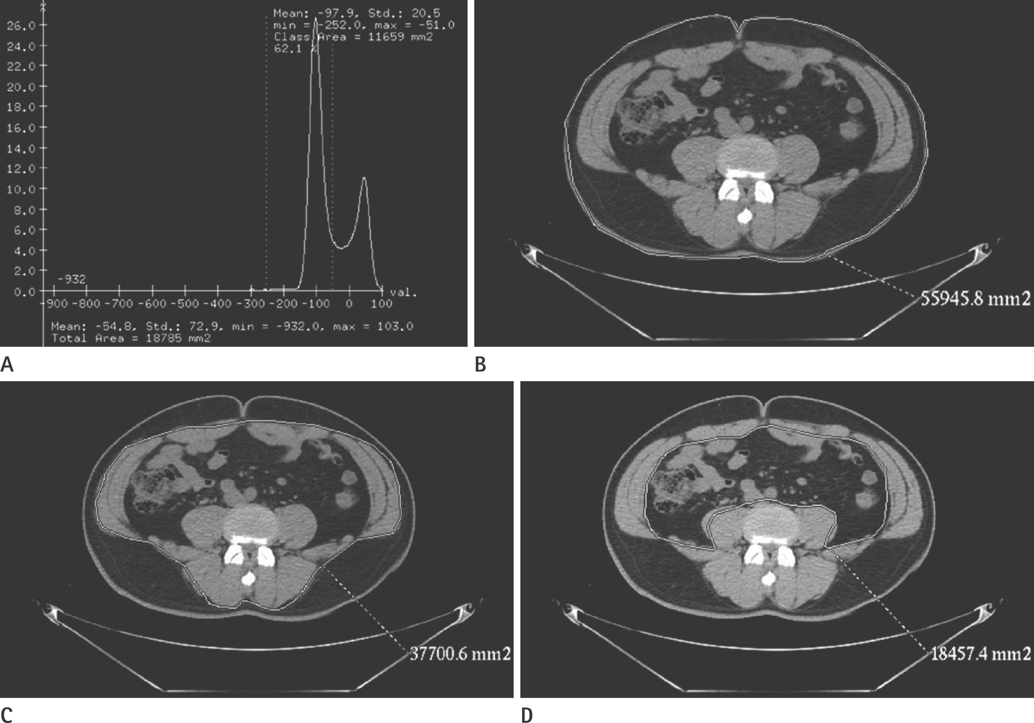
Fig. 2.
ROC curves of VF (A) and SF (B) for predicting metabolic syndromes in men. A. The AUC of VF was 0.762 and the result was statistically significant (p < 0.001). B. The AUC of SF was 0.681 and the result was statistically significant (p < 0.01). AUC = area under the curve, ROC = receiver operating characteristic, SF = subcutaneous fat, VF = visceral fat
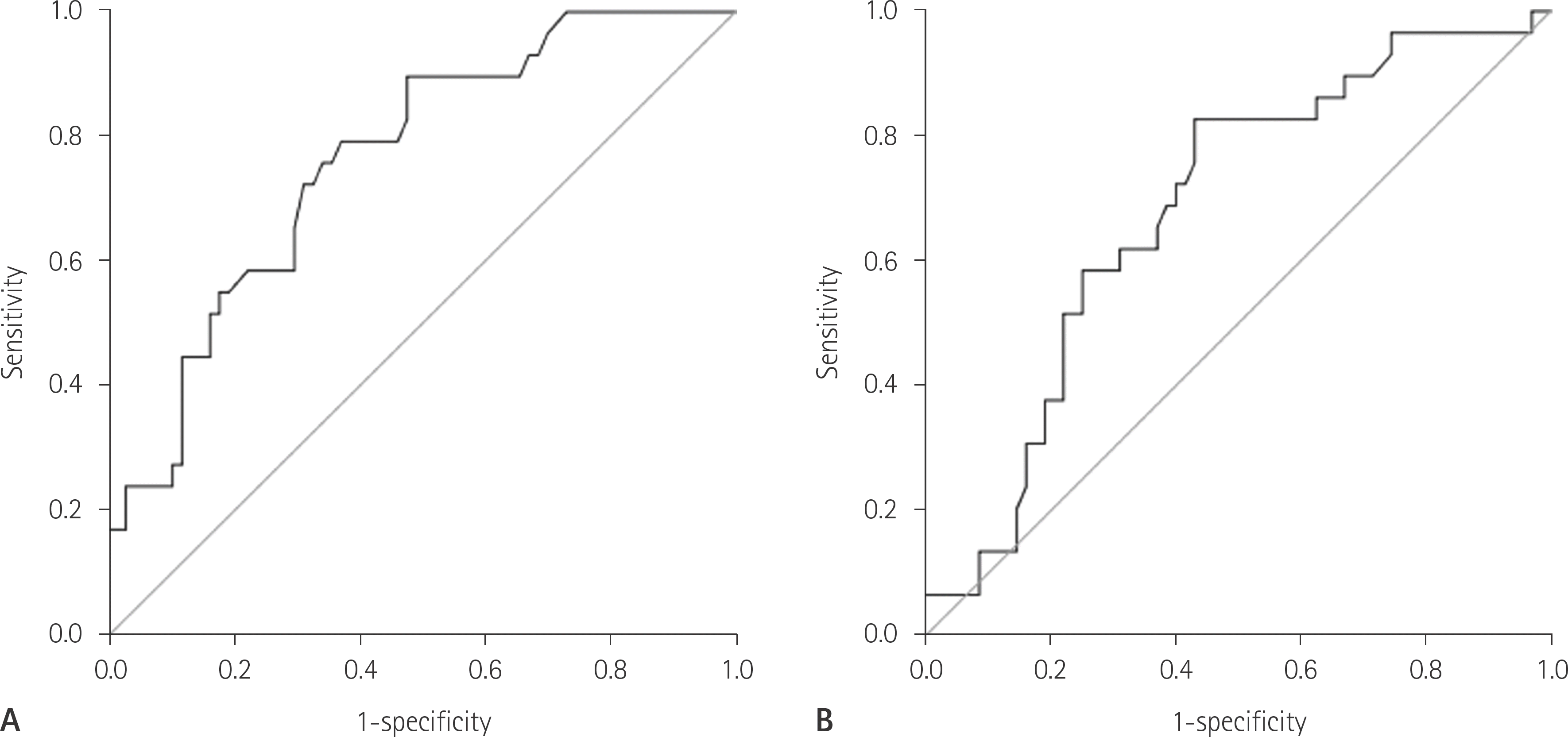
Fig. 3.
A ROC curve of VF for predicting metabolic syndrome in wom-en. The AUC of VF was 0.909, and the result was statistically significant (p < 0.05). AUC = area under the curve, ROC = receiver operating characteristic, VF = visceral fat
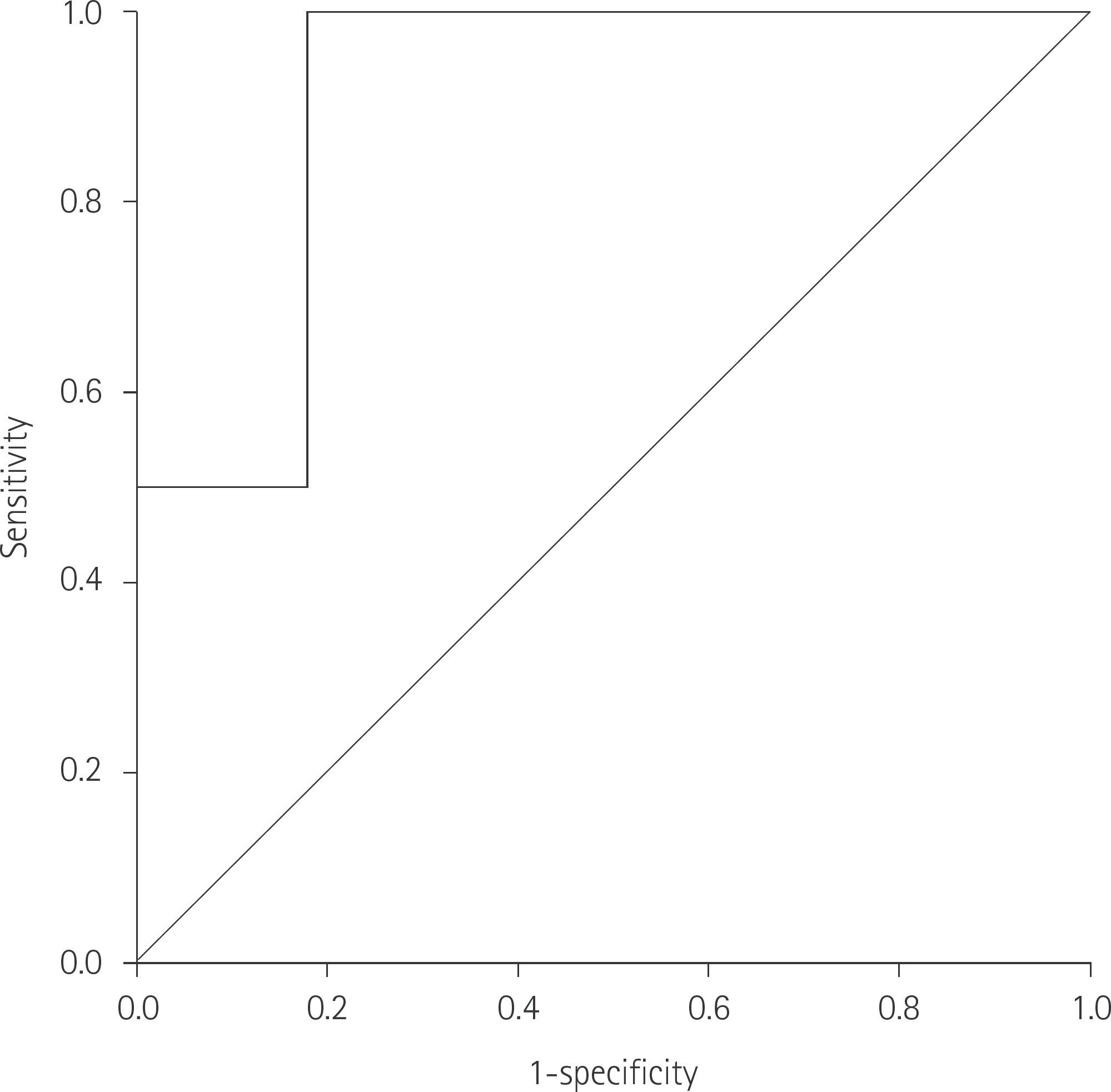
Table 1.
Baseline Characteristics
Table 2.
Pearson Correlation Coefficients between Indicators of Fat Measured by Abdominal Fat CT and MS, Cardiovascular Risk Factors in Men
Table 3.
Pearson Correlation Coefficients between Indicators of Fat Measured by Abdominal Fat CT and MS, Cardiovascular Risk Factors in Women
Table 4.
Relationships between Indicators of Fat and MS
 XML Download