

 PDF
PDF ePub
ePub Citation
 Print
Print


INTRODUCTION
Chondroblastomas are rare benign cartilaginous neoplasms found in young patients. These tumors typically arise in the epiphysis or apophysis of a long bone, such as the humerus, femur, or tibia. Additionally, 10–15% of chondroblastomas are accompanied by an aneurysmal bone cyst. In a previous report, chondroblastomas arising in the skull and facial bones are described as extremely rare entities. In this report, we describe a rare case of a patient presenting with chondroblastoma with aneurysmal bone cyst in the sphenoid sinus that mimicked invasive sinusitis or malignant bone tumor. We also include a review of the literature, focusing on radiologic imaging findings of chondroblastomas arising in the skull and facial bones.
CASE REPORT
A 13-year-old boy with right-sided visual disturbance was referred to the ophthalmic outpatient department. A sudden onset of symptoms was noted one week ago, and no visual abnormality was found during a routine visual test performed six months ago. In the general physical examination performed at the outpatient ophthalmology clinic, right-sided visual acuity was decreased, and only gross hand movement was recognizable. The slit-lamp microscope examination result was normal. Mild swelling and injection were found in the right optic nerve head on examination of the fundus. Although neither signs of infection nor abnormal laboratory test results were found, the clinician suspected optic neuritis. The patient was referred to the neurology clinic, and brain computed tomography and magnetic resonance imaging were performed.
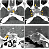
On the CT scan, a localized multilobulated, multiseptated cystic lesion was found in the sphenoid sinus. The mass was estimated to be about 35 × 40 × 40 mm in size and it involved both sphenoid sinuses, both posterior ethmoid sinuses, and both frontal base areas. Irregular-shaped high-attenuation foci [about 140 Hounsfield unit (HU)] suggesting chondroid calcification were identified in the central portion of the mass by pre-contrast enhanced CT. Additionally, a non-enhanced increased attenuation lesion (about 40 HU), which suggested a hemorrhagic portion, was accompanied by the calcified lesion. The mass extended to the right optic foramen, and local invasion of the anterior cavernous sinus area was suspected. Bony erosion of the right ethmoid roof and sellar floor was obvious on CT images (Fig. 1).
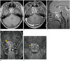
On sequentially performed MRI, a similar lesion correlating with the CT findings was observed. The internal contents of the cystic lesion showed high T2 signal intensity, and fluid-fluid levels were identified. These findings suggested internal hemorrhage in the cystic portion and they also correlated with a non-enhancing increased attenuation portion of the lesion on CT images. The walls of the internal septa and dura mater in both frontal bases were strongly enhanced. Furthermore, suspected invasion of the optic foramen became clear on CT, and obvious invasion was identified by MRI (Fig. 2).
Bone tumor such as chondroblastoma or giant cell tumor was suspected on first inspection because of its enhancing solid and cystic portion with peritumoral bony destruction and intratumoral chondroid calcifications. Additionally, blood-fluid levels implied aneurysmal bone cysts that often accompany chondroblastoma. However, the radiologist could not make confirmative diagnosis because of its rare presentation in the skull base. Therefore, an infectious condition like highly virulent or invasive sinusitis needs to be included in the differential diagnosis due to the abrupt onset of the symptom.
The symptoms were aggravated despite conservative management, and the neurosurgeon performed decompressive excision of the lesion. The surgery was performed via the trans-sphenoidal approach. A yellowish soft mass was detected immediately after the sphenoid sinus was opened. The mass was multiseptated, and hemorrhagic necrosis was also observed in the mass. Tumoral invasion of the planum sphenoidale, clivus, and the anterior clinoid process was observed, but no direct invasion of the dura mater was suspected in the imaging evaluation. All visible tumor masses were resected.
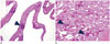
Pathologic findings showed a characteristic fine network of pericellular “chicken wire” calcifications and osteoclast-like giant cells. In addition, cystic walls and septae lined by fibroblasts, myofibroblasts, and histiocytes were observed. These findings indicate chondroblastoma with secondary aneurysmal bone cyst components (Fig. 3).
The patient underwent reoperation for complete resection of the tumor, and the visual symptoms gradually improved. On follow-up MRI 1 year later, there was no evidence of local recurrence.
DISCUSSION
Chondroblastomas are rare neoplasms that represent less than 1% of all bone tumors (1). Most typical chondroblastomas are diagnosed without any controversy due to their characteristic location in long bones, such as the humerus, femur, or tibia, and they affect those aged less than 20 years (2). However, chondroblastoma arising from the skull and facial bones is extremely rare. Although some cases have been reported, in most of them, the tumor arose from the temporal bone or condyle of the mandible (13). Only two cases have been reported in which the tumor arose from the sphenoid bone (45).
The general features of chondroblastoma in long bones include solid periosteal reaction, internal calcification, and cortical breach. Additionally, endosteal scalloping can be seen on CT or plain radiography (67). On MRI, the surrounding bone marrow and soft tissue edema are seen in a large proportion of the reported cases (8). Also, secondary aneurysmal bone cysts may complicate more than 33% of all chondroblastomas (1). Consequently, when there is an accompanying aneurysmal bone cyst, multiseptated cysts with fluid-fluid levels are observed (9).
On the other hand, radiologic features of chondroblastoma that arises from the skull have not been established. Our case of skull base chondroblastoma showed usual features of chondroblastoma arising from a long bone, including internal scattered chondroid calcifications and adjacent bony erosion. Multiseptated cysts with blood-fluid levels identified on MRI also corresponded with typical findings of usual secondary aneurysmal bone cysts in chondroblastoma arising from a long bone. Although these morphologic features exist, it is difficult to form an impression initially when it arises from the skull due to its rarity. An accompanying aneurysmal bone cyst can be a diagnostic clue for chondroblastoma of the skull (45).
In our case, both CT and MRI evaluation were helpful for diagnosis of the lesion. CT showed superior accuracy in the evaluation of bony involvement. The shape, margins, and subtle sclerotic edges of the bony lesion were well identified by CT in our case. However, there are limitations in diagnostic evaluation of lesional extent and local invasion, and components of the mass itself only by CT. MRI can provide more accurate information about these features (3). As shown in a previously reported case by Wang and Zhou (5), MRI has superiority in identifying typical blood-fluid levels in aneurysmal bone cysts. Thus, dual combined modalities of CT and MRI might be necessary for the diagnosis of chondroblastoma accompanied by aneurysmal bone cysts.
Despite the pathologic benign features, chondroblastoma can be locally aggressive due to its high growth rate and osteolytic capacity. Occasionally, pathologic fractures and malignant transformation can be observed. Additionally, the tumor may expand to adjacent structures and may rarely metastasize to other organs. The general treatment option for this chondroblastoma arising from the skull is surgical excision. The usual recurrence rate after resection of typical chondroblastoma is approximately 10% (10).
There is no consensus on the standard treatment option for chondroblastoma arising from the skull. According to two reported cases, wide surgical resection showed lower recurrence rates and better prognosis (45). Thus, early radiologic diagnosis of chondroblastoma arising from the skull is considered necessary for implementing treatment and achieving better prognosis. Radiologists need to include chondroblastoma as a differential diagnosis when they interpret masses arising from the skull.
 XML Download