INTRODUCTION
Percutaneous biopsy is a less invasive technique for establishing histologic diagnosis than laparoscopic biopsy or exploratory laparotomy. This minimally invasive technique helps to determine whether a lesion is benign or malignant. However, retroperitoneal lymph nodes are difficult to access because many critical organs are intervened in the biopsy pathway. The purpose of this case report was to show how to perform hydrodissection and how to use semi-automated devices for safe biopsy of a metastatic common iliac lymph node.
CASE REPORT
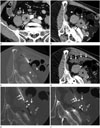
A 68-year-old man presented with right-sided flank pain. He had undergone hormonal and radiation therapy for treatment of prostate adenocarcinoma (T3bN1M0) 10 years ago. His latest prostate-specific antigen level was 0.21 ng/mL. Computed tomography (Aquilion; Toshiba Medical Systems Corp., Otawara, Japan) demonstrated an enlarged lymph node which was surrounded by the right ureter and common iliac vessels (Fig. 1A). Percutaneous biopsy was recommended to differentiate prostate cancer metastasis from other diseases after multi-department discussion.
Approximately 10 cc of lidocaine was percutaneously injected for local anesthesia and sedation was not performed. He was positioned in the right anterior oblique position to displace the ascending colon which was located in the biopsy needle pathway. However, the bowel loop was not displaced despite the change in the patient's position (Fig. 1B). Therefore, a total of 200 cc 5% dextrose water was instilled under CT guidance (Fig. 1C) so that the ascending colon was adequately displaced away from the biopsy pathway (Fig. 1D). Unenhanced CT scans were performed to determine how far away the bowel loops moved from the biopsy pathway every time 50–100 cc dextrose water was instilled. An 18 gauge biopsy needle (SH5-1815G; M.I. Tech, Seoul, Korea) was safely placed just proximal to the lesion. Sampling one core tissue included the following two steps: the first step was that an internal needle was manually introduced approximately 1 cm into the lesion to determine if the needle direction was safe (Fig. 1E). The second step was that the internal needle was completely pushed through the lesion after ensuring that the critical organs were not penetrated (Fig. 1F). The outer needle was fired to obtain a core tissue. A total of two cores were obtained and they were fixed with formalin. Co-axial biopsy was not performed and each biopsy core was percutaneously obtained. The histologic diagnosis was confirmed as metastatic small cell carcinoma, which was likely to have originated from the previous prostate cancer. He was scheduled to undergo chemotherapy and radiation therapy.
DISCUSSION
Laparoscopic biopsy is one of the options for sampling a metastatic lymph node around the critical organs. However, this technique is difficult to perform because of adhesion, which frequently occurs after radiation therapy for treatment of prostate carcinoma (1). Exploratory laparotomy is another option for sampling a metastatic lymph node, but it may be associated with a high risk of post-operative morbidity or mortality in case of post-radiation adhesion (1).
Percutaneous biopsy is a minimally invasive technique for sampling the cores from tumors arising from various organs. However, it is very difficult to target a deep-seated lymph node around the major vessels or the ureter. Moreover, small or large bowel loops are frequently encountered in the biopsy needle pathway. Hydrodissection has been used to avoid bowel injury during percutaneous renal tumor ablation (2). This technique can also be applied to target a para-aortic lymph node (3). Sampling a common iliac lymph node may cause injury to the bowel loops, the ureter, and the common iliac artery and vein. Hydrodissection can displace the bowel loops, but not the other organs. This technique is frequently performed for treating renal or hepatic tumors which are located close to the bowel loops. The use of dextrose water is preferred for radiofrequency ablation because the fluid is not ionic. Which fluid is optimal for hydrodissection in percutaneous biopsy has been not established. However, any kind of ionic or non-ionic fluid seems to be acceptable.
For safe biopsy, the needle should be placed appropriately within the lesion. Semi-automatic biopsy devices contain an inner needle which is manually introduced into the lesion. The outer needle is fired after ensuring that the lesion has been appropriately targeted with the inner needle. Therefore, semi-automatic firing helps reduce the risk of damage to the vasculature or other organs (4). However, automatic biopsy devices, which are used for prostate biopsy, are more likely to cause damage to the critical organs around the common iliac lymph node because both inner and outer needles are fired simultaneously.


 PDF
PDF ePub
ePub Citation
 Print
Print


 XML Download