

 PDF
PDF ePub
ePub Citation
 Print
Print


INTRODUCTION
Unilateral pulmonary artery hypoplasia occurs due to an embryologic developmental failure in the bud of the sixth aortic arch (123). Unilateral pulmonary artery hypoplasia is frequently associated with cardiovascular anomalies (3), but unilateral pulmonary artery hypoplasia without a cardiovascular anomaly is called ‘isolated’ unilateral pulmonary artery hypoplasia and this entity is much less common (24). Common presentations include shortness of breath, recurrent pulmonary infections, and hemoptysis (5); however, some patients are asymptomatic. Due to the nonspecific nature of the symptoms and imaging findings, it is easy to misdiagnose lung parenchymal change induced by unilateral pulmonary artery hypoplasia as other lung parenchymal diseases or pneumonia. There are only a few reports focusing on the radiologic findings in the pulmonary parenchyma, such as parenchymal bands caused by unilateral pulmonary artery agenesis (6). Therefore, this report describes the computed tomography (CT) findings in the pulmonary parenchyma due to isolated unilateral pulmonary artery hypoplasia.
CASE REPORT
A 36-year-old male was admitted to our hospital for the second time due to recurrent hemoptysis. He was an ex-smoker with a smoking history of 10 pack-years. He visited our hospital due to an episode of hemoptysis. The initial chest CT showed multifocal parenchymal bands in the left lung with patchy ground glass opacities, which were considered to be caused by aspirated blood. Initially it was thought that the parenchymal bands occurred due to previous infectious sequelae, and a definite diagnosis was not made at that time. He underwent bronchial artery embolization, which improved his symptoms.
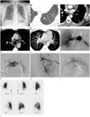
During his second visit to our hospital, chest radiography (Fig. 1A) showed decreased left-sided pulmonary vascularity, especially in the hilar area, and subtle elevation of the left hemidiaphragm, but no definite mediastinal shifting. A chest CT showed parenchymal bands and peripheral linear opacities in the left lung, which might be due to chronic lung infarction. Serrated pleural thickening was seen in the left hemithorax, corresponding to dilatation of the intercostal arteries. Left pulmonary artery diameter was small with decreased left-sided pulmonary vasculature. There were multifocal hypertrophied bronchial artery collateral vessels. Maximum intensity projection image showed a hypoplastic left pulmonary artery (Fig. 1B-E).
Pulmonary arteriography showed hypoplasia of the left pulmonary artery. Angiography showed that multifocal collateral arteries had arisen from the left subclavian artery and the left intercostal arteries (Fig. 1F-I). The patient received embolization of the collateral arteries via the left bronchial and left upper intercostal arteries, with PVA particles. Hemoptysis was completely resolved after embolization.
The patient was not aware of any childhood pulmonary infections or any specific family history of pulmonary disease. Transthoracic echocardiography (TTE) and a lung perfusion scan were performed for further evaluation of unilateral pulmonary artery hypoplasia. TTE revealed a normal left ventricular systolic function with normal valvular morphology. TTE did not show any septal defects or other cardiovascular anomalies or any evidence of pulmonary arterial hypertension. The lung perfusion scan showed a near total perfusion defect in the left lung (Fig. 1J). Hemoptysis was controlled and the patient was discharged from the hospital.
DISCUSSION
Unilateral pulmonary artery hypoplasia or agenesis is a rare congenital anomaly with an incidence of 1 in 300000. It occurs due to an embryologic developmental failure in the bud of the sixth aortic arch (3). It is frequently associated with other congenital anomalies, such as tetralogy of Fallot, a ventricular septal defect, transposition of great vessels, and aortic arch anomalies (2). Unilateral pulmonary artery agenesis as an isolated phenomenon without congenital cardiovascular anomalies is even more rare (7).
Terminologies related to unilateral pulmonary artery hypoplasia such as agenesis, aplasia, complete absence, and/or stenosis have been used interchangeably. Angiographic evidence was used to diagnose unilateral pulmonary artery aplasia by confirming the absence of the ipsilateral pulmonary artery. However, failure to fill the ipsilateral pulmonary artery with contrast material does not always indicate the absence of the pulmonary artery. At thoracotomy or on postmortem examination, a patent and small pulmonary artery has been observed even when there was no filling of the pulmonary artery in the previous pulmonary arteriography. This might be caused by increased bronchial arterial flow (1). It is unclear whether unilateral pulmonary artery hypoplasia is congenital or acquired (1). However, certain imaging characteristics may be useful in differentiating between congenital absence and hypoplasia of the pulmonary artery. Patients with acquired unilateral hyperlucent lungs have bronchiectasis or other destructive processes in the lungs with markedly impaired ventilation and mediastinal swing during inspiration. Patients with congenital unilateral pulmonary artery hypoplasia show normal ventilation in the affected lung and none or minimal mediastinal swing. In addition, a patient with congenital unilateral pulmonary artery hypoplasia shows more abundant systemic collateral vessels throughout the affected lung than a patient with acquired unilateral hyperlucent lungs (1). Therefore, we believe that the imaging findings in our patient were likely caused by congenital abnormalities.
Common presentations of unilateral pulmonary artery hypoplasia include shortness of breath, recurrent pulmonary infections, and hemoptysis (5); however, some patients are asymptomatic. Hemoptysis has been reported in approximately 18–20% of patients with unilateral pulmonary artery hypoplasia (8). Because of the nonspecific symptoms and imaging findings, it is easy to misdiagnose parenchymal change induced by unilateral pulmonary artery hypoplasia as other lung parenchymal diseases or pneumonia. In our case, initially it was thought that the parenchymal bands occurred due to the previous infectious sequelae, which delayed the diagnosis of unilateral pulmonary artery hypoplasia. CT findings of chronic lung infarction may include parenchymal bands, wedge-shaped opacities or irregular peripheral linear opacities (8). In the study by Sakai et al. (6), in eight patients with unilateral pulmonary artery agenesis, CT findings such as serrated pleural thickening were observed in six patients (75%), subpleural parenchymal bands in five (63%), and mosaic attenuation in three (38%) on the affected lung. In our case, parenchymal bands, serrated pleural thickening, and peripheral linear opacities were seen on CT altogether. These findings were probably due to parenchymal changes induced by unilateral pulmonary artery hypoplasia.
There is no general consensus on the treatment of unilateral pulmonary artery hypoplasia (9). However, there are several case reports of unilateral pulmonary artery hypoplasia with hemoptysis that were successfully treated with variable therapeutic options, such as pneumonectomy, vasodilator therapy, and embolization of collateral arteries (10).
In conclusion, if a patient presents with parenchymal findings, such as parenchymal bands, as well as hypertrophied collateral vessels, the possibility of unilateral pulmonary artery hypoplasia should be considered.
 XML Download