Abstract
A 31-year-old female with systemic lupus erythematosus (SLE) presented with fever, headache, seizures and mental status changes. Brain MRI showed T2 hyperintense lesions in the cerebellum and frontal white matter and a lesion in the cerebellum exhibited hemorrhagic changes and peripheral ring enhancement. The MRI features of listerial encephalitis are difficult to differentiate from those of neuropsychiatric SLE and various other diseases. Here, we report a case of hemorrhagic listerial encephalitis in a patient with SLE.
Central nervous system (CNS) infection is uncommon in patients with systemic lupus erythematosus (SLE), occurring in approximately 0.53–2.25% of patients (1). Patients with SLE are vulnerable to CNS infections due to disease-related immune dysfunction and immunosuppressive medication use (2). Since CNS infections remain a major cause of morbidity and mortality in patients with SLE, early and accurate diagnosis of these infections is important in affected patients (1).
Listerial infection occurs mainly in immunocompromised hosts. There have been some reports of listerial infections manifesting as meningitis, meningoencephalitis and brain abscesses in patients with SLE (1). Listerial encephalitis is rare; thus, it is difficult to differentiate the radiologic features of this disease from those of neuropsychiatric SLE (NPSLE) in patients with SLE. However, because the treatments for listerial infection differ from those for NPSLE, it is essential to accurately identify the former disease condition in affected patients.
Here, we describe the clinical and MR findings in a 31-year-old woman with SLE who was successfully treated for listerial encephalitis.
A 31-year-old woman presented to the emergency department with mental status changes and seizures. She had been diagnosed with SLE 9 years ago and lupus nephritis 7 years ago. Her lupus nephritis had worsened within the last month; hence, her oral prednisolone dose was increased from 10 mg/day to 60 mg/day. She was also taking tacrolimus (3 mg/day) and mycophenolate (MMF, 1 g/day). She complained of a 2-day history of headache, fever, vomiting, and diarrhea and reported having suffered a 1-minute tonic-clonic seizure 4 hours earlier. On neurologic examination, she appeared stuporous but exhibited normal brainstem reflexes without focal or lateralizing signs.
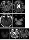
Initial computed tomography imaging revealed a focal low-density lesion in the upper cerebellum. T2-weighted brain (T2WI) magnetic resonance imaging (MRI) revealed nodular hyperintense lesions in the cerebellum and right frontal subcortical white matter (Fig. 1A, B). The cerebellar lesion exhibited a low-intensity signal blooming on susceptibility-weighted imaging due to hemorrhage (Fig. 1C). Diffusion-weighted imaging (DWI) and apparent diffusion coefficient (ADC) map showed no evidence of restricted diffusion (Fig. 1D, E). Although these MRI findings were nonspecific, they were concerning for bacterial or viral encephalitis or NPSLE.
Laboratory examination revealed an elevated WBC count (12400/mm3), as well as increased CRP (34.17 mg/dL) and procalcitonin levels (30.63 ng/dL) and a decreased platelet count (48000/mm3). Cerebrospinal fluid (CSF) examination was significant for an elevated opening pressure (570 mmH2O) and an increased WBC count (190/mm3) with a polymorphonuclear pr-edominance (78%), as well as elevated protein (223.9 mg/dL) and decreased glucose levels (4 mg/dL) compared with the serum glucose level (77 mg/dL). Although the CSF Gram stain was negative, bacterial meningoencephalitis was suspected, and empirical antibiotic therapy with vancomycin (1 g/day), ertapenem (2 g/day) and ampicillin (6 g/day) was initiated. Levetiracetam (1 g/day) was also initiated for seizure control. On hospital day 2, the patient became alert and exhibited no neurologic deficits. Follow-up CSF studies on hospital day 3 revealed a normal opening pressure (180 mmH2O) and an increased WBC count (430/mm3) with a polymorphonuclear predominance (70%), as well as an elevated protein level (197.1 mg/dL) and a decreased glucose level (128 mg/dL) compared to the serum glucose level (325 mg/dL). On hospital day 4, growth of Listeria monocytogenes in CSF culture was reported. Blood cultures were negative. The patient's antibiotics were subsequently changed to high-dose ampicillin (6 g/day) and ceftriaxone (4 g/day) for CNS listeriosis.
A follow-up MRI performed on hospital day 14 showed significant reductions in the severity of the lesions noted previously on T2WI (Fig. 2A, B). The hemorrhagic central core of the cerebellar lesion was hyperintense on T2-, T1-, and susceptibility-weighted images (Fig. 2A, C, D) and exhibited restricted diffusion on DWI and ADC map (Fig. 2E, F), as well as peripheral ring enhancement on contrast-enhanced T1WI (Fig. 2G). The combination of these focal cystic changes and restricted diffusion in the hemorrhagic portion of the lesion was suggestive of late subacute hemorrhage. A follow-up CSF study on hospital day 21 was negative. The patient's symptoms improved, and she was discharged without any neurological deficits.
In this case, a young female patient with SLE who was receiving immunosuppressive therapy presented with fever, headache, seizures and mental status changes. CSF studies revealed a greatly increased opening pressure, pleocytosis with polymorphonuclear predominance, an elevated protein level and a decreased glucose level. MRI showed T2 hyperintense lesions with hemorrhagic changes in the cerebellum and right frontal subcortical white matter. The patient was diagnosed with listerial encephalitis on the basis of her CSF culture results and MR findings. The aforementioned T2 hyperintense lesions exhibited improvement on a follow-up MR scan after ampicillin and ceftriaxone treatment, and the patient was discharged without any neurological deficits.
Listeria monocytogenes is a gram-positive, anaerobic intracellular bacterium (3); thus, host resistance is mainly mediated by cell-mediated immunity (13). As patients with SLE usually exhibit abnormal humoral- and cell-mediated immunity, they are susceptible to Listeria monocytogenes infection (23).
Horta-Baas et al. (1) studied 26 cases of CNS listerial infection in patients with SLE, including 22 cases of meningitis (85%), 5 cases of brain abscesses (19%) and 2 cases of meningoencephalitis (8%). One of the patients with meningoencephalitis presented with communicating hydrocephalus, meningeal enhancement and ventriculitis, and the other patient exhibited increased signal intensity in the temporal lobe white matter (56). There have been several case reports of listerial meningoencephalitis in non-SLE patients. In these cases, the brain stem and cerebellum are generally involved (78). There have also been some reports briefly mentioning petechial hemorrhagic changes in the setting of listerial encephalitis (7). These hemorrhages may be associated with histopathological findings, including perivascular leukocyte cuffing, small vessel fibrinoid necrosis, and vasculitis (8). Additionally, bleeding induced by high-dose steroid therapy and decreased platelet counts may contribute to the development of hemorrhagic changes in patients with SLE. There have been reports of listerial rhombencephalitis characterized by restricted diffusion in the brainstem (9), which may be due to abscess formation or hemorrhage (9).
The MR findings of listerial infection can be difficult to differentiate from those of NPSLE (3), which is a diagnosis of exclusion achieved on a case-by-case basis using clinical, laboratory, and imaging data. NPSLE is commonly characterized by cerebral atrophy and multiple small subcortical and deep white matter hyperintense lesions on T2-weighted imaging/FLAIR, as well as arterial infarctions, intracranial hemorrhages, dural venous sinus thromboses, and posterior reversible encephalopathy syndrome (4). CSF analysis can differentiate listerial encephalitis from NPSLE. Our patient exhibited clinical symptoms and CSF findings suggestive of CNS infection, as well as MRI findings of cerebellar lesions, which are unusual in NPSLE, and T2 hyperintense lesions that improved significantly with antibiotic therapy within a short period of time. Therefore, CNS infection was suspected to a greater degree than NPSLE.
There are many differential diagnoses in addition to NPSLE, including abscesses, viral encephalitis, vasculitis, tuberculosis, multiple sclerosis, and lymphoma (710). Abscesses typically exhibit restricted diffusion and ring enhancement. However, the diagnosis of an abscess was excluded in our patient because no restricted diffusion was observed initially. Viral encephalitis can be diagnosed on the basis of CSF pleocytosis with lymphocytic predominance. Varicella-zoster virus, in particular, is known to commonly cause cerebellitis and may cause hemorrhage. Behçet's disease typically manifests as T2 hyperintense lesions in the midbrain, pons, cerebral peduncles, thalamus, and basal ganglia (4). However, the possibility of Behçet's disease was less likely in our patient because of the atypical location of her lesions and the absence of oral and genital aphthous ulcers. However, Behçet's disease cannot be excluded based on radiologic findings alone; thus, it should be included in the differential diagnosis. Tuberculous meningitis primarily causes leptomeningeal abnormalities, especially in the basal cistern. Our patient did not exhibit any leptomeningeal abnormalities. Multiple sclerosis can involve the infratentorial area; however, periventricular supratentorial lesions are usually located at or near the callososeptal interface. The diagnosis of multiple sclerosis was excluded in our patient because her clinical course was not consistent with the disease. Neoplasms, such as lymphoma or glial tumors, were excluded because the aforementioned lesions rapidly improved with antibiotic therapy.
Listerial infection treatment comprises ampicillin and aminoglycosides. Hydrocephalus can be a complication of listerial encephalitis and is associated with a poor prognosis (10). Listerial encephalitis is often fatal (the mortality rate ranges from 24–52%) if it is not treated during its early stages, and survivors commonly experience critical neurological sequelae (4). Furthermore, because patients with SLE are prone to developing immunosuppressive conditions, early diagnosis of this disease is essential for deciding whether to administer antibiotics or immunosuppressive agents. CSF studies and brain MRI can be helpful in differentiating listerial encephalitis from NPSLE (7).
In conclusions, listeria monocytogenes-induced CNS infection is uncommon but often fatal in patients with SLE. It can be difficult to differentiate listerial encephalitis from NPSLE, but imaging studies and CSF examinations can be helpful in differentiating these two conditions. Hemorrhage can occur in listerial encephalitis.
Figures and Tables
Fig. 1
Listerial encephalitis in a 31-year-old woman with systemic lupus erythematosus.
T2-weighted axial images (A, B) show mass-like hyperintense lesions in the cerebellum and right frontal subcortical white matter. Low signal intensity of the central portion on T2-weighted axial image is correlated with hemorrhage on susceptibility weighted image (C). Diffusion weighted image (D) and apparent diffusion coefficient (E) map show no definite restricted diffusion.

Fig. 2
Follow up MR imaging on hospital day 14 after treatment with ampicillin and ceftriaxone.
T2-weighted axial images (A, B) show a significant decrease in the extent of mass-like hyperintense lesions in the cerebellum and right frontal subcortical white matter. T1-weighted (C) and susceptibility-weighted (D) axial images show hypersignal intensity of the hemorrhagic central core, consistent with late subacute hemorrhage.
Diffusion weighted image (E) and apparent diffusion coefficient map (F) show restricted diffusion of the hemorrhagic core in the cerebellar lesion. A contrast-enhanced T1-weighted image (G) shows peripheral ring enhancement in the cerebellum.

References
1. Horta-Baas G, Guerrero-Soto O, Barile-Fabris L. Central nervous system infection by Listeria monocytogenes in patients with systemic lupus erythematosus: analysis of 26 cases, including the report of a new case. Reumatol Clin. 2013; 9:340–347.
2. Yang CD, Wang XD, Ye S, Gu YY, Bao CD, Wang Y, et al. Clinical features, prognostic and risk factors of central nervous system infections in patients with systemic lupus erythematosus. Clin Rheumatol. 2007; 26:895–901.
3. Lee MC, Wu YK, Chen CH, Wu TW, Lee CH. Listeria monocytogenes meningitis in a young woman with systemic lupus erythematosus. Rheumatol Int. 2011; 31:555–557.
4. Osborn AG. Osborns brain: imaging, pathology, and anatomy. Salt Lake City, UT: Amirsys;2013.
5. McCaffrey LM, Petelin A, Cunha BA. Systemic lupus erythematosus (SLE) cerebritis versus Listeria monocytogenes meningoencephalitis in a patient with systemic lupus erythematosus on chronic corticosteroid therapy: the diagnostic importance of cerebrospinal fluid (CSF) of lactic acid levels. Heart Lung. 2012; 41:394–397.
6. López Montes A, Andrés Mompeán E, Martínez Villaescusa M, Hernández Belmonte A, Mateos Rodríguez F, Abad Ortiz L, et al. [Meningoencephalitis by Listeria in the lupus disease]. An Med Interna. 2005; 22:379–382.
7. Alper G, Knepper L, Kanal E. MR findings in listerial rhombencephalitis. AJNR Am J Neuroradiol. 1996; 17:593–596.
8. Just M, Krämer G, Higer HP, Thömke F, Pfannenstiel P. MRI of Listeria rhombencephalitis. Neuroradiology. 1987; 29:401–402.
9. Hatipoglu HG, Gurbuz MO, Sakman B, Yuksel E. Diffusion-weighted magnetic resonance imaging in rhombencephalitis due to Listeria monocytogenes. Acta Radiol. 2007; 48:464–467.
10. Pelegrín I, Moragas M, Suárez C, Ribera A, Verdaguer R, Martínez-Yelamos S, et al. Listeria monocytogenes meningoencephalitis in adults: analysis of factors related to unfavourable outcome. Infection. 2014; 42:817–827.


 PDF
PDF ePub
ePub Citation
 Print
Print


 XML Download