

 PDF
PDF ePub
ePub Citation
 Print
Print


INTRODUCTION
Synovitis, acne, pustulosis, hyperostosis, and osteitis (SAPHO) syndrome is an inflammatory clinical condition featuring aseptic bone lesions and characteristic skin manifestations (1). The association between skeletal lesions and skin lesions was first reported in 1961 (2). Subsequently, various terms were used to describe the disease entity until introduction of the SAPHO acronym (1). The age at presentation ranges from childhood to adulthood and the clinical and imaging features include a wide spectrum (3). Prevalence is estimated at no greater than 1 in 10000, which is probably an underestimate (4). The low incidence rate as well as the wide range of etiology and manifestations of this syndrome can complicate the diagnosis.
Herein, we reported a case of SAPHO syndrome showing typical radiologic features. This case report was approved by our Institutional Review Board and the requirement for the patient's informed consent was waived.
CASE REPORT
A 63-year-old woman visited our institute with chronic right buttock pain. She had a long history of vague pain around the lower back for about 30 years and had already visited a few other hospitals. She was taking oral analgesics and tender point injections, but the symptom was intractable.
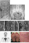
Blood testing revealed elevated erythrocyte sedimentation rate (73 mm/hour) and C-reactive protein (16.86 mg/L), with other results in the normal range. She underwent a general work-up including conventional radiographs of spine and pelvis. The radiographs revealed multifocal sclerotic lesions in the vertebral bodies and right ilium near the sacroiliac joint (Fig. 1A, B).
For further evaluation, magnetic resonance imaging (MRI) of the thoracic and lumbar spine was conducted. In the lumbar spine, L1, L2 and L3 vertebral bodies showed high-signal intensities in a fat-saturated T2 weighted image and low-signal intensities in a T1 weighted image, which suggested bone marrow edema (Fig. 1C). The lumbar vertebral body lesions were confined to the anterior corner of the vertebral bodies. The corner lesions showed MRI findings that were similar to those of Romanus lesion found with spondyloarthorpathies including ankylosing spondylitis. But, unlike spondyloarthropathies, these corner lesions progressed to adjacent vertebral end plates and involved contiguous vertebrae. Prevertebral soft tissue thickening was present, which also lessened the possibility of spondyloarthropathies.
In the thoracic spines, cortical erosions and subchondral sclerosis were seen in T5, T6 and T7 vertebrae combined with prevertebral soft tissue thickening with enhancements (Fig. 1D), which resembled infection. But, unlike infection, disc involvement, abscess formation, epidural involvement, or large paravertebral soft-tissue masses were absent.
The broad spectrum of imaging features involving various sites strongly suggested SAPHO syndrome. In addition, multiple hot uptake was evident on bone scintigraphy, corresponding with CT and MRI findings. Moreover, marked uptake was apparent in the manubrium, bilateral sternoclavicular joints, and adjacent claviculae known as the bullhead sign (Fig. 1E). Although the bullhead sign strongly indicated SAPHO, bone biopsy from the lumbar and thoracic vertebrae was done to confirm the diagnosis. Culture of the specimen was negative for microorganisms and pathology revealed only fibrous degeneration.
Skin inspection conducted immediately after suspecting SAPHO syndrome from radiologic findings revealed pustulosis of both feet, which was present for 9 years (Fig. 1F). The patient was regularly visiting the dermatologist and using topical steroids; however, the pustulosis was never correlated with the chronic buttock pain prior to evaluation of the radiologic findings. The patient was started on steroid therapy and her symptoms are now under control.
DISCUSSION
Clinical features of SAPHO are still unclear due to the rarity of reported cases. Prevalence of SAPHO has been estimated at no greater than 1 in 10000, which is possibly an underestimate due to low awareness of the disease (4). It can occur in patients with a wide age range from childhood to adulthood (5), and is characterized by repeated episodes of remission and recurrence (6). Symptoms can vary, but patients mostly complain of musculoskeletal symptoms including pain, tenderness, and swelling. Multiple sites of involvement in different osteoarticular sites are common. The anterior chest wall and spine are the most commonly affected sites in adults (3). Mild fever can be present but other systemic manifestations are rare. Laboratory results are usually in the normal range, except for possible elevation in the Erythrocyte sedimentation rate and C-reactive protein level (6).
Skin manifestations might not be present, which can make diagnosis even more challenging. If present, the skin manifestations appear as severe acne and/or pustulosis of palms and soles (3). The skin lesions may occur before, simultaneously, or after the onset of skeletal symptoms. Clinicians should be aware that the skin and osteoarticular manifestations of SAPHO syndrome do not necessarily co-exist (7).
Osteoarticular manifestations include synovitis, hyperostosis, osteitis, arthropathy, and enthesopathy (3). Hyperostosis and osteitis is characterized by increased sclerosis of the bone involving the cortex and medulla, respectively (3). Hyperostosis appears radiologically as cortical thickening and narrowing of the medullary cavity, whereas osteitis appears as increased sclerosis of the medullary cavity (8). Arthritis is associated with joint space narrowing, periarticular osteopenia, and bone erosions (8). It usually affects axial joints, such as sternoclavicular joints, sacroiliac joint, costochondral joints, and symphyses (9).
The anterior chest wall is the most common site of involvement in adults (9). In the early stage, it can be evident as soft tissue swelling around the costoclavicular ligaments. However, in the late stage, hyperostosis, osteosclerosis, and hypertrophy of the medial ends of clavicles is evident (8). These lesions characteristically show hot uptake on bone scintigraphy and may resemble the contour of bull's head, but this finding is not entirely definitive (10).
The spine is the second most commonly involved site in patients of all ages (6). The six main radiological manifestations include vertebral body corner lesions, spondylodiscitis, osteodestructive lesions, osteosclerotic vertebral lesions, paravertebral ossification, and sacroiliitis (3). In the present case, vertebral body corner lesions, spondylodiscitis, osteodestructive lesions, osteosclerotic vertebral lesions, and sacroiliitis were present.
SAPHO can be difficult to diagnose because it can involve any skeletal site and has variable appearances on imaging (4). Diagnostic criteria for SAPHO have been proposed by several authors, but none have been validated. In 1988, Benhamou et al. (1) proposed that, after excluding an infective cause, palmo-plantar keratodermia, diffuse idiopathic skeletal hyperostosis, and manifestations of retinoid therapy, the presence of only one of the four inclusion criteria is sufficient for the diagnosis of SAPHO. Inclusion criteria include osteoarticular manifestations of acne conglobata, acne fulminans, or hirsadenitis suppurativa, (2) osteoarticular manifestations of palmoplantar pustulosis, (3) hyperostosis involving either the anterior chest wall, spine, or limbs with or without dermatosis, and (4) recurrent multifocal chronic osteomyelitis with or without dermatosis (1). Kahn updated these criteria at the 67th annual scientific meeting of the American College of Rheumatology in 2003. Suggested inclusion criteria are (1) bone and/or joint involvement associated with palmoplantar pustulosis and pustular psoriasis, (2) bone and/or joint involvement associated with severe acne, (3) isolated sterile hy-perostosis/osteitis in adults, (4) chronic recurrent multifocal osteomyelitis in children and, (5) bone and/or joint involvement associated with chronic bowel diseases. Exclusion criteria are (1) infectious osteitis, (2) tumoral conditions of bone and, (3) non-inflammatory condensing lesions of bone. Meeting one criteria is sufficient for the diagnosis of SAPHO. In our case, both criteria were met because plantar pustulosis and oateoarticular manifestatations were present at the same time. However the diagnosis can be difficult in patients with a solitary symptomatic skeletal lesion without skin lesions, which is a common presentation of SAPHO. The time interval between the onset of skin and osteoarticular manifestations can be of up to 40 years (9). In such cases, the radiologist needs to take an active role in considering the diagnosis, since awareness of the imaging features facilitates early diagnosis and initiating appropriate treatment.
 XML Download