

 PDF
PDF ePub
ePub Citation
 Print
Print


Abstract
Purpose
This study was performed to compare paranasal sinus tomosynthesis with computed tomography (CT) imaging as a radiologic tool to evaluate the paranasal sinuses, using measurement of the soft tissue thickness of the maxillary sinus.
Materials and Methods
A total of 114 patients with sinusitis who underwent both paranasal sinus digital tomosynthesis (DT) and CT were enrolled in this retrospective study. Two observers independently assessed soft tissue thickness in both maxillary sinus chambers using both DT and CT images.
Results
The mean difference in soft tissue thickness measured by each observer was -0.31 mm on CT and 0.15 mm on DT. The mean differences in soft tissue thickness measured with DT and CT were -0.15 by observer 1 and -0.31 by observer 2. Evaluation of the agreement in measurement of soft tissue thickness in the maxillary sinus using DT and CT showed a high intraclass correlation, with the 95% limit of agreement ranging from -3.36 mm to 3.06 mm [intraclass correlation coefficient (ICC), 0.994: p < 0.01] for observer 1 and from -5.56 mm to 4.95 mm (ICC, 0.984: p < 0.01) for observer 2.
Go to : 
Index terms
Radiography, Maxillary Sinus, Sinusitis, Tomography, Radiation DosageREFERENCES
1. Chadha NK, Chadha R. 10-minute consultation: sinusitis. BMJ. 2007; 334:1165.

2. Gliklich RE, Metson R. The health impact of chronic sinusitis in patients seeking otolaryngologic care. Otolaryngol Head Neck Surg. 1995; 113:104–109.
3. McQuillan L, Crane LA, Kempe A. Diagnosis and management of acute sinusitis by pediatricians. Pediatrics. 2009; 123:e193–e198.
4. Momeni AK, Roberts CC, Chew FS. Imaging of chronic and exotic sinonasal disease: review. AJR Am J Roentgenol. 2007; 189(6 Suppl):S35–S45.
5. Hoang JK, Eastwood JD, Tebbit CL, Glastonbury CM. Multi-planar sinus CT: a systematic approach to imaging before functional endoscopic sinus surgery. AJR Am J Roentgenol. 2010; 194:W527–W536.
6. Aal⊘kken TM, Hagtvedt T, Dalen I, Kolbenstvedt A. Conven-tional sinus radiography compared with CT in the diagnosis of acute sinusitis. Dentomaxillofac Radiol. 2003; 32:60–62.
7. Dobbins JT 3rd, Godfrey DJ. Digital X-ray tomosynthesis: current state of the art and clinical potential. Phys Med Biol. 2003; 48:R65–R106.
8. Johnsson AA, Vikgren J, Svalkvist A, Zachrisson S, Flinck A, Boijsen M, et al. Overview of two years of clinical experience of chest tomosynthesis at Sahlgrenska University Hospital. Radiat Prot Dosimetry. 2010; 139:124–129.
9. Aoki T, Fujii M, Yamashita Y, Takahashi H, Oki H, Hayashida Y, et al. Tomosynthesis of the wrist and hand in patients with rheumatoid arthritis: comparison with radiography and MRI. AJR Am J Roentgenol. 2014; 202:386–390.
10. Dalbeth N, Gao A, Roger M, Doyle AJ, McQueen FM. Digital tomosynthesis for bone erosion scoring in gout: comparison with plain radiography and computed tomography. Rheumatology (Oxford). 2014; 53:1712–1713.
11. Roth RG, Maidment AD, Weinstein SP, Roth SO, Conant EF. Digital breast tomosynthesis: lessons learned from early clinical implementation. Radiographics. 2014; 34:E89–E102.
12. Shim SS, Oh YW, Kong KA, Ryu YJ, Kim Y, Jang DH. Pulmo-nary nodule size evaluation with chest tomosynthesis and CT: a phantom study. Br J Radiol. 2015; 88:20140040.
13. Machida H, Yuhara T, Tamura M, Numano T, Abe S, Sabol JM, et al. Radiation dose of digital tomosynthesis for sinonasal examination: comparison with multidetector CT. Eur J Radiol. 2012; 81:1140–1145.
14. Yoo JY, Chung MJ, Choi B, Jung HN, Koo JH, Bae YA, et al. Digital tomosynthesis for PNS evaluation: comparisons of patient exposure and image quality with plain radiography. Korean J Radiol. 2012; 13:136–143.
15. Machida H, Yuhara T, Ueno E, Yoda K, Sunose H, Kita K, et al. Detection of paranasal sinus opacification with digital tomosynthesis radiography: a clinical pilot study. J Comput Assist Tomogr. 2013; 37:252–256.
16. Huda W, Ogden KM, Khorasani MR. Converting dose-length product to effective dose at CT. Radiology. 2008; 248:995–1003.
17. Gomi T. X-ray digital tomosynthesis imaging-comparison of reconstruction algorithms in terms of a reduction in the exposure dose for arthroplasty. In. Bagaria V, editor. Arthroplasty - a comprehensive review. Croatia: InTech;2016.
18. Czechowski J, Janeczek J, Kelly G, Johansen J. Radiation dose to the lens in sequential and spiral CT of the facial bones and sinuses. Eur Radiol. 2001; 11:711–713.
19. Maillet M, Bowles WR, McClanahan SL, John MT, Ahmad M. Cone-beam computed tomography evaluation of maxillary sinusitis. J Endod. 2011; 37:753–757.
20. Rak KM, Newell JD 2nd, Yakes WF, Damiano MA, Luethke JM. Paranasal sinuses on MR images of the brain: signifi-cance of mucosal thickening. AJR Am J Roentgenol. 1991; 156:381–384.
21. Low DE, Desrosiers M, McSherry J, Garber G, Williams JW Jr, Remy H, et al. A practical guide for the diagnosis and treatment of acute sinusitis. CMAJ. 1997; 156(Suppl 6):S1–S14.
22. Ha AS, Lee AY, Hippe DS, Chou SH, Chew FS. Digital tomosynthesis to evaluate fracture healing: prospective comparison with radiography and CT. AJR Am J Roentgenol. 2015; 205:136–141.
Go to :
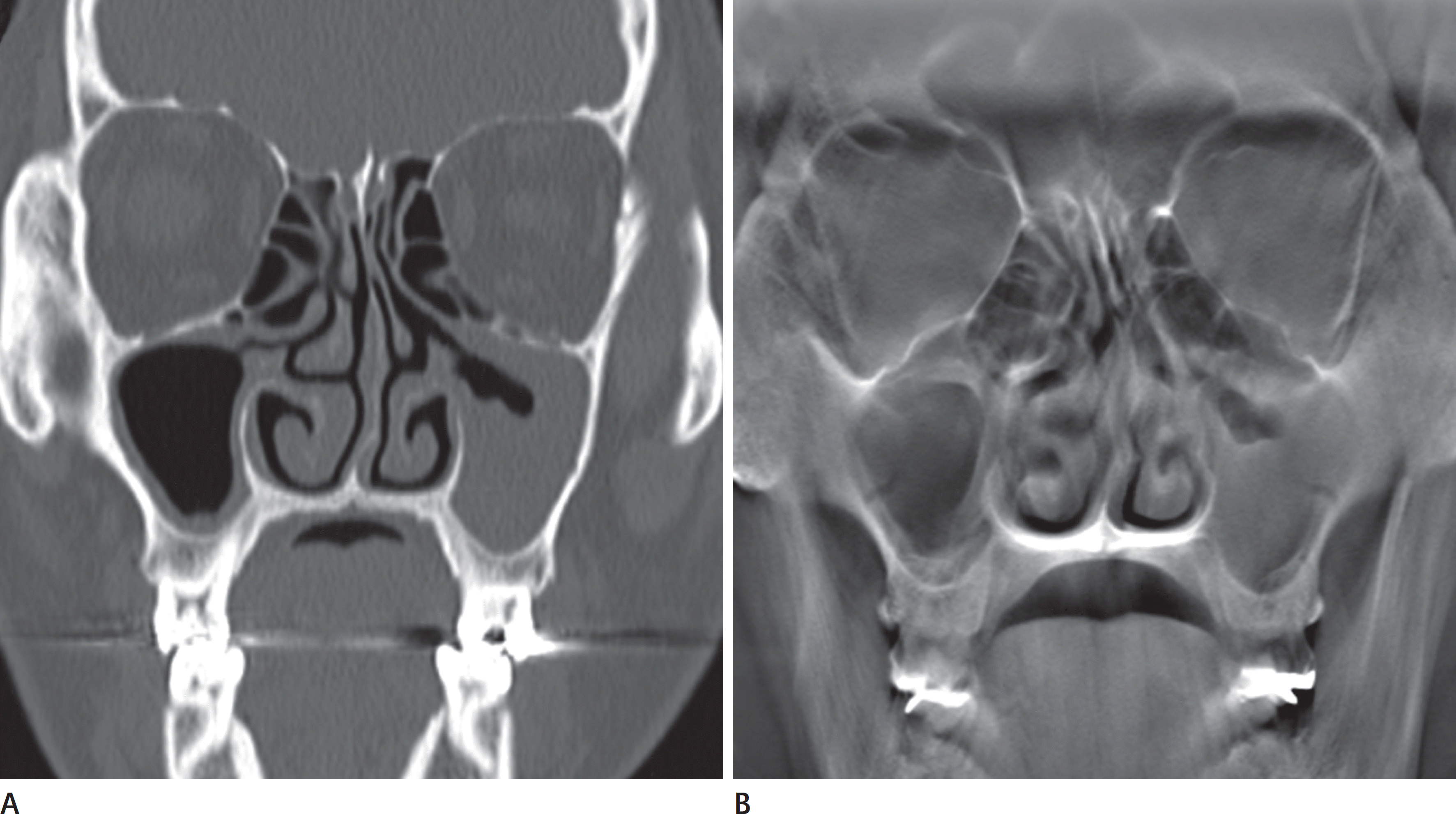 | Fig. 1.DT (A) and MDCT (B) MPR coronal images of the paranasal sinuses acquired on the same day in a 52-year-old man with clinically suspected sinusitis. (A) DT shows abnormal mucosal thickening of both maxillary sinuses which is comparable to that shown by MDCT (B). DT = digital tomosynthesis, MDCT = multidetector computed tomography, MPR = multiplanar reconstruction |
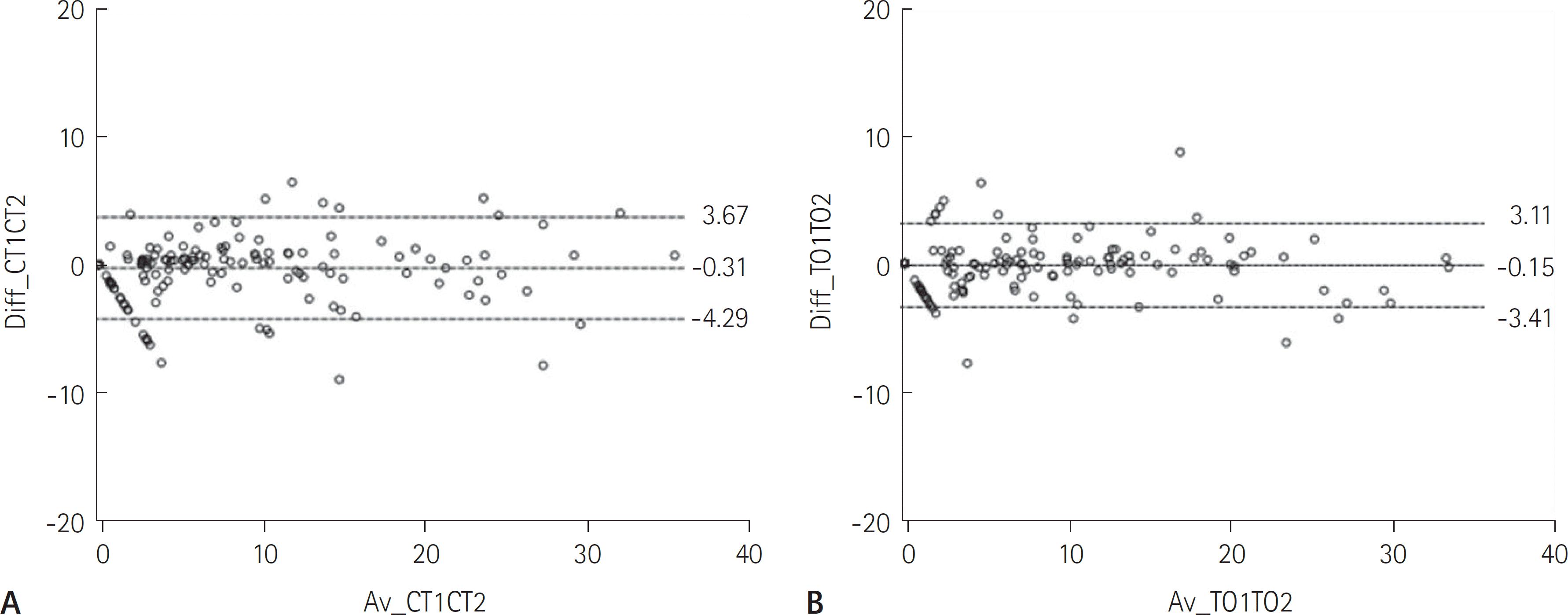 | Fig. 2.Bland-Altman plots showing the difference in mean soft tissue thickness measurements between the observers’ measurements as a function of the mean CT (A) and DT values (B). The dashed middle line represents the bias (mean difference between measurements). The dashed lower and upper lines represent the limits of agreement (mean ± 1.96 SD). CT = computed tomography, DT = digital tomosynthesis, SD = standard deviation |
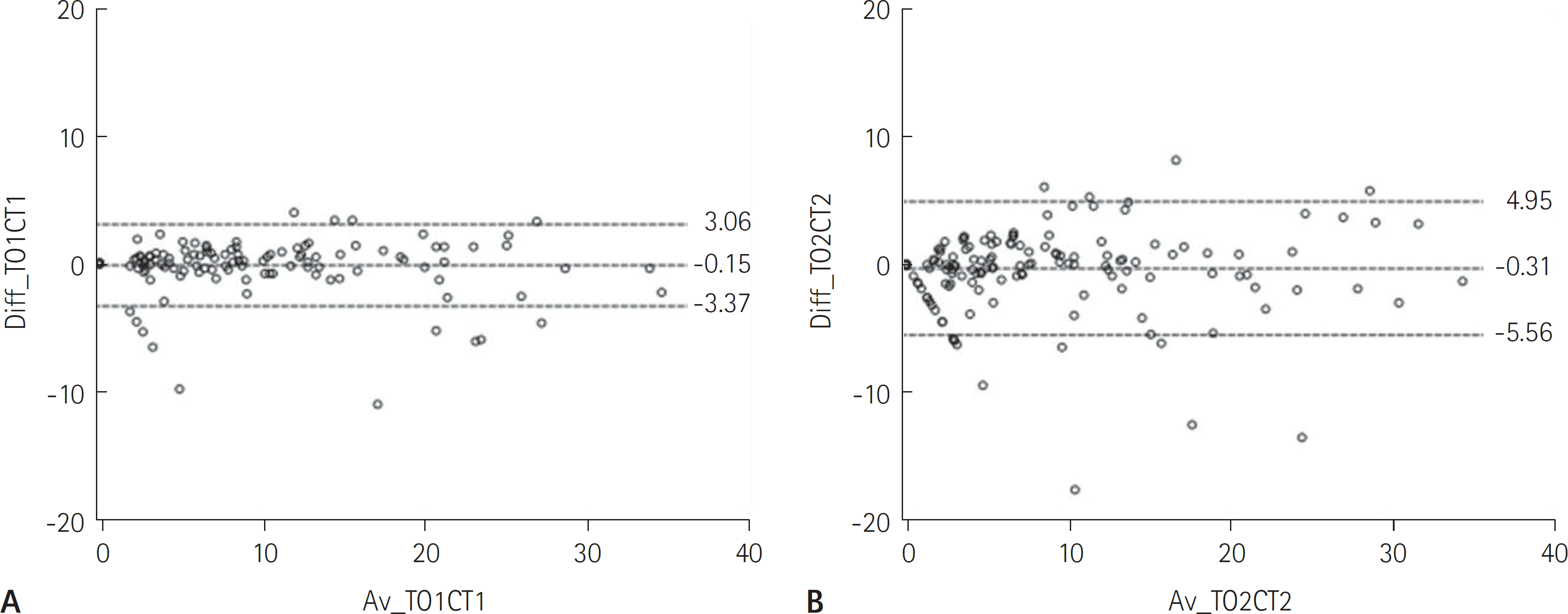 | Fig. 3.Bland-Altman plots showing the difference in measurements of soft tissue thickness between DT and CT as a function of the mean measurement according to the two modalities obtained by observer 1 (A) and observer 2 (B). The dashed middle line represents the bias (mean difference between measurements). The dashed lower and upper lines represent the limits of agreement (mean ± 1.96 SD). CT = computed tomography, DT = digital tomosynthesis, SD = standard deviation |
Table 1.
Measurement of Maxillary Sinus Soft Tissue Thickness with Both DT and CT (mm)
| DT | CT | |||
|---|---|---|---|---|
| Observer 1 | Observer 2 | Observer 1 | Observer 2 | |
| Mean measurement | 12.67 | 12.82 | 12.82 | 13.13 |
| Standard deviation | 15.00 | 14.96 | 15.02 | 14.83 |
Table 2.
Mean Maxillary Sinus Soft Tissue Thickness Measurement Error between the Two Observers according to Modality and Interobserver Agreement (mm)
| Difference in Mean Measurement | Standard Deviation | Lower LOA | Upper LOA | ICC (95% CI) | |
|---|---|---|---|---|---|
| DT | −0.15 | 1.66 | −3.41 | 3.11 | 0.994 (0.992–0.995) |
| CT | −0.31 | 2.03 | −4.29 | 3.67 | 0.991 (0.988–0.993) |
Table 3.
Mean Error for Maxillary Sinus Soft Tissue Thickness Measurements Using DT Compared to CT for All Observers with Intraobserver Agreement (mm)
| Difference in Mean Measurement | Standard Deviation | Lower LOA | Upper LOA | ICC (95% CI) | |
|---|---|---|---|---|---|
| Observer 1 | −0.15 | 1.64 | −3.37 | 3.06 | 0.994 (0.992–0.995) |
| Observer 2 | −0.31 | 2.68 | −5.56 | 4.95 | 0.984 (0.979–0.987) |
 XML Download