

 PDF
PDF ePub
ePub Citation
 Print
Print


INTRODUCTION
Uterine tumor resembling ovarian sex-cord tumor (UTROSCT) is a very rare uterine neoplasm. It presents as a polypoid or nodular uterine mass and usually shows benign behavior. In 1976, Clement and Scully (1) proposed the concept of sex-cord differentiation of uterine tumors. Although approximately 70 case reports have been found, most of these reports mainly focused on the pathologic and clinical presentation of this tumor. All of the reported cases only confirmed the diagnosis after a pathologic review, and UTROSCT was not initially included in the differential diagnosis.
Franco et al. (2) described ultrasonographic findings, and Suzuki et al. (3), and Okada et al. (4) presented MRI findings of a UTROSCT. However, only a few cases have described its imaging features and have provided limited radiologic findings. Consequently, no characteristic features of this tumor have been established. It showed almost similar findings to those of leiomyoma of the uterus in previous reports. Here, we describe our experience of a UTROSCT.
CASE REPORT
A 50-year-old woman presenting with vaginal bleeding visited the gynecology outpatient department. The patient was post-menopausal without a remarkable past medial history, and gynecologic examination had not been performed recently. Vaginal bleeding continued for 2–3 weeks.
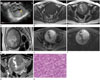
Trans-vaginal ultrasonography was performed initially and an approximately 8 × 7 cm, smooth-margined, round, heterogeneous echoic solid mass with internal multi-loculated anechoic cystic portions was found in the anterior wall of the uterus (Fig. 1A). Neither calcifications nor remarkable vascularity on color Doppler images was identified in the mass. Both ovaries were normal. A large uterine leiomyoma with cystic degeneration was suspected upon initial trans-vaginal ultrasonography. On pelvic magnetic resonance imaging, an approximately 8.7 cm, well-defined, round solid mass with central multi-loculated cystic portions was observed in the right anterolateral wall of the uterus (Fig. 1B-G). Its solid portion showed iso-signal intensity compared to the myometrium on T1-weighted images, and heterogeneous high signal intensity was observed on T2-weighted images. It showed heterogeneously dense enhancement except in the central cystic portion on gadolinium-enhanced T1-weighted images. In addition, multi-loculated central cystic portions showed low signal intensity on T1-weighted images and bright signal intensity on T2-weighted images. These foci were considered cystic or necrotic changes, and they were correlated with anechoic portions on ultrasonography. These MRI findings suggested uterine leiomyoma with cystic degeneration, and they corresponded with the ultrasonographic findings. However, its solid potion showed high signal intensity on high b-value diffusion weighted images (b = 1000), and low signal intensity on ADC maps. Although a completely hyaline degenerated leiomyoma can exhibit diffusion restriction, the lesion rarely enhances on post-contrast study. Therefore, good enhancement with diffusion restriction implied the possibility of highly cellularity. Thus, we suggested the possibility of a cellular leiomyoma. But, a malignant tumor, such as leiomyosarcoma, could not be completely excluded from the differential diagnosis, although the mass was smoothly marginated and there was lack of intramural necrosis or hemorrhage.
The patient underwent total abdominal hysterectomy with bilateral salpingo-oophorectomy. The cut surfaces of the mass had a slightly yellowish color, but it grossly seemed to be uterine leiomyoma with degenerative changes. Upon histologic examination, the tumor showed microfollicular, macrofollicular, and glandular architectural patterns, and neoplastic cells showed rare nuclear grooves, and they were small with round and angulated nuclei, hyperchromasia, clumped chromatin, indistinct nucleoli, and little cytoplasm (Fig. 1H). These findings were completely different from pathologic findings of uterine leiomyoma, and the final pathologic diagnosis of the mass was a UTROSCT. The patient recovered and was discharged one week after surgery without any remarkable complications.
DISCUSSION
UTROSCT was first reported by Morehead and Bowman in 1945 (5). Clement and Scully established the concept of sex-cord differentiation of uterine tumors and categorized them into two subtypes in 1976. The first subtype was termed as endometrial stromal tumor with sex cord-like elements (ESTSCLE) and it showed similar features to those of traditional endometrial stromal tumors with focal sex-cord differentiation. The second subtype comprised tumors entirely composed of elements resembling sex-cord tumors of the ovary and it was named UTROSCT (1). The gross pathology of these two subtypes of tumors is similar, but they present very different clinical and molecular genetic features. While UTROSCT shows benign behavior, ESTSCLE manifests malignant behavior with a much higher recurrence rate and metastasis (6).
Although UTROSCT is known to show benign behavior, some cases have reported metastasis or recurrence. Therefore, the optimal treatment for a patient with UTROSCT in the peri-menopausal period is abdominal hysterectomy and bilateral salpingo-oophorectomy. In case of younger patients in the premenopausal state, fertility conserving treatment can be attempted with a close follow-up. Three cases of UTROSCT patients who received fertility conserving treatment have been reported (7). However, due to the limited number of cases and lack of information on the long-term follow-up results, fertility conserving treatments remain con-troversial.
Since the first case report in 1976, case reports of UTROSCT have been presented very rarely and specific radiologic findings are extremely limited. On reviewing the previously reported cases, UTROSCT was confirmed in all the cases either after biopsy or a post-operative pathologic review. All clinicians did not include UTROSCT in their primary differential diagnosis because there are no imaging features or potential clues for the radiological diagnosis or suspicion of UTROSCT prior to surgical resection or biopsy. Previously, Franco et al. (2) reported that UTROSCT presented as an endometrial polypoid mass-like lesion. Suzuki et al. (3) initially reported the MRI findings of UTROSCT. In this case of UTROSCT located in the cervix, the mass was observed as a round mass with low signal intensity on T1-weighted images, and intermediate signal intensity with small areas of high signal intensity on T2-weighted images. In another case of a UTROSCT located in the myometrium reported by Okada et al. (4), MRI demonstrated a mass with slightly high signal intensity on T2-weighted images and intermediate signal intensity compared with the myometrium on T1-weighted images. In another case reported by Calisir et al. (8), UTROSCT was identified as an endometrial mass with homogeneous high signal intensity on T2-weighted images and intermediate signal intensity compared to the myometrium on T1-weighted images.
In our case, UTROSCT was observed as a well-defined round mass with internal cystic portions in the myometrium. The solid portion of the mass showed high signal intensity on T2-weighted images and intermediate signal intensity on T1-weighted images. In previous case reports presented by Okada et al. (4) and Calisir et al. (8), UTROSCT cases showed similar MR findings; iso-signal intensity on T1-weighted images and high signal intensity on T2-weighted images. However, these findings overlapped with MR findings of ordinary leiomyomas. Some different imaging findings were observed in the previously reported cases. Okada et al. (4) described intratumoral central hypointense foci on T2-weighted images, which were confirmed to be proliferated smooth muscle cells with rich collagen. Also, the mass showed less enhancement compared with the adjacent myometrium. In our case, unlike the other previously reported cases, intratumoral cystic foci were observed, which mimicked a cystic degenerated leiomyoma.
To date, there is no description of diffusion restriction in UTROSCT. We obtained diffusion-weighted images and ADC maps, and this tumor revealed diffusion restriction in contrast to the usual uterine leiomyoma. In the previous pathologic reports, UTROSCT showed high cellularity, and this seems to be reflected in diffusion-weighted images and ADC maps.
Typically, a non-degenerated uterine leiomyoma shows low signal intensity on T2-weighted images, whereas cellular leiomyoma, which comprises compact smooth muscle cells without collagen, shows high signal intensity on T2-weighted images (9). In addition, cellular leiomyoma and leiomyosarcoma show diffusion restriction, which reflects their dense cellularity (10). Therefore, these imaging findings of cellular leiomyoma or leiomyosarcoma overlap with those of UTROSCT. Thus, restricted diffusion and high signal intensity on T2-weighted images of a uterine mass are not specific imaging findings for UTROSCT.
Further study which correlates the pathologic features with radiologic findings is needed, and radiologists need to gain a thorough understanding of the clinical features of UTROSCT including metastasis or recurrence in order to evaluate it properly.
 XML Download