

 PDF
PDF ePub
ePub Citation
 Print
Print


Abstract
Purpose
To compare the characteristics and outcomes of multimodal endovascular therapy (EVT) in patients with acute basilar artery occlusion (BAO) with and without underlying intracranial atherosclerotic stenosis (ICAS).
Materials and Methods
We retrospectively analyzed the data from 50 patients with acute BAO who were treated with EVT. The baseline characteristics and outcomes of patients with and without ICAS were compared. Patients with ICAS underwent intracranial angioplasty or stenting after mechanical thrombectomy.
Results
Thirty percent of the patients (15/50) had underlying ICAS at the occlusion site. On pretreatment diffusion-weighted imaging (DWI), bilateral thalamic infarction was less frequently found in patients with ICAS (0% vs. 25.7%, p = 0.03). Occlusion in the proximal segment of the basilar artery was more common in patients with ICAS (60% vs. 5.7%, p < 0.001), whereas occlusion in the distal segment of the basilar artery was more common in patients without ICAS (26.7% vs. 91.4%, p < 0.001). There were no significant differences in the rates of successful revascularization, 3-month modified Rankin Scale scores of 0-2, symptomatic hemorrhage, and mortality between the two groups.
Go to : 
Index terms
Brain Infarction, Intracranial Arteriosclerosis, Basilar Artery, Mechanical Thrombolysis, Endovascular ProceduresREFERENCES
1. Mattle HP, Arnold M, Lindsberg PJ, Schonewille WJ, Schroth G. Basilar artery occlusion. Lancet Neurol. 2011; 10:1002–1014.

2. Mortimer AM, Bradley M, Renowden SA. Endovascular therapy for acute basilar artery occlusion: a review of the literature. J Neurointerv Surg. 2012; 4:266–273.
3. Yeung JT, Matouk CC, Bulsara KR, Sheth KN. Endovascular revascularization for basilar artery occlusion. Interv Neurol. 2015; 3:31–40.
4. Baek JM, Yoon W, Kim SK, Jung MY, Park MS, Kim JT, et al. Acute basilar artery occlusion: outcome of mechanical thrombectomy with Solitaire stent within 8 hours of stroke onset. AJNR Am J Neuroradiol. 2014; 35:989–993.
5. Yoon W, Kim SK, Heo TW, Baek BH, Lee YY, Kang HK. Predic-tors of good outcome after stent-retriever thrombectomy in acute basilar artery occlusion. Stroke. 2015; 46:2972–2975.
6. Gory B, Eldesouky I, Sivan-Hoffmann R, Rabilloud M, Ong E, Riva R, et al. Outcomes of stent retriever thrombectomy in basilar artery occlusion: an observational study and system-atic review. J Neurol Neurosurg Psychiatry. 2016; 87:520–525.
7. Bang OY. Intracranial atherosclerosis: current understand-ing and perspectives. J Stroke. 2014; 16:27–35.
8. Yoon W, Kim SK, Park MS, Kim BC, Kang HK. Endovascular treatment and the outcomes of atherosclerotic intracranial stenosis in patients with hyperacute stroke. Neurosurgery. 2015; 76:680–686. discussion 686.
9. Samuels OB, Joseph GJ, Lynn MJ, Smith HA, Chimowitz MI. A standardized method for measuring intracranial arterial stenosis. AJNR Am J Neuroradiol. 2000; 21:643–646.
10. Archer CR, Horenstein S. Basilar artery occlusion: clinical and radiological correlation. Stroke. 1977; 8:383–390.
11. Zaidat OO, Yoo AJ, Khatri P, Tomsick TA, von Kummer R, Saver JL, et al. Recommendations on angiographic revascularization grading standards for acute ischemic stroke: a consensus statement. Stroke. 2013; 44:2650–2663.
12. Tei H, Uchiyama S, Usui T, Ohara K. Posterior circulation AS-PECTS on diffusion-weighted MRI can be a powerful mark-er for predicting functional outcome. J Neurol. 2010; 257:767–773.
13. Song YM. Topographic patterns of thalamic infarcts in as-sociation with stroke syndromes and aetiologies. J Neurol Neurosurg Psychiatry. 2011; 82:1083–1086.
14. Kang DH, Kim YW, Hwang YH, Park SP, Kim YS, Baik SK. Instant reocclusion following mechanical thrombectomy of in situ thromboocclusion and the role of low-dose in-tra-arterial tirofiban. Cerebrovasc Dis. 2014; 37:350–355.
15. Lee JS, Hong JM, Lee KS, Suh HI, Choi JW, Kim SY. Primary stent retrieval for acute intracranial large artery occlusion due to atherosclerotic disease. J Stroke. 2016; 18:96–101.
16. Gao F, Lo WT, Sun X, Mo DP, Ma N, Miao ZR. Combined use of mechanical thrombectomy with angioplasty and stenting for acute basilar occlusions with underlying severe intracranial vertebrobasilar stenosis: preliminary experience from a single chinese center. AJNR Am J Neuroradiol. 2015; 36:1947–1952.
17. Kim SK, Yoon W, Moon SM, Park MS, Jeong GW, Kang HK. Outcomes of manual aspiration thrombectomy for acute ischemic stroke refractory to stent-based thrombectomy. J Neurointerv Surg. 2015; 7:473–477.
18. Lee SH, Lee BH, Hwang YJ, Kim SY, Lee JY, Hong KS, et al. Mechanical thrombectomy using a solitaire stent in acute ischemic stroke: the relationship between the visible ante-grade flow on first device deployment and final success in revascularization. J Korean Soc Radiol. 2015; 72:313–318.
19. Lazzaro NA, Wright B, Castillo M, Fischbein NJ, Glastonbury CM, Hildenbrand PG, et al. Artery of percheron infarction: imaging patterns and clinical spectrum. AJNR Am J Neuroradiol. 2010; 31:1283–1289.
20. Li X, Agarwal N, Hansberry DR, Prestigiacomo CJ, Gandhi CD. Contemporary therapeutic strategies for occlusion of the artery of Percheron: a review of the literature. J Neurointerv Surg. 2015; 7:95–98.
Go to :
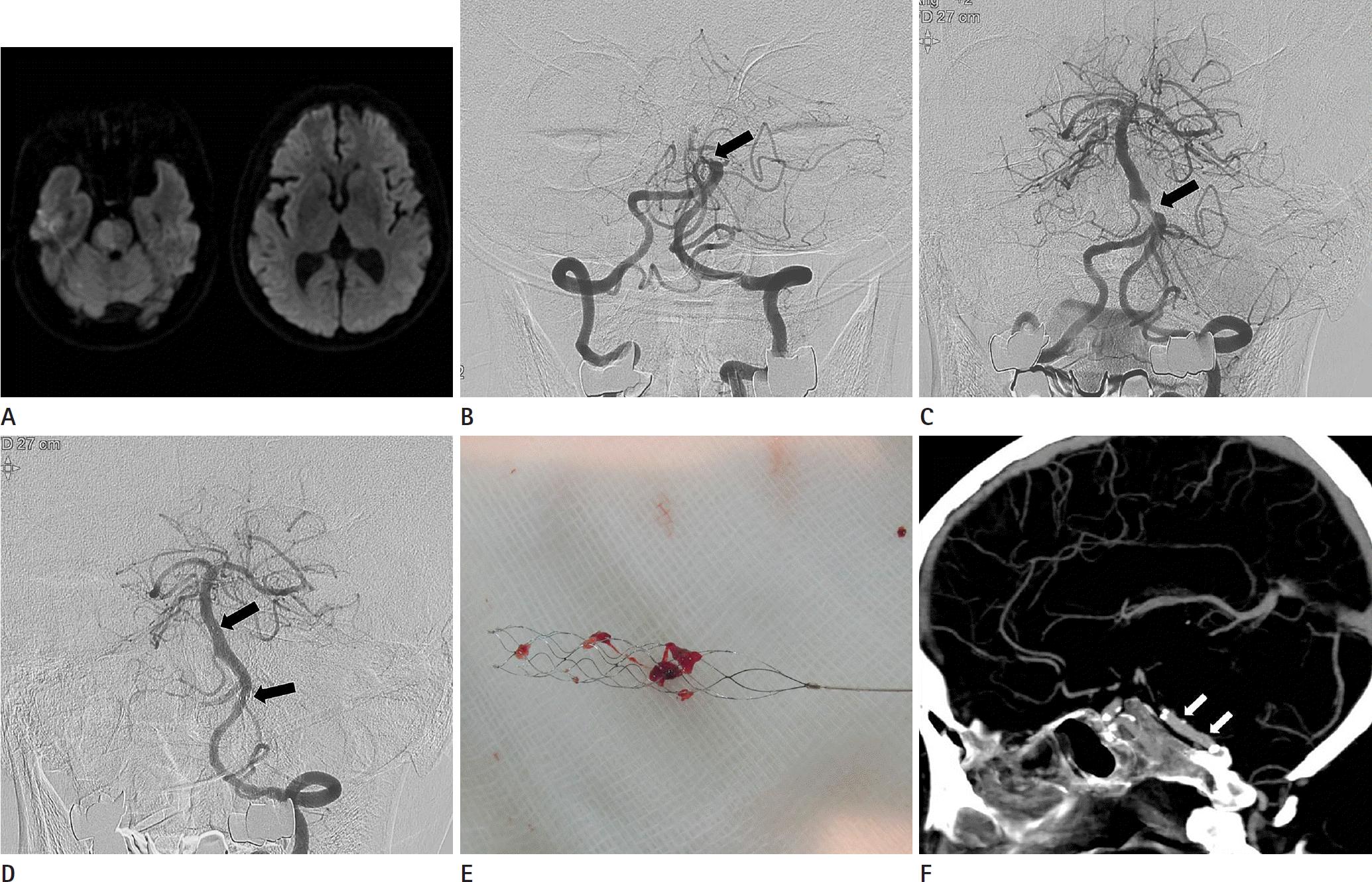 | Fig. 1.A 74-year-old patient with acute stroke due to acute basilar artery occlusion. A. DWI images show a high signal intensity lesion involving the right sided pons. There is no evidence of acute infarction in bilateral thalami. B. Left vertebral artery angiography shows an occlusion (arrow) in the proximal segment of the basilar artery. C. Angiography obtained after single passage of the stent-retriever reveals a severe underlying atherosclerotic stenosis (arrow) at the previous occlusion site. D. Left vertebral angiography obtained after intracranial angioplasty and stenting shows complete recanalization of the basilar artery with good distal perfusion. Arrows indicate the proximal and distal ends of the Wingspan stent. E. Photograph shows small fragmented thrombi retrieved with a Solitaire stent. F. Maximum-intensity-projection image of the followup CT angiography 1 week after the procedure shows good patency of the stented segment (arrows) of the basilar artery. DWI = diffusion-weighted imaging |
Table 1.
Baseline Characteristics of Study Population
Table 2.
Outcomes after Endovascular Therapy in 50 Patients with Acute Stroke due to Basilar Artery Occlusion
 XML Download