

 PDF
PDF ePub
ePub Citation
 Print
Print


INTRODUCTION
Central venous catheterization (CVC) is commonly used for hemodynamic monitoring, rapid large-volume replacement, antibiotic administration, and hemodialysis. However, inadvertent arterial puncture or arterial placement frequently occurs during insertion of a central venous catheter (1). Iatrogenic arterial injury at a non-compressible site can lead to serious complications, including fatal hemorrhage, while the catheter is removed. This complication can be treated by traditional surgical approaches or endovascular treatment. We report a rare case of inadvertent puncture of the subclavian artery during CVC that was successfully managed with a FemoSeal (St. Jude Medical Systems, Uppsala, Sweden) closure device. This study was approved by the Institutional Review Board. The requirement for obtaining informed consent was waived because of the retrospective nature of the study.
CASE REPORT
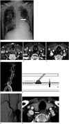
A 73-year-old man was admitted with peritonitis secondary to small bowel perforation. Following laparotomic small bowel resection, attempts were made to place a central line in the intensive care unit. A 7 French (Fr) catheter was introduced via the right subclavian approach. Post-procedural chest radiography revealed that the catheter projected to the left of the intended position, suggesting inadvertent arterial placement (Fig. 1A), as well as right pneumothorax. Following chest tube placement for pneumothorax, CT angiography (CTA) of the neck was undertaken to confirm the presence of the inadvertently inserted catheter and its relation to the subclavian vessels (Fig. 1B-E). CTA revealed that the catheter was placed in the right subclavian artery near the ostium of the right internal mammary artery, just below the origin of the vertebral artery, with the tip lying in the descending aorta. The internal jugular vein, the brachiocephalic vein and lymph nodes were not involved. No intramuscular hemorrhage was seen at the site surrounding the catheter. Also, no intraluminal thrombus or occlusion was noted within any of the great vessels arising from the aortic arch.
The catheter was left in situ without performing immediate catheter removal or direct external compression due to the concern of uncontrollable arterial bleeding that might result from the pull or pressure technique. As the patient remained hemodynamically stable, the clinical team consulted the interventional radiology team for removal of the 7 Fr catheter. Femoral artery access was gained, and an aortic arch angiogram was performed, which confirmed that the catheter was situated in the right subclavian artery with extension into the descending aorta. If covered-stent deployment was performed, there was a risk of occluding the dominant vertebral artery because the inadvertently inserted catheter was in close relation to the origin of the right vertebral artery; hence, a vascular closure device (VSD) was chosen for treatment.
A 0.035-inch guide-wire was placed through the misplaced 7 Fr central catheter and the catheter was exchanged for a 6 Fr sheath. Then, a 0.038-inch FemoSeal guide-wire was inserted through the existing 6 Fr sheath to achieve a position inside the right subclavian artery. After removing the procedural 6 Frsheath, a threaded FemoSeal unit, consisting of a sheath and a dilator, was advanced over the guide-wire into the right subclavian artery. While slowly retracting the FemoSeal Safety Catch, we removed the Safety Catch with the dilator. The intra-arterial positioning of the FemoSeal device was confirmed by visualization of the blood flow into the transparent window at the proximal portion of the sheath. At a distance, the button was pressed twice, first for the deployment of the Inner Seal and then for the Outer Locking Disc. We removed the FemoSeal Unit and applied tension to the suture as it was cut below the skin level. It took approximately 2 minutes to position and deploy the FemoSeal Unit in the subclavian artery (Fig. 1F).
Final angiography obtained immediately after the deployment of the FemoSeal device showed preservation of the major arteries, no signs of contrast medium extravasation or pseudoaneurysm, and confirmed procedural success (Fig. 1G). During his hospital stay, the patient experienced no neurological deficits. Follow-up CTA performed 3 weeks later demonstrated that the right subclavian arterial lumen was patent and there was fatty infiltration in the surrounding vessel without any vascular complications (Fig. 1H).
DISCUSSION
Insertion of a central venous catheter is a commonly performed procedure for critically ill patients in real-world practice. The increasing use of central venous catheters has been associated with a proportionate increase in the frequency of iatrogenic mechanical complications, including the formation of an arteriovenous fistula, arterial dissection or pseudoaneurysm, airway obstruction due to a cervical hematoma, shock from hemothorax, stroke from arterial thrombosis or cerebral emboli, or death (1). When a catheter has been inadvertently inserted into an artery at a compressible site, the misplacement can be easily corrected by removal and manual compression. However, if catheter removal is attempted at a non-compressible site, there is a greater propensity for serious complications such as uncontrolled hemorrhage and death.
The management of iatrogenic subclavian arterial injury is challenging because it is located at a non-compressible site. The incidence of inadvertent subclavian arterial cannulation during placement of a central venous catheter is low, accounting for 2.7–4.9% of all cases (12). It is easy to recognize this misplacement, but it is difficult to correct it because of a potentially lethal situation during removal of the catheter.
Several options have been reported for the management of inadvertent subclavian arterial cannulation. Surgical treatment usually requires partial removal of the 1st rib or a thoracotomy on the condition that the patient's vital signs are stable without the presence of coagulopathy. With recent technical and device advancements, endovascular approaches have been increasingly used and they have been reported to be successful in managing subclavian artery injury (3). Endovascular management appears to be safe, minimally invasive, and relatively inexpensive. There are various endovascular treatment options such as covered stents, VSDs, tract embolization, or gradual downsizing of transarterial catheters. The choice for these options depends on multiple factors such as location of the arterial injury.
The use of a stent-graft is a good choice for patients who have lateral subclavian arterial injury and are on antiplatelet therapy. In our case, there was a risk of occluding the dominant vertebral artery after stent deployment due to the fact that the right subclavian artery is in close relation to the origin of the right vertebral artery. Balloon angioplasty is required during management with tract embolization using Gelfoam in order to minimize the risk of Gelfoam migration into the artery, with the concern of controlling any bleeding that might occur from the injured artery. Gradual downsizing of transarterial catheters takes a long time with the need for prolonged common femoral arterial access for angiographic evaluation at the time of each downsizing procedure, and therefore, it is not appropriate for life-threatening subclavian arterial cannulation. If the length of the tract from the skin entry site to the arteriotomy is less than 5 cm, a VSD may be considered, although it is contraindicated at bifurcations and in small vessels (3).
FemoSeal is a new non-collagen, resorbable VSD that contains two resorbable polymer discs with an intra-arterial anchor (10 × 5 mm) and an extra-arterial cap (5 mm) (4), which are held together by a resorbable multifilament. The advantages of FemoSeal include a significantly lower rate of large hematoma formation and faster hemostasis compared to closure by manual compression (5). Time to hemostasis is less than 125 seconds in all cases with excellent deliverability by an experienced practitioner (6). According to Leijdekkers et al. (7), the maximum length of the tract needed to close the arteriotomy site using a VCD depends on the maximum working length of the VCD. The FemoSeal might be considered as long as the length of the tract is less than 10 cm, which is the maximum working length of a FemoSeal. In our case, the length of the tract from the skin entry site was 6 cm. Hence, we successfully used the FemoSeal.
In conclusion, endovascular management of inadvertent subclavian artery injury induced by CVC using a FemoSeal VCD may be safe and effective without the need for surgical management. These results are expected to be helpful in the management of inadvertent subclavian artery cannulation.
 XML Download