

 PDF
PDF ePub
ePub Citation
 Print
Print


INTRODUCTION
The paravertebral sympathetic ganglia are divided into 3 cervical, 11 thoracic, 5 lumbar, and 5 sacral ganglia. The superior cervical sympathetic ganglion (SCSG), which is the most cranial and largest of the 3 cervical ganglia, coordinates and transfers sympathetic outputs to specific targets in the head and neck, including the blood vessels, iris, and salivary glands (1). One cadaveric study determined that the SCSG is usually located between the internal carotid artery and the longus capitis muscle at the level of the C2 vertebra (2). The SCSG appears as an elongated oval structure connected to a thin sympathetic trunk, and has a mean width of 8.1 ± 2.8 mm and a mean length of 33.0 ± 6.2 mm. Accordingly, a hyperplastic SCSG can be large enough to mimic retropharyngeal lymph node metastasis in patients with head and neck cancer due to its location between the internal carotid artery and longus capitis muscle, just lateral to the retropharyngeal space.
Here, we describe a case of an enlarged SCSG that mimicked a retropharyngeal metastatic lymph node after surgery in a 42-year-old man diagnosed with oral tongue cancer.
CASE REPORT
A 42-year-old man, diagnosed with squamous cell carcinoma of the left lateral surface of the oral tongue, underwent a partial glossectomy with supraomohyoid neck dissection of the ipsilateral neck. Complete removal of the tumor with an adequate resection margin was achieved, and surgical staging of T1N0M0 was reported.
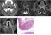
Follow-up magnetic resonance (MR) imaging was performed 6 months after surgery. An enlarged mass, measuring approximately 1.2 cm in the axial plane, which had not been identified on the preoperative CT, was observed in the left retropharyngeal area of the suprahyoid neck, medial to the internal carotid artery and lateral to the longus capitis muscle (Fig. 1). This mass exhibited high signal intensity on axial T2-weighted images, and strong contrast enhancement, mainly in the periphery, on axial contrast-enhanced T1-weighted images. In addition, dot-like fluid signal intensity was observed within the mass on axial T2-weighted images. Furthermore, a relatively poorly enhanced area, believed to be internal necrosis within the presumed metastatic lymph node, was visible at the center of the mass on axial contrast-enhanced T1-weighted images.
The retropharyngeal mass was dissected using a transoral approach, and the dissected mass was sent to the pathology department to determine whether it represented a metastatic lymph node requiring the patient to undergo additional treatment. Unexpectedly, the final histopathologic examination revealed a hyperplastic ganglion and hypertrophic nerves without evidence of malignancy. The patient developed left-sided Horner syndrome, including miosis, ptosis, and anhidrosis with ipsilateral vocal cord palsy after surgery, but symptoms improved upon further follow-up.
DISCUSSION
In the suprahyoid neck, a large percentage of the retropharyngeal space consists of retropharyngeal lymph nodes located in the far lateral aspect of the retropharyngeal space immediately medial to the internal carotid artery (3). Therefore, the primary pathologies involving this space are tumors or infections affecting the lymph nodes. These nodes are primary lymphatic drainage sites for the nasopharynx and the posterior wall of the oro-hypopharynx. The retropharyngeal space, therefore, always should be carefully examined in patients with head and neck cancer, especially since lymph node metastases involving the retropharyngeal space are often clinically occult. However, the lymph nodes may be barely enlarged in patients with oral cavity cancer (4).
In the general population, the SCSG is frequently identified as a benign retropharyngeal lymph node without careful observation on cross-sectional imaging studies. Anatomically, the cervical sympathetic chain spans along the medial margin of the carotid sheath, which is separated from the medially located retropharyngeal space (5). Therefore, it is important for radiologists to recognize that pathologies of the sympathetic chain can mimic masses arising from the retropharyngeal space due to their close proximity.
Yuen et al. (6) reported a case of enlarged SCSG after radiotherapy in a patient with nasopharyngeal carcinoma, and suggested that irradiation might contribute to hyperplasia of the cervical sympathetic ganglion. However, our patient had no history of irradiation. We believed that prior neck dissection might have influenced the growth of the ipsilateral SCSG as a compensatory reactive response after surgical injury to the complex neural plexus in the neck. Unfortunately, the exact reason for the enlargement of the cervical sympathetic ganglion was not clear in this case.
As for imaging characteristics distinguishing the SCSG from retropharyngeal pathology, both the SCSG and retropharyngeal lymph node show high signal intensity on T2-weighted MR images and avid contrast enhancement. However, pathology originating from the SCSG shows a fusiform or elongated oval shape in the longitudinal dimension in the typical location. Therefore, careful observation of the overall shape of masses in the retropharyngeal area in the coronal plane might help distinguish the SCSG from a retropharyngeal lymph node, as was seen in our case. Moreover, an internal, poorly enhanced lesion was observed in the central portion of the SCSG on axial contrast-enhanced T1-weighted MR images in our patient. A lesion such as this might be misinterpreted as necrotic or cystic changes of the metastatic lymph node in patients with head and neck cancer. Although the poorly enhanced intraganglionic lesion could not be correlated precisely with histopathologic findings, one possible explanation is that clustered intraganglionic ectatic venules might have contributed to these imaging findings, as seen in Fig. 1E. Further studies are necessary to verify this hypothesis.
In conclusion, radiologists should be aware that an enlarged SCSG can mimic a metastatic lymph node in the retropharyngeal space on cross-sectional imaging studies. Radiologists should thoroughly examine the elongated oval-shaped structure of the SCSG in the coronal plane to prevent unnecessary injury to the SCSG and avoid additional morbidity in patients with head and neck cancer.
 XML Download