

 PDF
PDF ePub
ePub Citation
 Print
Print


INTRODUCTION
Tracheal bronchus is a congenital anomaly of bronchial division. A variety of bronchial anomalies originate from the trachea or main bronchus and are directed to the upper lobe territory (1). Tracheal bronchus usually originates from the main bronchus or trachea within 2 cm from the carina, and it supplies the entire upper lobe or apical segment of the upper lobe (234). The incidence of tracheal bronchus has been reported to range from 0.001% to 2% based on a bronchoscopy, autopsy, or radio-logical study. Tracheal bronchus may also be associated with other congenital anomalies such as Down's syndrome and congenital heart disease (5). Pig bronchus is an anomaly of the entire right upper lobe bronchus arising from the trachea. Tracheal bronchus can be divided into displaced type or supernumerary type, with the displaced type being more frequent than the supernumerary type. The supernumerary bronchi may end blindly. They are called tracheal diverticulum (12345678). In tracheal bronchus, an anomaly that originates only from the trachea is called true tracheal bronchus (TTB), which has a great clinical significance. It can be easily confirmed by surgery or bronchoscopy (2). Therefore in this study, we only focused on TTB.
Recently, CT findings of TTB have been studied using multi-detector computed tomography (MDCT) with multiplanar reconstruction and three-dimensional images (3589). Most articles on CT findings of TTB have focused on its incidence, classification, or accompanying anomalies. To the best of our knowledge, studies illustrating the anatomical relationship between TTB and the surrounding structures have been rarely reported (369). Therefore, the purpose of this study was to classify TTB radiologically using multiplanar reconstruction and three-dimensional images obtained by MDCT and to evaluate the anatomical relationship between TTB and the surrounding structures. Because it is difficult to perform precise differentiation of displaced type or supernumerary type at the lung periphery only by CT, we tried a new subspecialized classification of TTB according to the existence of right upper lobe bronchus originating from the right main bronchus, and the number of segmental bronchi originating from the trachea in this study.
MATERIALS AND METHODS
This retrospective study was approved by the Institutional Review Board of our hospital. The requirement for obtaining informed patient consent was waived.
Patients
This study included 44 patients (2–80 years old, 24 males and 20 females; mean age of 51.3 years) who underwent MDCT examination between January 2010 and August 2015 at a single tertiary center in Korea.
CT Analysis
All chest CTs were performed with a 64-channel multidetector scanner (LightSpeed VCT; GE Medical Systems, Milwaukee, WI, USA) and a 256-channel multidetector scanner (Brilliance iCT; Philips Medical Systems, Cleveland, OH, USA). CT images were restored as DICOM files after being reconstructed to a slice thickness of 1.25 mm and an interval of 1.25 mm. Using three-dimensional image analysis program (Portal workstation V2.6.0.32, Philips Medical Systems), we acquired coronal views, minimum intensity projection, and three-dimensional volume rendering (VR) images. CT images were reviewed by two radiologists (K.Y.T and K.H.J) with 26 and 3 years of experience in interpreting thoracic CT in consensus. We did not formally assess interobserver agreement.
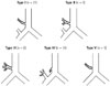
In this article, TTB was defined as an anomalous bronchus arising from the trachea and directed to the right upper lobe. TTB was classified by analyzing axial, coronal and minimum intensity projection images, multi-planar reformation images, and VR images. We classified TTB into five types (Type I to Type V) according to the existence of right upper lobe bronchus originating from the right main bronchus and the number of branches of segmental bronchus originating from TTB (Fig. 1).
We evaluated the sites of origin and running directions of TTB based on its anatomical relationship with surrounding structures. The sites of origin of TTB were classified as follows: above the aortic arch, at the level of the aortic arch, below the aortic arch, above the azygos arch, at the level of azygos arch, and below the azygos arch. The associations between the types of TTB and gender, age, or the anatomical relationships were determined. We also reviewed ancillary findings such as changes in the diameter of the trachea and main bronchus.
Statistical Analysis
The patient group for each type was too small to perform a statistical analysis of the anatomical relationship between each type and the surrounding structures. However, we were able to analyze the relationship with gender and age. We used Fisher exact test for gender and Kruskal-Wallis test for age. Statistical significance was attained when p value was less than 0.05. Type 5 was excluded from the statistical analysis because there was only one such case.
RESULTS
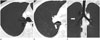
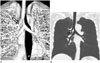
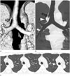
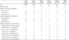
Based on the imaging classification of TTB, Type I occurred at a frequency of 47.7% (n = 21/44) (Fig. 2). Type II occurred at a frequency of 13.6% (n = 6/44) (Fig. 3). Type III occurred at a frequency of 11.4% (n = 5/44) (Fig. 4). Type IV occurred at a frequency of 25.0% (n = 11/44) (Table 1, Fig. 5). Type V occurred at a frequency of 2.0% (n = 1/44) (Fig. 6).
According to the site of origin of TTB, below the aortic arch (52.3%, n = 23/44) and at the level of the aortic arch (43.1%, n = 19/44) were the two main sites of origin. The frequency of the sites of origin above the azygos arch, at the level of the azygos arch, and below the azygos arch was 27.3% (n = 12/44), 38.6% (n = 17/44) and 34.1% (n = 15/44), respectively. In 10 out of the 11 cases of Type IV TTB, the site of origin was above the azygos arch (Fig. 5).
Considering both aortic and azygos arches, below the aortic arch and below the azygos arch were the most common sites of origin (27.3%). In Type I TTB, at the level of the aortic arch and at the level of the azygos arch were the most common sites of origin. Below the aortic arch and below the azygos arch were the most common sites of origin in Type III TTB (Fig. 4). At the level of the aortic arch and above the azygos arch were the most common sites of origin in Type IV TTB (Fig. 5).
With respect to different anatomical relationships between the running directions of TTB and the surrounding structures, in all cases, TTB passed below the azygos arch to the right upper lobe.
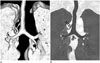
There was no statistically significant (p > 0.05) association between gender or age and the types of TTB. The diameter of the trachea was decreased in two cases (one case of Type II TTB and one case of Type III TTB) (Fig. 4). The diameter of the right main bronchus was lesser than that of the left main bronchus in two cases (one case of Type I TTB and one case of Type V TTB) (Fig. 6). One case showed luminal narrowing of the TTB and bronchiectasis at the distal portion. A highly located azygos arch above the aortic arch was observed in two cases (Fig. 2).
DISCUSSION
Tracheal bronchus, an anomaly arising from the right tracheal wall to upper lobe, was first described in 1785 (1). Although it is inappropriate anatomically, tracheal bronchus encompasses bronchial anomalies originating from the trachea or main bronchus (9). When any bronchus originates from the trachea, it is called TTB. In animals such as sheep, goats, camels, giraffes, and swans, the right upper lobe bronchus normally originates from the right trachea. The entire right upper lobe bronchus originating from the trachea is called pig bronchus. It is uncommon in humans with a frequency of 0.1% to 3%. Tracheal diverticuli vary in shape and location. They can be divided into congenital and acquired types. The supernumerary type of TTB may end blindly. It is called tracheal diverticulum in some studies. The diagnostic sensitivity of MDCT for TTB is 100% (348101112). Recognizing the presence of a tracheal anomaly is helpful while performing tracheal intubation and unilateral lung ventilation (1013). Also, most of these procedures are performed in the trachea rather than the main bronchus. In this study, we only focused on TTB since it is very significant clinically (2).
There are a few hypotheses about the pathogenesis of tracheal bronchus. Bremer (14) proposed that tracheal bronchus is caused by a failure of regression of tracheal buds in utero. He found an incidence of aberrant tracheal buds of 5% in 80 human embryos. This incidence is much higher than that in the general population. Most tracheal buds regress. Only a few tracheal buds will develop into tracheal bronchi or diverticuli. Another theory suggested that tracheal bronchus occurs as a result of disruption of normal embryogenesis (15). Alescio and Cassini (16) induced the development of tracheal buds by transplanting bronchial mesenchyme into tracheal epithelium.
In general, tracheal bronchus is subdivided into the displaced type or supernumerary type. An ectopic bronchus is supernumerary if the right upper lobe bronchus has normal trifurcation into apical, posterior, and anterior segmental bronchi. Tracheal bronchus is defined as the displaced type when the normal right upper lobar bronchus or segment is missing. In some studies, pig bronchus has been defined as a different type of TTB (51017). However, it is difficult to perform precise differentiation of the displaced type or supernumerary type at the lung periphery only by CT. Therefore, in this study, we tried another classification of TTB, according to the existence of right upper lobe bronchus originating from the right main bronchus and the number of segmental bronchi originating from the trachea. Our results revealed that Type I TTB was the most frequent (47.7%) type followed by Type IV TTB (25%). Suzuki et al. (3) reported that in 17 cases, a displaced TTB supplied all segments of the upper lobe (eight cases), apical segment (eight cases), and both apical and anterior segments (one case). TTB supplying the apical segment was classified as Type I TTB in this study. TTB supplying the apical and anterior segments was classified as Type II TTB. Pig bronchus, a TTB supplying all segments of the upper lobe, was classified as Type III. Suzuki et al. (3) reported the same frequency of Type I and III TTBs. However, in this study, Type I TTB was the most frequent. The frequencies of Type II and III TTBs were similar to each other. In this study, there was only one case of Type V TTB with one segmental bronchus originating from the trachea and absence of the right upper lobe bronchus originating from the right main bronchus. Since the right minor fissure was absent in this case, we suggested that it could be hypoplasia or lobar agenesis of the right upper lobe. To the best of our knowledge, such a case has not yet been reported.
In this study, the sites of origin of TTB were mostly below the aortic arch (52.3%) and at the level of the aortic arch (43.1%). Only two cases had sites of origin above the aortic arch. The sites of origin of TTB were not related to the location of the azygos arch. In two cases, the azygos arch was unusually highly located above the aortic arch (Fig. 2). Considering both aortic and azygos arches, below the aortic arch and below the azygos arch were the most common sites of origin.
In this study, the running direction of TTB was not related to the sites of origin. In every case, tracheal bronchus passed below the azygos arch to the right upper lobe. In utero, the trachea bifurcates at 4–6 weeks and branches progressively to form airways, while the azygos system develops generally at 6–7 weeks. Since the azygos vein starts to migrate from the lung apex after the development of tracheal bronchus, the downward migration of the azygos vein is blocked by TTB. Then, the azygos arch lies above the TTB. If TTB originates above the aortic arch, the azygos arch will also be located higher (3571819). In two cases included in this study, TTB passed below the highly located azygos arch (Fig. 2). The relationships between TTB and the surrounding structures have not yet been reported. This study provides useful information that will enable us to understand a variety of CT findings of TTB and offers a more subspecialized classification of TTB by using MDCT. This CT evaluation based on the anatomical relationship with the surrounding structures would result in increasing the success rate of surgery or intubation and decreasing the potential complications.
We could not obtain statistical information on the anatomical relationships between the types of TTB and the surrounding structures due to the small number cases of each type. Further study with more cases will be helpful to evaluate the anatomical relationship among types of TTB. Also, this more subspecialized classification could be helpful in clinical application.
There was no statistically significant association between gender or age and the types of TTB.
TTB is usually asymptomatic. Occasionally, it is the cause of relapsing cough and bronchitis as a result of retained secretion due to structural problems (3). In addition, there have been reports about bronchial stenosis, bronchiectasis, collapse, and accessory lobe related to TTB (310). Luminal narrowing of the TTB and bronchiectasis in the distal lung were found in one case included in this study, suggesting that they might have been caused by recurrent or chronic infection. Another case showed focal luminal narrowing of the trachea just proximal to the TTB. It might be associated with inflammation. In the study by Suzuki et al. (3), there was no case of tracheal stenosis. However, there were 10 cases of luminal narrowing of the TTB and there was one case of collapse with bronchiectasis of the right upper lobe. In two cases included in our study, the diameter of the right main bronchus was smaller than that of the left main bronchus (Fig. 6). Doolittle and Mair (5) reported one case of pig bronchus with luminal narrowing of the right main bronchus. They explained that right main bronchial stenosis might have occurred embryologically when the right upper lobe bronchus no longer contributes to the growth or the development of the main bronchus. In one case included in our study, distal tracheal stenosis after branching of TTB was found in a 49-year-old woman (Fig. 4) who had a history of recurrent bronchitis or chronic cough since childhood. Gower et al. (17) reported distal tracheal stenosis after branching of TTB in a 6-month-old boy who presented with cough and noisy breathing since 5 weeks of age. Distal tracheal stenosis after branching of TTB also suggests that the right upper lobe bronchus might have affected the growth of the trachea embryologically.
There were some limitations to our study. First, the classification used in this study was based on a new radiological approach. The existing classification divides TTB into the supernumerary and the displaced types. CT was unable to accurately detect the existence of normal bronchus in the peripheral potion of the lung by applying the classification proposed by Ghaye et al. (1). Second, there was a possibility of sample selection bias because we excluded patients who had only axial images. Third, although this study investigated more patients than previous studies, less than ten patients with different types of TTB led to difficulty in discussing the differences among types.
In conclusion, we tried to perform imaging classification of tracheal bronchus based on MDCT. In addition, we determined the anatomical relationships between TTB and the surrounding structures. This study will help improve our understanding of various imaging features and embryologic development of TTB. Also, the importance of evaluation of airway including the trachea should be emphasized in thoracic imaging.

 XML Download