

 PDF
PDF ePub
ePub Citation
 Print
Print


INTRODUCTION
Epidermal inclusion cysts are lined by stratified squamous epithelium that contains a granular layer and lamellated keratin. Usually, they develop due to proliferation and implantation of epidermal elements within a circumscribed space in the dermis (1). They most commonly occur on the face, scalp, skin of the neck and trunk. Epidermal inclusion cyst of the breast is an uncommon benign condition. Furthermore, epidermal inclusion cyst presenting as a large palpable mass in the male breast is an extremely rare clinical entity. To the best of our knowledge, only seven cases have been reported in the English-language literature (234), and a case of giant epidermal inclusion cyst in the male breast has not been reported in Korean literature. Here, we present the sonographic images, computed tomography (CT), and pathological findings of a giant epidermal inclusion cyst in the breast of a male patient.
CASE REPORT
A 63-year-old man presented with a growing palpable mass in the right breast of nine months' duration. The patient suffered from right breast pain. The patient was not taking any medication and he did not have any history of trauma or previous surgery. Physical examination revealed an approximately 6 × 7 cm sized mass in the upper central portion of the right breast, which appeared to be a firm, well-defined mass attached to the skin.
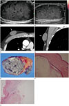
The patient subsequently underwent several studies for establishing the diagnosis. Abnormal laboratory findings were not observed and complete blood count was within the normal range, suggesting a low possibility of an infectious condition or abscess. Mammography was not performed in this patient because he did not want to undergo the procedure due to severe pain. Breast sonography (IU22 unit; Philips Medical Systems, Bothell, WA, USA) showed a 5.2 cm sized, oval shaped, circumscribed, and complex cystic and solid mass with posterior acoustic enhancement in the 12 o'clock direction of the right breast (Fig. 1A). There were some linear echogenic reflectors and filiform anechoic areas in the mass, suggestive of the pseudotestis pattern, as described later (Fig. 1B). The mass showed no vascularity on color Doppler (Fig. 1B). There was no abnormal ultrasonography (US) finding in the left breast or the axillary area. The lesion was oval, circumscribed, and had parallel orientation, which are usually benign features, but it was a growing, palpable, complex cystic and solid mass; hence, we classified the lesion into the BIRADS category 4a (Low suspicious malignancy) and recommended US-guided core-needle biopsy to exclude malignancy (5). As the patient was very anxious about the increasing size of the breast mass, surgical excision without preoperative diagnosis was planned. Before surgical excision, CT (Somatome Sensation 64; Siemens, Forchheim, Germany) was performed to identify the exact location of the lesion and its relationship to the adjacent tissue. On CT, a breast mass measuring 5.2 × 2.3 × 4.1 cm was noted (Fig. 1C, D). The lesion was a well-defined, homogeneous, low density mass without enhancement in the upper central portion of the right breast. Surgical excision was performed, and histopathological examination revealed an intradermal cyst lined by keratinizing stratified squamous epithelium with layers of desquamated keratin within the cyst (Fig. 1E, F). There were cholesterol clefts surrounded by multinucleated giant cells, lymphocytes and histiocytes, suggesting a microruptured cyst (Fig. 1G).
DISCUSSION
Epidermal inclusion cyst is a small, oval shaped, smooth, slightly compressible, cystic benign lesion of the skin. This cyst most commonly occurs on the scalp and on the skin of the neck and back, while its presentation as a large breast mass in males is very rare (23). Epidermal inclusion cysts are usually slow-growing masses ranging from a few millimeters to a few centimeters in diameter. If there is an epidermal inclusion cyst which is more than 5 cm in diameter, we consider it as a giant epidermal inclusion cyst (6). The common age group of presentation is young adult males during the third or fourth decade of life. The previously reported cases of giant epidermal inclusion cyst of the male breast showed that the cyst occurred during the second to fifth decade of life in males, while in our case the patient was 63 years old.
There is no established theory of how epidermal inclusion cysts actually develop; however, a few hypotheses on their etiology have been postulated. These include obstruction of the hair follicle, resulting in proliferation of epidermal cells within a confined area of the skin. Trauma, insect bite, surgery, reduction mammoplasty or breast augmentation, needle biopsy are also the etiological factors. It has been reported that they rarely arise after squamous metaplasia of normal columnar cells possibly found in the ductal epithelium (278). However in our case, no specific cause could be established; therefore, it appears that obstruction of the hair follicle was the cause of its origin.
Most breast epidermal inclusion cysts occur in the skin layer, and physical examination reveals a firm, well-demarcated small mass. Epidermal inclusion cysts in the breast typically appear as well-circumscribed masses with homogeneously increased density on mammography. On sonography, epidermal inclusion cysts appear as superficial well-circumscribed, oval masses, showing a complex or heterogeneous appearance. Huang et al. (9) described the features of epidermal inclusion cyst as the pseudotestis pattern, and it appears as a ovoid nodule with homogeneous low to medium echoes simulating a testicle. In their research on cysts, frequent findings of epidermal inclusion cysts are described as the pseudotestis pattern with intralesional bright echogenic reflectors (defined as strongly hyperechoic intralesional foci) and filiform anechoic areas (defined as intralesional curvilinear or branchlike hypoechoic intralesional foci with a width of < 0.3 cm). They suggested that the intralesional bright echogenic reflectors may be related to randomly distributed cholesterol, sebaceous foci, or calcifications, whereas scattered fragments of packed keratin lamellae lead to the filiform anechoic areas (9). Epidermal inclusion cysts have another specific sonographic feature of an onion skin appearance, corresponding to lamellated keratin with alternating concentric hyperechoic and hypoechoic rings. They often have extension into the dermis, contiguous to the skin, and occasionally a tract may be seen leading from the lesion toward the skin (410). In several previous case reports of giant epidermal inclusion cysts, they showed extension into the dermis, parallel linear echos (tram-track appearance), or hypoechoic mass with posterior enhancement. In our case, the lesion showed the pseudotestis pattern with echogenic reflectors, filiform anechoic areas and posterior acoustic enhancement, among the specific features described above. On CT, it appears as a small and unilocular lesion having similar attenuation to the skeletal muscle. No appreciable enhancement of the lesion should be seen. Magnetic resonance imaging shows a fluid-like high signal with variable low-signal components on T2-weighted images. Peripheral rim enhancement may be seen on gadolinium-enhanced images (7).
The diagnosis is not difficult when the epidermal inclusion cyst presents as a small nodule in the subcutaneous tissue. However, giant epidermal inclusion cyst occurring in the breast can occasionally be misdiagnosed on imaging alone. In the female breast, mammographic and sonographic images of epidermal inclusion cyst may show features that mimic other benign tumors such as fibroadenoma or phyllodes tumor, or even a malignant breast lesion with benign features such as mucinous carcinoma (1). In the male breast, differential diagnosis of a palpable mass includes benign lesions such as gynecomastia, lipoma, pseudoangiomatous stromal hyperplasia, and very rarely, fibroadenoma. Also, differentiation from malignant tumors is required (10).
Also, epidermal inclusion cysts can rupture and cause serious sequelae. Rupture releases non absorbable keratin, irritating the surrounding tissue, which can lead to secondary foreign body reactions, granulomatous reactions, or abscess formation (7). In our case, the cyst had ruptured and it showed foreign body giant cells on pathologic examination, but there was no abscess formation. Even though epidermal inclusion cysts are known to be benign, they occasionally have malignant potential to transform into squamous cell carcinoma. But, the incidence of malignant potential is extremely variable (0.045 to 19%) and the true incidence is uncertain (7).
Treatment decision depends upon the condition of the lesion. Asymptomatic small-sized lesions do not require treatment; biopsy is unnecessary, and follow-up imaging suffices if typical sonographic, mammographic and clinical findings are found. In the case of giant epidermal inclusion cyst, biopsy is obligatory to exclude malignancy. Symptomatic cases presenting with an enlarging palpable breast mass, especially when the patient is not physically and psychologically comfortable, require surgical excision (7). Entire cyst wall removal is recommended for pathologic confirmation and for prevention of recurrence, inflammation and malignant change.
In summary, although giant epidermal inclusion cyst in the male breast is extremely rare, radiologists should consider it in the differential diagnosis of a large palpable mass in the male breast.
 XML Download