

 PDF
PDF ePub
ePub Citation
 Print
Print


Abstract
Purpose
The purpose of this study was to evaluate the usefulness of MR imaging and to describe the MR imaging findings of pregnancy-associated breast cancer.
Materials and Methods
From 2006 to 2013, MR images of 23 patients with preg-nancy-associated breast cancer were retrospectively evaluated. MR images were re-viewed to evaluate lesion detection and imaging findings of pregnancy-associated breast cancer. MR images were analyzed by using the Breast Imaging Reporting and Data System and an additional MR-detected lesion with no mammographic or sonographic abnormality was determined.
Results
MR imaging depicted breast cancer in all patients, even in marked background parenchymal enhancement. Pregnancy-associated breast cancer was seen as a mass in 20 patients and as non-mass enhancement with segmental distribution in 3 patients. The most common features of the masses were irregular shape (85%), non-circumscribed margin (85%), and heterogeneous enhancement (60%). An additional site of cancer was detected with MR imaging in 5 patients (21.7%) and the type of surgery was changed.
Go to : 
Index terms
Breast Neoplasm, Pregnancy Complications, Neoplastic, Lactation Magnetic Resonance ImagingREFERENCES
1. Anderson JM. Mammary cancers and pregnancy. Br Med J. 1979; 1:1124–1127.

2. DiFronzo LA, O'Connell TX. Breast cancer in pregnancy and lactation. Surg Clin North Am. 1996; 76:267–278.
3. Liberman L, Giess CS, Dershaw DD, Deutch BM, Petrek JA. Imaging of pregnancy-associated breast cancer. Radiology. 1994; 191:245–248.
4. Gemignani ML, Petrek JA. Pregnancy-associated breast cancer: diagnosis and treatment. Breast J. 2000; 6:68–73.
5. Hogge JP, De Paredes ES, Magnant CM, Lage J. Imaging and management of breast masses during pregnancy and lactation. Breast J. 1999; 5:272–283.
6. Andersson TM, Johansson AL, Hsieh CC, Cnattingius S, Lambe M. Increasing incidence of pregnancy-associated breast cancer in Sweden. Obstet Gynecol. 2009; 114:568–572.
7. Beadle BM, Woodward WA, Middleton LP, Tereffe W, Strom EA, Litton JK, et al. The impact of pregnancy on breast cancer outcomes in women<or=35 years. Cancer. 2009; 115:1174–1184.
8. Sickles EA, D'Orsi CJ, Bassett LW, Appleton CM, Berg WA, Burnside ES, et al. ACR BI-RADS® mammography. In American College of Radiology. ACR BI-RADS® atlas: breast imaging reporting and data system. Reston, VA: American College of Radiology. 2013; 13–120.
9. Mendelson EB, Böhm-Vélez M, Berg WA, Whitman GJ, Feldman MI, Madjar H, et al. ACR BI-RADS® ultrasound. In American College of Radiology. ACR BI-RADS® atlas: breast imaging reporting and data system. Reston, VA: American College of Radiology. 2013; 35–120.
10. Morris EA, Comstock C, Lee C, Lehman CD, Ikeda DM, Newstead GM, et al. ACR BI-RADS® magnetic resonance imaging. In American College of Radiology. ACR BI-RADS® atlas: breast imaging reporting and data system. Reston, VA: American College of Radiology. 2013; 23–124.
11. Liberman L, Morris EA, Dershaw DD, Abramson AF, Tan LK. MR imaging of the ipsilateral breast in women with percutaneously proven breast cancer. AJR Am J Roentgenol. 2003; 180:901–910.
12. Wallack MK, Wolf JA Jr, Bedwinek J, Denes AE, Glasgow G, Kumar B, et al. Gestational carcinoma of the female breast. Curr Probl Cancer. 1983; 7:1–58.
13. Barnes DM, Newman LA. Pregnancy-associated breast cancer: a literature review. Surg Clin North Am. 2007; 87:417–430. x.
14. Ahn BY, Kim HH, Moon WK, Pisano ED, Kim HS, Cha ES, et al. Pregnancy- and lactation-associated breast cancer: mammographic and sonographic findings. J Ultrasound Med. 2003; 22:491–497. quiz 498-499.
15. Yang WT, Dryden MJ, Gwyn K, Whitman GJ, Theriault R. Imaging of breast cancer diagnosed and treated with che-motherapy during pregnancy. Radiology. 2006; 239:52–60.
16. Foxcroft LM, Evans EB, Porter AJ. The diagnosis of breast cancer in women younger than 40. Breast. 2004; 13:297–306.
17. Webb JA, Thomsen HS, Morcos SK. Members of Contrast Media Safety Committee of European Society of Urogeni-tal Radiology (ESUR). The use of iodinated and gadolinium contrast media during pregnancy and lactation. Eur Radiol. 2005; 15:1234–1240.
18. American College of Radiology. ACR manual on contrastmedia. Web site.http://www.acr.org/~/media/ACR/Documents/PDF/QualitySafety/Resources/Contrast%20Manual/2016_Contrast_Media.pdf/#page=105. Accessed December 22,. 2015.
19. Talele AC, Slanetz PJ, Edmister WB, Yeh ED, Kopans DB. The lactating breast: MRI findings and literature review. Breast J. 2003; 9:237–240.
20. Espinosa LA, Daniel BL, Vidarsson L, Zakhour M, Ikeda DM, Herfkens RJ. The lactating breast: contrast-enhanced MR imaging of normal tissue and cancer. Radiology. 2005; 237:429–436.
21. Lehman CD, Gatsonis C, Kuhl CK, Hendrick RE, Pisano ED, Hanna L, et al. MRI evaluation of the contralateral breast in women with recently diagnosed breast cancer. N Engl J Med. 2007; 356:1295–1303.
22. Holland R, Veling SH, Mravunac M, Hendriks JH. Histologic multifocality of Tis, T1-2 breast carcinomas. Implications for clinical trials of breast-conserving surgery. Cancer. 1985; 56:979–990.
23. Taylor D, Lazberger J, Ives A, Wylie E, Saunders C. Reducing delay in the diagnosis of pregnancy-associated breast cancer: how imaging can help us. J Med Imaging Radiat Oncol. 2011; 55:33–42.
Go to :
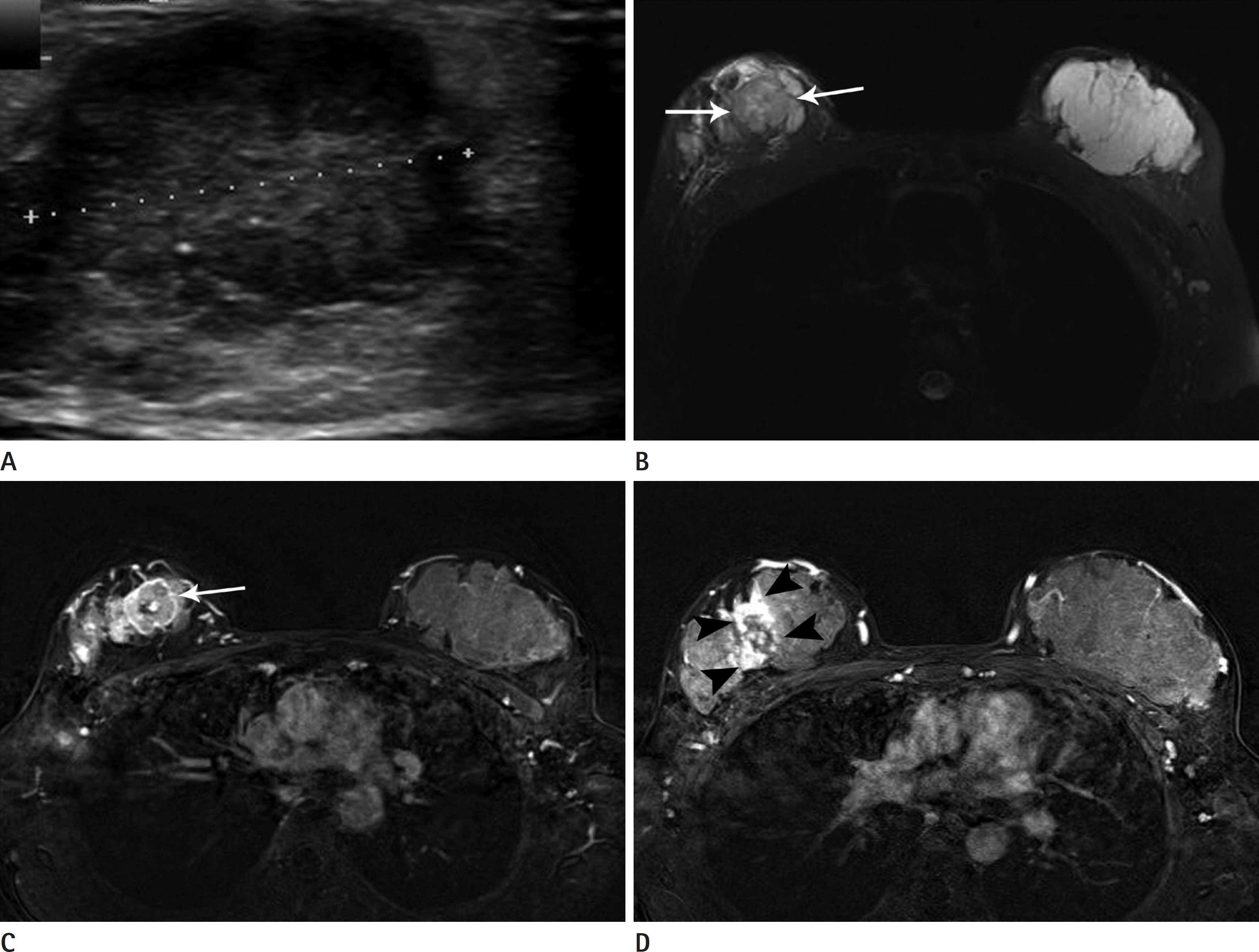 | Fig. 1.A 33-year-old lactating woman with invasive carcinoma of no special type in the right breast. A. Ultrasound image shows an irregular hypoechoic mass in the palpable area of the right breast. An invasive ductal carcinoma was confirmed by an ultrasound-guided core needle biopsy. B. An axial fat-suppressed T2-weighted fast spin-echo MR image shows a large mass with low signal intensity (arrows). Note the extreme fibroglandular tissue and high signal intensity of the contralateral normal lactating breast. C, D. An axial contrast-enhanced fat-suppressed subtraction T1-weighted MR image shows a rim-enhancing mass (arrow in C) in the upper right breast. In addition, segmental non-mass enhancement is seen in the upper outer right breast (arrowheads in D) that extends more than 4.0 cm, which was confirmed to be ductal carcinoma in situ on pathologic examination. Note the marked background parenchymal enhancement of the contralateral breast. |
Table 1.
Histopathologic Characteristics
| Characteristics | Number of Lesions (n = 23) | % |
|---|---|---|
| Histologic type | ||
| Invasive carcinoma NST | 18 | 78.3 |
| DCIS | 2 | 8.7 |
| Medullary carcinoma | 2 | 8.7 |
| Metaplastic carcinoma | 1 | 4.3 |
| Pathological tumor size† | ||
| Tis | 1 | 4.3 |
| T1 | 11 | 47.8 |
| T2 | 8 | 34.8 |
| T3 | 1 | 4.3 |
| Pathological nodal status† | ||
| Positive | 9 | 39.1 |
| Negative | 12 | 52.2 |
| ER status∗ | ||
| Positive | 11 | 47.8 |
| Negative | 11 | 47.8 |
| PR status∗ | ||
| Positive | 7 | 30.4 |
| Negative | 15 | 65.2 |
| HER-2 status∗ | ||
| Positive | 6 | 26.1 |
| Negative | 16 | 69.6 |
| Molecular subtype∗ | ||
| Luminal A | 8 | 34.8 |
| Luminal B | 3 | 13.0 |
| HER-2 | 3 | 13.0 |
| Triple-negative | 8 | 34.8 |
Table 2.
Sonographic Findings of Pregnancy-Associated Breast Cancer
Table 3.
Breast Parenchymal Features on MRI
| Features | Number of Lesions (n = 23) | % |
|---|---|---|
| Amount of FGT | ||
| Almost entirely fat | 0 | 0 |
| Scattered | 1 | 4.4 |
| Heterogeneous | 7 | 30.4 |
| Extreme | 15 | 65.2 |
| BPE | ||
| Minimal | 2 | 8.7 |
| Mild | 4 | 17.4 |
| Moderate Marked | 4 | 17.4 |
| Marked | 13 | 56.5 |
Table 4.
MR Imaging Findings of Pregnancy-Associated Breast Cancer
| Findings | Number of Lesions (n = 23) | % |
|---|---|---|
| Mass | 20 | 87.0 |
| Shape | ||
| Oval/round | 3 | 15.0 |
| Irregular | 17 | 85.0 |
| Margin | ||
| Circumscribed | 3 | 15.0 |
| Not circumscribed | 17 | 85.0 |
| Internal enhancement | ||
| Homogeneous | 2 | 10.0 |
| Heterogeneous | 12 | 60.0 |
| Rim | 6 | 30.0 |
| Non-mass enhancement | 3 | 13.0 |
| Kinetic curve∗ | ||
| Initial phase | ||
| Fast | 22 | 100 |
| Delayed phase | ||
| Persistent | 0 | 0 |
| Plateau | 6 | 27.3 |
| Washout | 16 | 72.7 |
 XML Download