

 PDF
PDF ePub
ePub Citation
 Print
Print


INTRODUCTION
Multiple myeloma (MM), or plasma cell myeloma, is a debilitating malignancy. It is characterized by proliferation of malignant plasma cells and subsequent production of a large amount of specific immunoglobulin. The clinical presentation may vary from asymptomatic to severely symptomatic, with complications that require emergent treatment. Aggressive myeloma, which invades organs other than the bone marrow, lymph notes, or reticuloendothelial system (1), is treated with high-dose chemotherapy and total body irradiation, although the prognosis is very poor. Of the extramedullary involvements of MM, peritoneal involvement with ascites is extremely rare and very few cases have been reported (23). Here, we report a case of myelomatous ascites detected on abdominal computed tomography (CT) as an initial presentation of extramedullary involvement of MM, which progressed to multiple extramedullary sites, including the testis, bowel, and retroperitoneum.
CASE REPORT
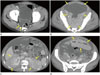
A 39-year-old male was admitted for fatigue and tremor with swelling of both legs. His past medical history and family history were unremarkable. Laboratory finding on presentation revealed pancytopenia: hemoglobin of 3.7 g/dL, hematocrit of 12.5%, white blood cell count of 3.78 × 10⁹/L (granulocytes 42%, lymphocytes 33%, monocytes 12%, basophils 1%, and eosinophils 1%), and platelet could of 66 × 109/L. His serum sodium, potassium, calcium, blood urea nitrogen, and creatinine levels were normal. Other laboratory rest results were as follows: total protein of 6.4 g/dL (normal, 6.6–8.0), albumin of 3.3 g/dL (normal, 3.3–5.2), and total bilirubin of 1.40 mg/dL (normal, 0.2–1.2). Alkaline phosphatase and other liver function tests were normal. Parameters involving connective tissue disorders, with ANA, anti dsDNA, c-ANCA, p-ANCA, and all other evaluated parameters were normal. Quantitative serum immunoglobulin levels were also normal: IgG < 300 mg/dL (normal, 408–1788), IgA 19 mg/dL (normal, 70–400), and IgM < 40 mg/dL (40–230). Plasma protein electrophoresis showed an M band in the gamma region. A serum immunoassay was positive for monoclonal gammopathy in the lambda region. His free kappa chain concentration was 8.10 mg/L (normal, 3.3–19.4), free lambda chain concentration was 5460 mg/L (normal, 5.71–26.3), and the ratio of free kappa to lambda was 0.00 (normal, 0.26–1.65). A urinary Bence-Jones protein test was positive. A bone marrow biopsy showed near 100% cellularity with diffuse infiltration by plasma cells (Fig. 1A). The plasma cells showed moderate nuclear pleomorphism, eccentric nuclei, abundant amphophilic cytoplasm, and occasional prominent nucleoli. Immunohistochemical stains for kappa and lambda light chain revealed lambda monoclonality, consistent with plasma cell myeloma. Initial CT of the abdomen taken to investigate the leg swelling revealed a small number of ascites with mild peritoneal thickening. Perfusion of both kidneys seemed to be normal, without focal lesions (Fig. 2A). Based on these findings, the patient was diagnosed with MM and began chemotherapy with bortezomib and dexamethasone. While being treated, his pancytopenia worsened as a possible side effect of the drugs. Therefore, the medication was stopped. About one month after discontinuing the medication, the patient was readmitted for abdominal distension with right scrotal pain. Contrast-enhanced abdominal CT showed a marked increased in the number of ascites with irregular thickening of the peritoneum as well as infiltrative perinephric soft tissue attenuation encasing both kidneys and ureters. Homogeneously enhancing mass-like wall thickening of small bowel loops and enhancing soft tissue lesions abutting the thickened diaphragm were also detected on the CT scan (Fig. 2B-D). A cytologic examination of ascitic fluid revealed numerous malignant-appearing plasma cells, consistent with myelomatous involvement (Fig. 1B). About five months after the initial detection of ascites on the CT scan, the patient expired.
DISCUSSION
This is a rare case presentation regarding a patient with MM that initially presented as myelomatous ascites with extramedullary manifestation and finally progressed to multiple extramedullary involvement of tumors. Approximately 70% of patients with plasma cell tumors have tumor cell infiltration outside the bone marrow (4). The most common sites for extramedullary involvement are the upper respiratory tract and liver. Ascites in myeloma patients are usually secondary to extensive liver infiltration by plasma cells. Other complication or comorbidities such as heart failure, renal damage, and infectious peritonitis can also contribute to the formation of ascites. In contrast, myelomatous ascites caused by peritoneal infiltration of tumor cells are very rarely encountered in patients with MM (56). They can occur either at presentation or, more frequently, develop during disease progression.
Several cases of myelomatous ascites and pleural effusion have been reported. These are considered initial symptoms of aggressive myeloma or late findings during the natural history of MM. Pleural plasmacytic effusions are more frequently reported and are occasionally described as a first sign of disease, in contrast to myelomatous ascites. As described above, myelomatous ascites and pleural effusion are highly associated with poor prognosis (57). Karp and Shareef (6) have reported that the median survival after the development of ascites was only 1.5 months in a total of 9 patients. Despite the poor prognosis, several cases of successful treatment have been reported (89). In order to increase patient survival with early intensive treatment, prompt diagnosis of myelomatous ascites is crucial. If there are newly detected ascites during the follow-up period in a patient with MM, the radiologist should consider the possibility of peritoneal infiltration by tumor cells and recommend cytologic examination of the ascites before the disease progresses further.
While the IgA isotype makes up one quarter of MM cases, 62.5% of patients with myelomatous ascites have IgA isotype myeloma. Higher plasma IgA levels are associated with extramedullary myeloma and high tumor burden (10). Lake et al. (10) have surmised that the cause is a higher concentration of IgA-producing lymphoid cells in cases of myeloma, which can disproportionally affect extramedullary sites. However, despite extensive extramedullary infiltration of tumor cells, the serum IgA level in our patient was within the normal range.
Whereas the imaging findings of peritoneal lymphomatosis and lymphoma involvement of intraabdominal solid organs have frequently been reported, those of peritoneal plasmacytoma and extramedullary intraabdominal involvement of MM have rarely been reported from a radiological point of view. However, as in our case, radiologic findings of extramedullary involvement of the peritoneum or intraabdominal organs by MM could closely resemble those conditions, and the underlying disease would be the only clue for differentiating between these two disease entities in such cases.
In conclusion, we report a case of myelomatous ascites with an initial extramedullary presentation of MM. Radiologists should be familiar with myelomatous ascites and should keep in mind the possibility of this disease entity in at-risk patients.

 XML Download