

 PDF
PDF ePub
ePub Citation
 Print
Print


Abstract
Purpose
Using standard mammographic phantom images, we compared the image quality obtained between a mammography dedicated 5 megapixel monitor (5M) and a UHD 4K (4K) monitor with digital imaging and communications in medicine display, to investigate the possibility of clinical application of 4K monitors.
Materials and Methods
Three different exposures (autoexposure, overexposure and underexposure) images of mammographic phantom were obtained, and six radiologists independently evaluated the images in 5M and 4K without image modulation, by scoring of fibers, groups of specks and masses within the phantom image. The mean score of each object on both monitors was independently analyzed, using t-test and interobserver reliability by intraclass correlation coefficient (ICC) of SPSS.
Results
The overall mean scores of fiber, group of specks, and mass in 5M were 4.25, 3.92, and 3.28 respectively, and scores obtained in 4K monitor were 3.81, 3.58, and 3.14, respectively. No statistical difference was seen in scores of fiber and mass between the two monitors at all exposure conditions, but the score of group of specks in 4K was statistically lower in the overall (p = 0.0492) and in underexposure conditions (p = 0.012). The ICC for interobserver reliability was excellent (0.874).
Conclusion
Our study suggests that since the mammographic phantom images are appropriate with no significant difference in image quality observed between the two monitors, the 4K monitor could be used for clinical studies. Since this is a small preliminary study using phantom images, the result may differ in actual mammographic images, and subsequent investigation with clinical mammographic images is required.
Figures and Tables
Fig. 1
ACR mammographic phantom image composed of fibers, groups of specks and masses. ACR = American College of Radiology
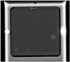
Fig. 2
Bar graph comparing object scores of mammographic phantom images between 5M and 4K monitors, according to different exposure conditions. Difference of mean scores of each object between 5M and 4K monitors are not significant (p > 0.05) in auto and overexposure conditions, but the score of group of specs in underexposure condition is significantly lower in 4K than in 5M monitor (p ≤ 0.012, asterisk). Auto = autoexposure, Over = overexposure, Under = underexposure, 5M = mammography dedicated 5 megapixel monitor, 4K = UHD 4K monitor
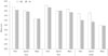
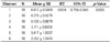
Table 1
Specification of 5M and 4K Monitors

Table 2
Overall Results of Comparison of Mammographic Phantom Images between 5M and 4K Monitors
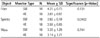
| Object | Monitor Type | N | Mean ± SD | Significance (p-Value) |
|---|---|---|---|---|
| Fiber | 5M | 18 | 4.25 ± 0.73 | 0.131 |
| 4K | 18 | 3.81 ± 0.97 | ||
| Specks | 5M | 18 | 3.92 ± 0.19 | 0.0492 |
| 4K | 18 | 3.58 ± 0.65 | ||
| Mass | 5M | 18 | 3.28 ± 1.29 | 0.741 |
| 4K | 18 | 3.14 ± 1.21 |
References
1. Lee EH, Park B, Kim NS, Seo HJ, Ko KL, Min JW, et al. The Korean guideline for breast cancer screening. J Korean Med Assoc. 2015; 58:408–419.
2. Kanal KM, Krupinski E, Berns EA, Geiser WR, Karellas A, Mainiero MB, et al. ACR-AAPM-SIIM practice guideline for determinants of image quality in digital mammography. J Digit Imaging. 2013; 26:10–25.
3. Kamitani T, Yabuuchi H, Soeda H, Matsuo Y, Okafuji T, Sakai S, et al. Detection of masses and microcalcifications of breast cancer on digital mammograms: comparison among hard-copy film, 3-megapixel liquid crystal display (LCD) monitors and 5-megapixel LCD monitors: an observer performance study. Eur Radiol. 2007; 17:1365–1371.
4. Yamada T, Suzuki A, Uchiyama N, Ohuchi N, Takahashi S. Diagnostic performance of detecting breast cancer on computed radiographic (CR) mammograms: comparison of hard copy film, 3-megapixel liquid-crystal-display (LCD) monitor and 5-megapixel LCD monitor. Eur Radiol. 2008; 18:2363–2369.
5. Kamitani T, Yabuuchi H, Matsuo Y, Setoguchi T, Sakai S, Okafuji T, et al. Diagnostic performance in differentiation of breast lesion on digital mammograms: comparison among hard-copy film, 3-megapixel LCD monitor, and 5-megapixel LCD monitor. Clin Imaging. 2011; 35:341–345.
6. Ong AH, Pitman AG, Tan SY, Gledhill S, Hennessy O, Lui B, et al. Comparison of 3MP medical-grade to 1MP office-grade LCD monitors in mammographic diagnostic and perceptual performance. J Med Imaging Radiat Oncol. 2011; 55:153–162.
7. Geijer H, Geijer M, Forsberg L, Kheddache S, Sund P. Comparison of color LCD and medical-grade monochrome LCD displays in diagnostic radiology. J Digit Imaging. 2007; 20:114–121.
8. Wu J, Wu TH, Han RP, Chang SJ, Shih CT, Sun JY, et al. Comparison of the commercial color LCD and the medical monochrome LCD using randomized object test patterns. PLoS One. 2012; 7:e37769.
9. Chen Y, James JJ, Turnbull AE, Gale AG. The use of lower resolution viewing devices for mammographic interpretation: implications for education and training. Eur Radiol. 2015; 25:3003–3008.
10. Diagnostic Display Monitor Management & QC. 2008. p. 75–81.
11. Son GG, Sung DW, Jeong JH, Kang HD, Lee JR, Jung HK. A study on the usefulness of the AAPM TG18 evaluation tool for diagnostic monitor QC. J Korean Radiol Soc. 2008; 58:631–638.
12. Song SE, Seo BK, Yie A, Ku BK, Kim HY, Cho KR, et al. Which phantom is better for assessing the image quality in full-field digital mammography?: American College of Radiology accreditation phantom versus digital mammography accreditation phantom. Korean J Radiol. 2012; 13:776–783.
 XML Download