

 PDF
PDF ePub
ePub Citation
 Print
Print


Abstract
Periurethral bulking agent injection (or transurethral submucosal injection) is a comparatively less invasive procedure for the treatment of stress urinary incontinence in patients who develop incontinence after radical prostatectomy, and who are more frequently being treated with transurethral submucosal injection. However, as the radiologic findings of bulking agents are not very well known, they can be mistaken for local recurrence in prostate cancer patients who have undergone prostatectomy. Unlike some of the literatures, in which the radiologic features of collagen injections have been reported, the radiologic findings of silicone injections are yet to be determined. Thus, it is our intention to report this case along with the lit-erature review as the authors have experienced an actual case of a silicone injection mistaken as local recurrence.
Index terms
Prostatic Neoplasms, Urinary Incontinence, Stress, Urinary Sphincter, ArtificialREFERENCES
1. King T, Almallah YZ. Post-radical-prostatectomy urinary incontinence: the management of concomitant bladder neck contracture. Adv Urol. 2012; 2012:295798.

2. Maki DD, Banner MP, Ramchandani P, Stolpen A, Rovner ES, Wein AJ. Injected periurethral collagen for postprosta-tectomy urinary incontinence: MR and CT appearance. Abdom Imaging. 2000; 25:658–662.
3. Kumar D, Kaufman MR, Dmochowski RR. Case reports: periurethral bulking agents and presumed urethral diver-ticula. Int Urogynecol J. 2011; 22:1039–1043.
4. Bridges MD, Petrou SP, Lightner DJ. Urethral bulking agents: imaging review. AJR Am J Roentgenol. 2005; 185:257–264.
5. Yablon CM, Banner MP, Ramchandani P, Rovner ES. Com-plications of prostate cancer treatment: spectrum of imaging findings. Radiographics. 2004; 24(Suppl 1):S181–S194.
6. Doherty R, Almallah Z. Urinary incontinence after treatment for prostate cancer. BMJ. 2011; 343:d6298.
7. Shekarriz B, Upadhyay J, Wood DP. Intraoperative, periop-erative, and longterm complications of radical prostatectomy. Urol Clin North Am. 2001; 28:639–653.
8. Tamanini JT, D'Ancona CA, Tadini V, Netto NR Jr. Macro-plastique implantation system for the treatment of female stress urinary incontinence. J Urol. 2003; 169:2229–2233.
9. Defreitas GA, Wilson TS, Zimmern PE, Forte TB. Three-di-mensional ultrasonography: an objective outcome tool to assess collagen distribution in women with stress urinary incontinence. Urology. 2003; 62:232–236.
10. Sella T, Schwartz LH, Swindle PW, Onyebuchi CN, Scardino PT, Scher HI, et al. Suspected local recurrence after radical prostatectomy: endorectal coil MR imaging. Radiology. 2004; 231:379–385.
Fig. 1.
CT and MR images of a 64-year-old male with prostate cancer treated by radical prostatectomy. A, B. Axial CT images show well-defined, peripheral enhancing nodular lesions around the vesicourethral anastomosis (arrows). C, D. Coronal CT images show well-defined, peripheral enhancing nodular lesions around the vesicourethral anastomosis (arrows). E, F. T2-weighted oblique axial images of prostate MRI show a relatively well-defined nodular lesion with irregular thick walls and internal contents with high signal intensity (arrows).

Fig. 1.
CT and MR images of a 64-year-old male with prostate cancer treated by radical prostatectomy. G, H. T2-weighted coronal images of prostate MRI show a relatively well-defined nodular lesion with irregular thick walls and internal contents with high signal intensity (arrows). I-K. T2-weighted sagittal images of prostate MRI show a relatively well-defined nodular lesion with irregular thick walls and internal contents with high signal intensity (arrows).

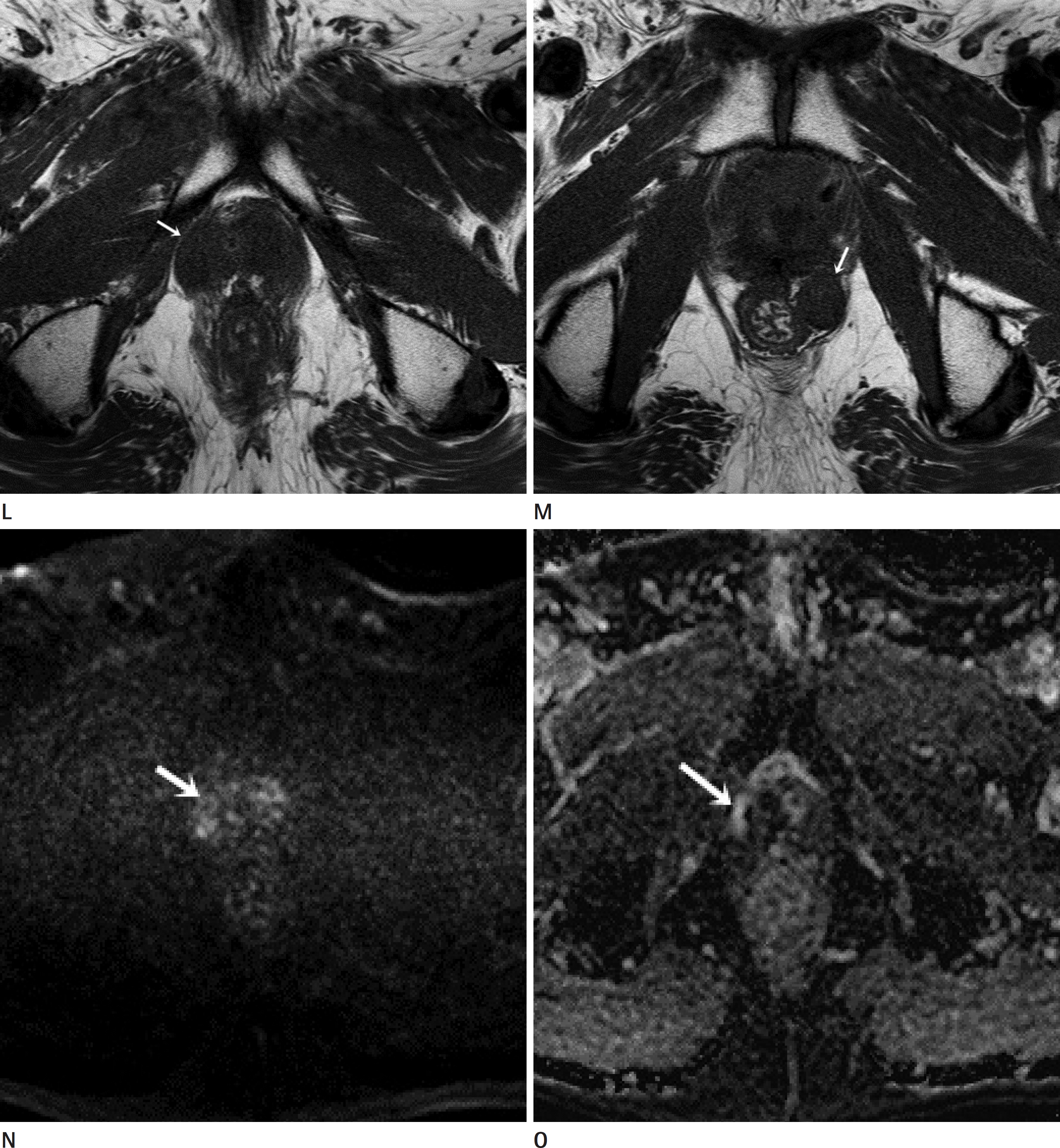
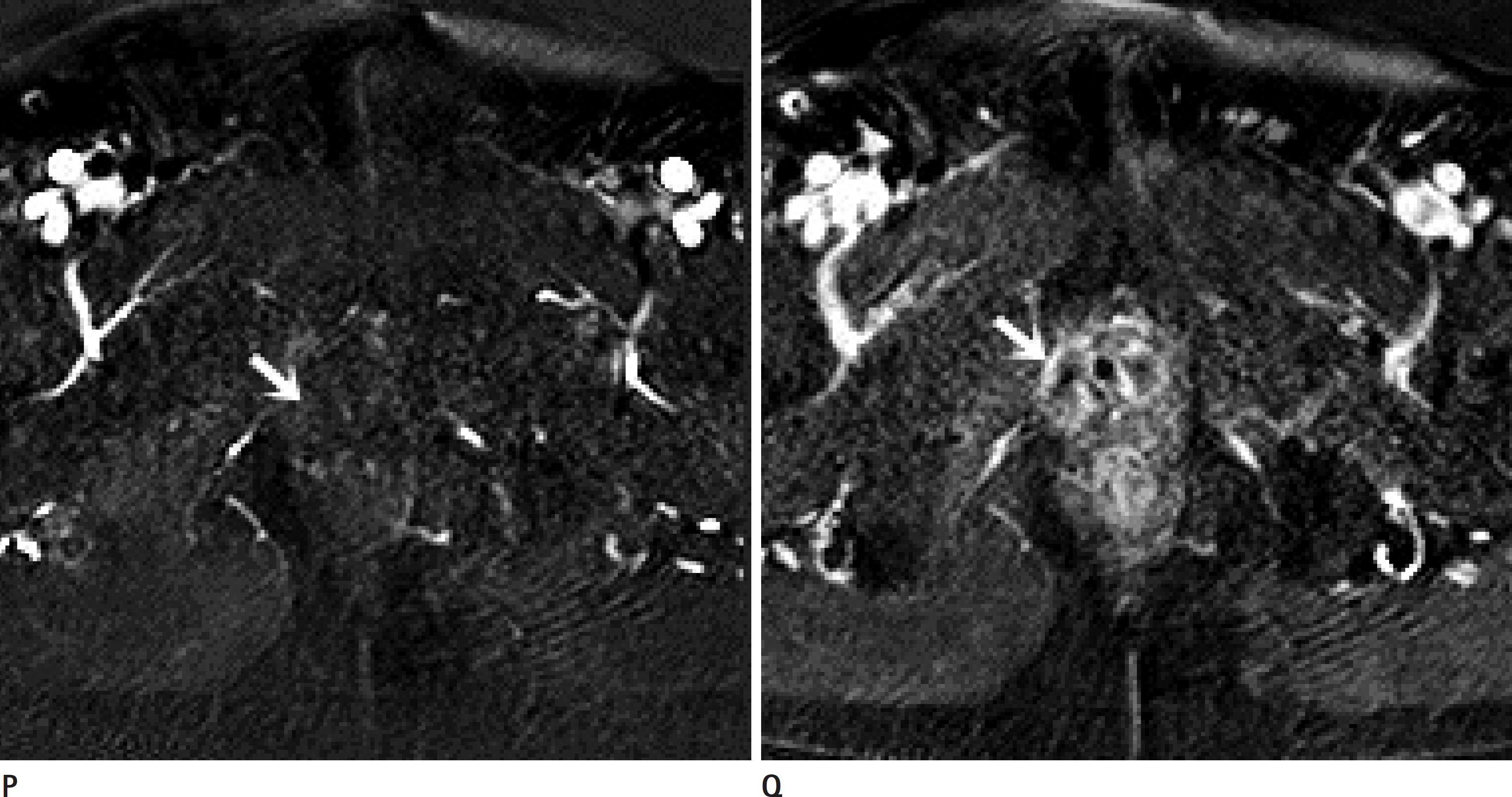
Fig. 1.
CT and MR images of a 64-year-old male with prostate cancer treated by radical prostatectomy. L, M. On T1-weighted oblique axial images, the nodular lesions show homogeneous iso to low signal intensity (arrows). N, O. Diffusion weighted images show no evidence of diffusion restriction (arrows).
 XML Download